

Mechanism of the chromosome-induced polar body extrusion in mouse eggs
- PMID: 21867530
- PMCID: PMC3179692
- DOI: 10.1186/1747-1028-6-17
Mechanism of the chromosome-induced polar body extrusion in mouse eggs
Abstract
Background: An oocyte undergoes two rounds of asymmetric division to generate a haploid gamete and two small polar bodies designed for apoptosis. Chromosomes play important roles in specifying the asymmetric meiotic divisions in the oocytes but the underlying mechanism is poorly understood.
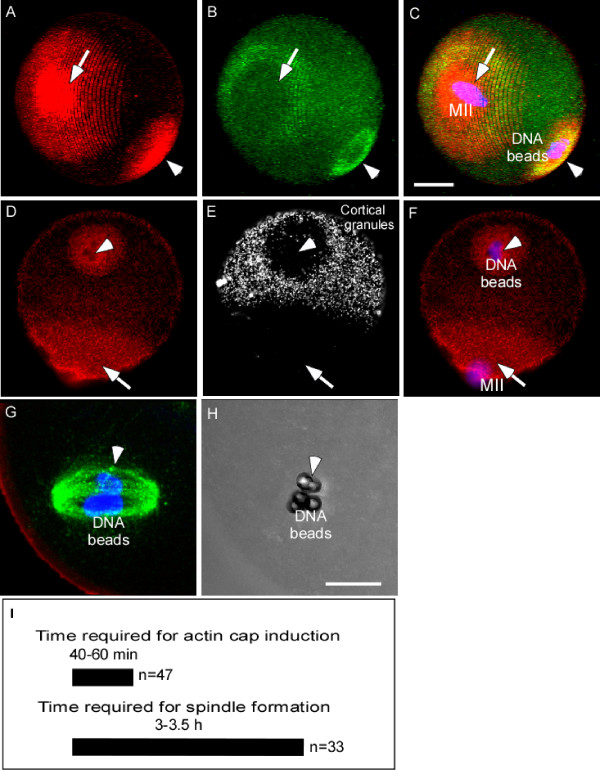
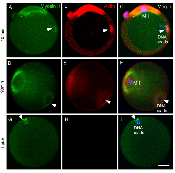
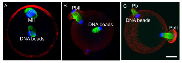
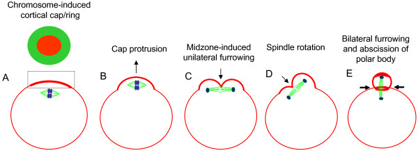
Results: Chromosomes independently induce spindle formation and cortical actomyosin assembly into special cap and ring structures in the cortex of the oocyte. The spindle and the cortical cap/ring interact to generate mechanical forces, leading to polar body extrusion. Two distinct force-driven membrane changes were observed during 2nd polar body extrusion: a protrusion of the cortical cap and a membrane invagination induced by an anaphase spindle midzone. The cortical cap protrusion and invagination help rotate the spindle perpendicularly so that the spindle midzone can induce bilateral furrows at the shoulder of the protruding cap, leading to an abscission of the polar body. It is interesting to note that while the mitotic spindle midzone induces bilateral furrowing, leading to efficient symmetric division in the zygote, the meiotic spindle midzone induced cytokinetic furrowing only locally.
Conclusions: Distinct forces driving cortical cap protrusion and membrane invagination are involved in spindle rotation and polar body extrusion during meiosis II in mouse oocytes.
Figures

Similar articles
-
Anillin localization suggests distinct mechanisms of division plane specification in mouse oogenic meiosis I and II.Gene Expr Patterns. 2015 Mar;17(2):98-106. doi: 10.1016/j.gep.2015.03.002. Epub 2015 Mar 26. Gene Expr Patterns. 2015. PMID: 25818309 Free PMC article.
-
Sperm chromatin-induced ectopic polar body extrusion in mouse eggs after ICSI and delayed egg activation.PLoS One. 2009 Sep 29;4(9):e7171. doi: 10.1371/journal.pone.0007171. PLoS One. 2009. PMID: 19787051 Free PMC article.
-
Role of PB1 Midbody Remnant Creating Tethered Polar Bodies during Meiosis II.Genes (Basel). 2020 Nov 24;11(12):1394. doi: 10.3390/genes11121394. Genes (Basel). 2020. PMID: 33255457 Free PMC article.
-
Mechanisms that prevent catastrophic interactions between paternal chromosomes and the oocyte meiotic spindle.Cell Cycle. 2018;17(5):529-534. doi: 10.1080/15384101.2018.1431495. Epub 2018 Feb 22. Cell Cycle. 2018. PMID: 29375006 Free PMC article. Review.
-
Actin cytoskeleton in cell polarity and asymmetric division during mouse oocyte maturation.Cytoskeleton (Hoboken). 2012 Oct;69(10):727-37. doi: 10.1002/cm.21048. Epub 2012 Aug 13. Cytoskeleton (Hoboken). 2012. PMID: 22753278 Review.
Cited by
-
RhoA- and Cdc42-induced antagonistic forces underlie symmetry breaking and spindle rotation in mouse oocytes.PLoS Biol. 2021 Sep 7;19(9):e3001376. doi: 10.1371/journal.pbio.3001376. eCollection 2021 Sep. PLoS Biol. 2021. PMID: 34491981 Free PMC article.
-
Unilateral Cleavage Furrows in Multinucleate Cells.Cells. 2020 Jun 18;9(6):1493. doi: 10.3390/cells9061493. Cells. 2020. PMID: 32570994 Free PMC article.
-
Anillin localization suggests distinct mechanisms of division plane specification in mouse oogenic meiosis I and II.Gene Expr Patterns. 2015 Mar;17(2):98-106. doi: 10.1016/j.gep.2015.03.002. Epub 2015 Mar 26. Gene Expr Patterns. 2015. PMID: 25818309 Free PMC article.
-
Cdk1 inactivation induces post-anaphase-onset spindle migration and membrane protrusion required for extreme asymmetry in mouse oocytes.Nat Commun. 2018 Oct 2;9(1):4029. doi: 10.1038/s41467-018-06510-9. Nat Commun. 2018. PMID: 30279413 Free PMC article.
-
Mechanisms of spindle positioning.J Cell Biol. 2013 Jan 21;200(2):131-40. doi: 10.1083/jcb.201210007. J Cell Biol. 2013. PMID: 23337115 Free PMC article. Review.
References
-
- Maro B, Johnson MH, Webb M, Flach G. Mechanism of polar body formation in the mouse oocyte: an interaction between the chromosomes, the cytoskeleton and the plasma membrane. J Embryol Exp Morphol. 1986;92:11–32. - PubMed
-
- Van Blerkom J, Bell H. Regulation of development in the fully grown mouse oocyte: chromosome-mediated temporal and spatial differentiation of the cytoplasm and plasma membrane. J Embryol Exp Morphol. 1986;93:213–238. - PubMed
LinkOut - more resources
Full Text Sources
Miscellaneous
