

Isolated angiitis of the central nervous system with tumor-like lesion, mimicking brain malignant glioma: a case report and review of the literature
- PMID: 21867556
- PMCID: PMC3178486
- DOI: 10.1186/1477-7819-9-97
Isolated angiitis of the central nervous system with tumor-like lesion, mimicking brain malignant glioma: a case report and review of the literature
Abstract
Background: Isolated angiitis of the central nervous system (IACNS) is a rare but severe vascular disease, which could present like an isolated inflammatory lesion on magnetic resonance imaging (MRI). To date, only a few such cases with tumor-like IACNS have been reported.
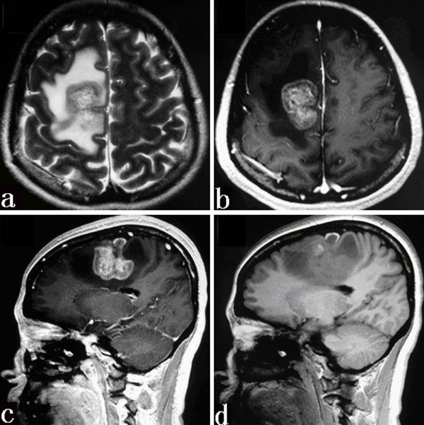
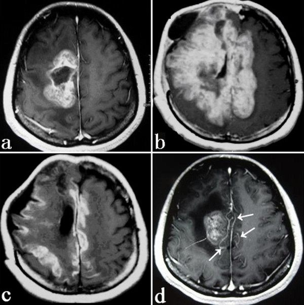
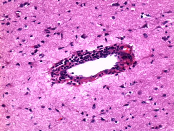
Case presentation: A 35-year-old woman presented with headache and left-sided weakness. MRI scans initially mislead us to a diagnosis of glioblastoma (GBM). Surgery was performed. The mass was sub-totally resected. Pathological examination confirmed a cerebral vasculitis. Radiological features, such as disproportionate mass effect, striped hemorrhage and abnormal enhancement of adjacent vessels, could be helpful to distinguish a tumor-like IACNS from a GBM. Single therapy with high doses of steroid did not improve the patient's condition. Combined therapy with prednisolone and cyclophosphamide showed great benefit to the patient. No relapse occurred during the period of 18 months follow-up.
Conclusions: Although a tumor-like IACNS has no established imaging features, a diagnosis of tumor-like IACNS should be suspected when MRI shows inappropriate presentations of a tumor. Greater awareness of this potential manifestation of IACNS may facilitate more prompt diagnosis and treatment.
Figures
Similar articles
-
A rare case of tumor-mimicking primary angiitis of the central nervous system.Mol Clin Oncol. 2016 May;4(5):827-829. doi: 10.3892/mco.2016.784. Epub 2016 Feb 18. Mol Clin Oncol. 2016. PMID: 27123289 Free PMC article.
-
Isolated angiitis of the CNS in children.Neurology. 2001 Apr 10;56(7):837-42. doi: 10.1212/wnl.56.7.837. Neurology. 2001. PMID: 11314698
-
Tumor-Like Presentation of Primary Angiitis of the Central Nervous System.Stroke. 2016 Sep;47(9):2401-4. doi: 10.1161/STROKEAHA.116.013917. Epub 2016 Jul 28. Stroke. 2016. PMID: 27470990
-
Delirium and isolated angiitis of the central nervous system: a case report and review.CNS Spectr. 2008 Mar;13(3):209-13. doi: 10.1017/s1092852900028455. CNS Spectr. 2008. PMID: 18323754 Review.
-
Primary central nervous system vasculitis mimicking brain tumour: case report and literature review.Rheumatol Int. 2009 Nov;30(1):127-34. doi: 10.1007/s00296-009-0914-7. Rheumatol Int. 2009. PMID: 19370353 Review.
Cited by
-
Imaging mimics of primary malignant tumors of the central nervous system (CNS).Curr Oncol Rep. 2014;16(8):399. doi: 10.1007/s11912-014-0399-8. Curr Oncol Rep. 2014. PMID: 24927848 Review.
-
Primary CNS vasculitis masquerading as glioblastoma: A case report and review.Asian J Neurosurg. 2017 Jan-Mar;12(1):69-71. doi: 10.4103/1793-5482.145187. Asian J Neurosurg. 2017. PMID: 28413538 Free PMC article.
-
Primary Angiitis of the Central Nervous System Mimicking Glioblastoma: A Case Report and Literature Review.Front Neurol. 2019 Nov 13;10:1208. doi: 10.3389/fneur.2019.01208. eCollection 2019. Front Neurol. 2019. PMID: 31798527 Free PMC article.
-
Intravascular lymphomatosis mimicking primary central nervous system lymphoma: a case report and literature review.Case Rep Neurol. 2014 Mar 29;6(1):101-8. doi: 10.1159/000362121. eCollection 2014 Jan. Case Rep Neurol. 2014. PMID: 24761147 Free PMC article.
-
Tumefactive Primary Central Nervous System Vasculitis: Imaging Findings of a Rare and Underrecognized Neuroinflammatory Disease.AJNR Am J Neuroradiol. 2020 Nov;41(11):2075-2081. doi: 10.3174/ajnr.A6736. Epub 2020 Sep 3. AJNR Am J Neuroradiol. 2020. PMID: 32883666 Free PMC article.
References
-
- MacLaren K, Gillespie J, Shrestha S, Neary D, Ballardie FW. Primary angiitis of the central nervous system: emerging variants. Q J Med. 2005;98:643–654. - PubMed
-
- Markus K, Peter B. Primary central nervous system vasculitis: clinical experiences with 21 new European cases. Rheumatol Int. 2009;31:463–472. - PubMed
-
- Newman W, Wolf A. Non-infectious granulomatous angiitis involving the central nervous system. Trans Am Neurol Assoc. 1952;56:114–117. - PubMed
Publication types
MeSH terms
Substances
LinkOut - more resources
Full Text Sources
Medical
