. 2011 Aug 25;71(4):632-9.
doi: 10.1016/j.neuron.2011.07.006.

Cadherin-6 mediates axon-target matching in a non-image-forming visual circuit

Affiliations

Cadherin-6 mediates axon-target matching in a non-image-forming visual circuit

Jessica A Osterhout et al. Neuron. .

Abstract

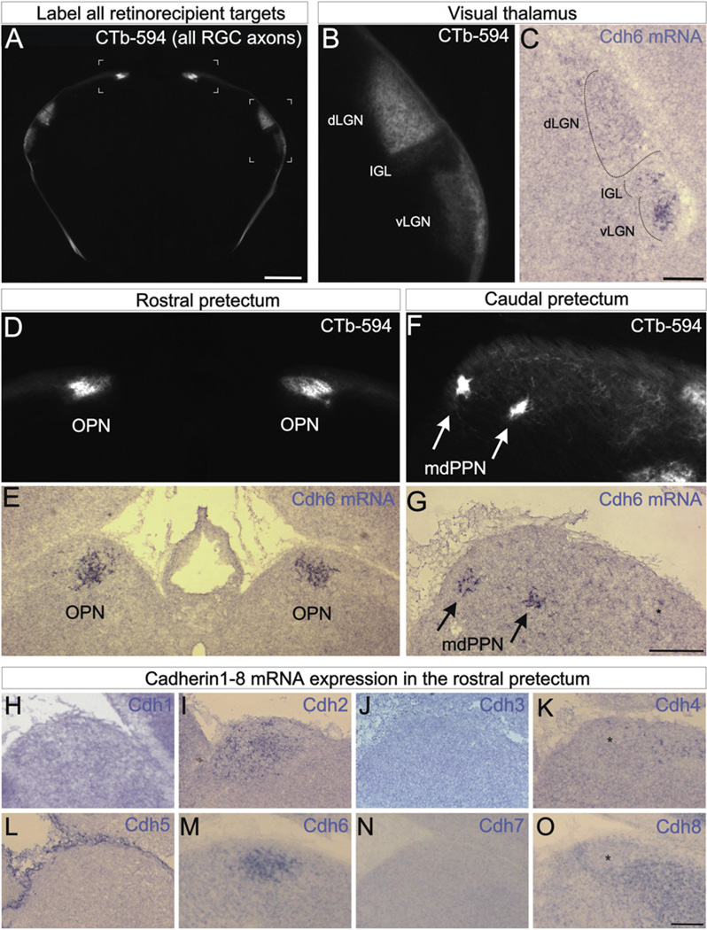
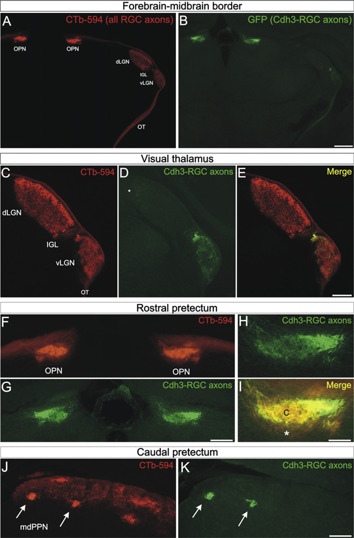
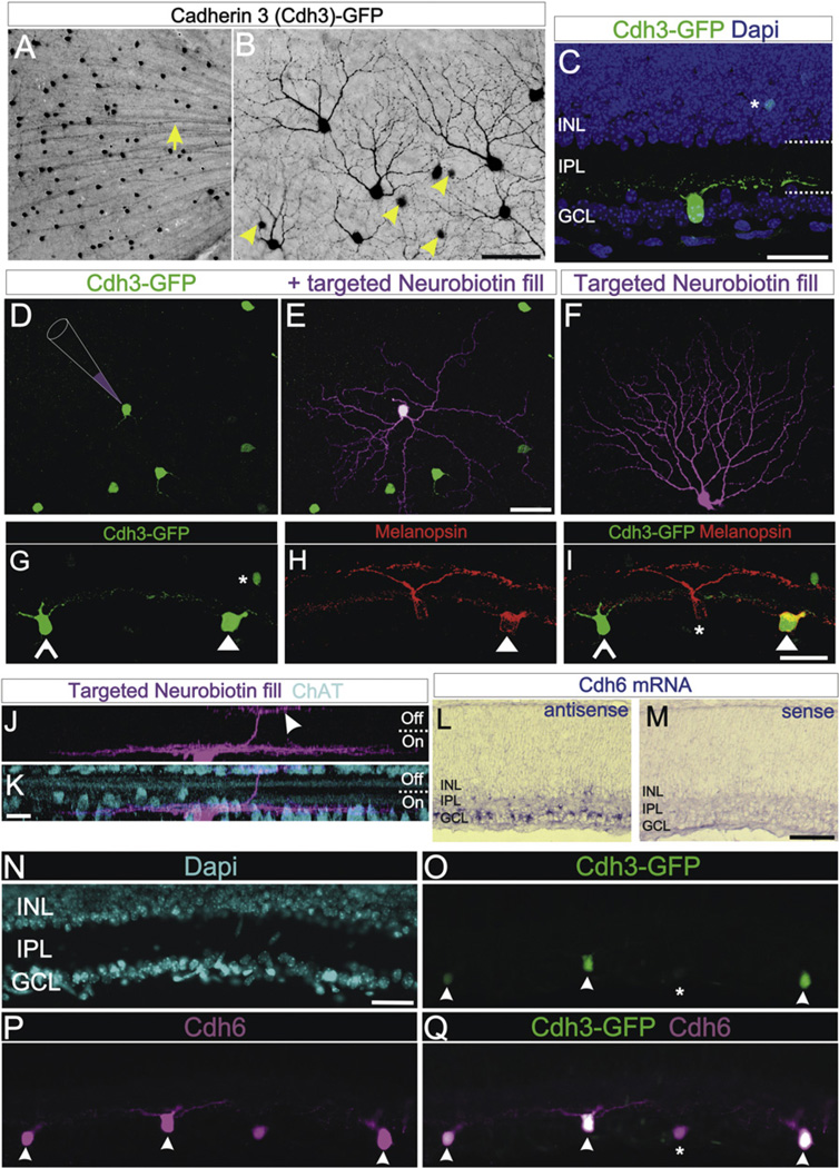
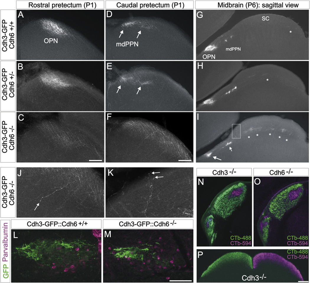
Neural circuits consist of highly precise connections among specific types of neurons that serve a common functional goal. How neurons distinguish among different synaptic targets to form functionally precise circuits remains largely unknown. Here, we show that during development, the adhesion molecule cadherin-6 (Cdh6) is expressed by a subset of retinal ganglion cells (RGCs) and also by their targets in the brain. All of the Cdh6-expressing retinorecipient nuclei mediate non-image-forming visual functions. A screen of mice expressing GFP in specific subsets of RGCs revealed that Cdh3-RGCs which also express Cdh6 selectively innervate Cdh6-expressing retinorecipient targets. Moreover, in Cdh6-deficient mice, the axons of Cdh3-RGCs fail to properly innervate their targets and instead project to other visual nuclei. These findings provide functional evidence that classical cadherins promote mammalian CNS circuit development by ensuring that axons of specific cell types connect to their appropriate synaptic targets.


Figures

Figure 1
Figure 1. Cadherin-6 Is Expressed in Specific Subcortical Visual Nuclei
(A) CTb-594 labeled RGC axons at the forebrainmidbrainborder of a postnatal day 2 (P2) mouse. Image is in coronal plane. Bracketed regions correspond to panels (B and D). Scale = 500 µm. (B) Pan-RGC axon labeling in the visual thalamus. The ventral lateral geniculate nucleus (vLGN), intergeniculate leaflet (IGL) and dorsal lateral geniculate nucleus (dLGN) contain RGC axons. (C) Cdh6 mRNA expressing cells in the IGL and vLGN. Scale = 100 µm. (D) CTb-594 labeled RGC axons in the rostral pretectum. The left and right olivary pretectal nuclei (OPN) are densely innervated. (E) Cdh6 mRNA expression in the OPN of a P1 mouse. (F) RGC axons in the caudal pretectum. The medial division of the posterior pretectal nucleus (mdPPN) of Scalia (1972) appear as two foci (arrows). (G) Cdh6 mRNA in the mdPPN (arrows) of a P1 mouse. Asterisk: a few Cdh6 expressing cell; these may correspond to the caudal-most OPN. Scale = (D)–(G), 250 µm. (H–O) Cdh1–8 antisense mRNA labeling in the rostral pretectum of the early postnatal mouse. Cdh2 (I) and Cdh6 (M) are expressed by the OPN, whereas Cdh4 (K) and Cdh8 (O) are expressed by cells nearby the OPN but not in the OPN itself (asterisks). Cdh1 (H), 3 (J), 5 (L), and 7 (N) are not expressed by the OPN or nearby nuclei but some of these (e.g., Cdh7) are expressed by other retinorecipient targets (not shown). (H–O) Scale = 150 µm. See also Figure S1.
Figure 2
Figure 2. Cdh3-RGCs Project to Cdh6-Expressing Visual Targets
(A) CTb-594 labeled RGC axons and (B) Cdh3-GFP+ RGC axons (“Cdh3-RGCs”) at the level of the forebrain-midbrain border (coronal plane). The optic tract (OT), vLGN, IGL, dLGN, and OPN are apparent from CTb-594 label. (B) Cdh3-RGC axons in the OT, IGL, vLGN and OPN. Scale = 500 µm. (C–E) CTb labeled RGC axons (C), and Cdh3-RGC axons (D), and their merge (E) in the visual thalamus. Except for sparse terminals in the dorsal cap of the dLGN (asterisk), Cdh3-RGC axons selectively terminate in the vLGN and IGL. Scale = 300 µm. (F–I) Rostral pretectum OPN with (F) CTb-labeled RGC axons and (G and H) Cdh3-RGC axons and (I) their merge. (I) The OPN core (“c”) is occupied by Cdh3-RGC axons, whereas the OPN “shell” (asterisk) is not and contains only CTb-594 label. Scale in (F) and (G) = 250 µm; scale in (H and I) = 100 µm. (J and K) The caudal pretectum with (J) CTb-labeled RGC axons and (I) Cdh3-RGCs axons. (J) The caudal pretectum includes many retinorecipient areas, including the mdPPN (arrows). (K) Cdh3-RGC axons selectively terminate in the mdPPN (arrows). (J and K) Scale = 175 µm. See also Figures S1–S3.
Figure 3
Figure 3. Cdh3-GFP and Cdh6 Expression in a Subset of Retinal Ganglion Cells
(A) GFP+ somas and axons in the ganglion cell layer (GCL) of a flat mounted Cdh3-GFP retina. Arrow: GFP+ axons. (B) GFP+ somas and dendrites of Cdh3-RGCs. GFP+ amacrine cell somas (arrowheads) are also seen deep to the plane of focus. Scale = 200 µm (A) and 50 µm (B). (C) Cdh3-RGC in a retinal section; the cell stratifies its dendrites proximal to the GCL, in the “On” sublamina of IPL. Dashed lines: IPL boundaries. Asterisk: a GFP+ amacrine. (D–F) Targeted intracellular injections with Neurobiotin (schematized in D; magenta label in E and F) reveal the morphology of individual Cdh3-RGCs. (D) Cdh3-RGCs without and (E) with injection. (E) A Cdh3-RGC with a symmetric, sparsely branched dendritic arbor. (F) A different filled Cdh3-RGC. This cell has a densely branched, asymmetric arbor (F). Scale = 50 µm (D–F). (G–I) A subset of Cdh3-RGCs express melanopsin. (G) Cdh3-RGCs (open and solid arrowheads) and an amacrine (asterisk). (H) Two RGCs and their dendrites expressing melanopsin (red). (I) Merge of (G) and (H). One of the Cdh3-RGCs expresses melanopsin (solid arrowhead) whereas the other does not (open arrowhead), a non-GFP+ melanopsin RGC is also present (asterisk) Scale = 50 µm. (J and K) Confocal z stack of a Neurobiotin-filled Cdh3-RGC whose dendrites stratify mainly in the On-sublamina of the IPL. The cell extends a secondary dendritic arbor into the Off-sublamina (arrowhead). (K) ChAT stained amacrine cell bodies and dendrites (blue). Scale = 10 µm (J and K). (L and M) Cdh6 mRNA is expressed in the GCL (L) Cdh6 antisense. (M) Cdh6 sense control. Scale = 100 µm. (N–Q) Retinal section with (N) Dapi+ nuclei (blue), (O) Cdh3-RGCs, and (P) Cdh6 immunoreactive cells. (Q) Merge of (O) and (P). All Cdh3-RGCs express Cdh6 (arrowheads); but some Cdh6-immunopositive cells are not Cdh3-GFP+. Scale = 50 µm. See also Figure S3.
Figure 4
Figure 4. Cdh3-RGCs Display Target Recognition Errors in Cdh6 Knockout Mice
(A–F) Coronal view of Cdh3-RGC axon projections to the rostral (A–C) and caudal (D–F) pretectum of P1 mice. (A and D) Cdh3-GFP:Cdh6+/+, (B and E) Cdh3-GFP:Cdh6+/−, (C and F) Cdh3-GFP:Cdh6−/− mice. In Cdh6+/+ and Cdh6+/− mice, an oval termination zone characteristic of the OPN is apparent, whereas in Cdh6−/−mice, Cdh3-RGC axons fail to terminate in the OPN region. In Cdh3-GFP:Cdh6+/+ mice (D) and Cdh3-GFP:Cdh6+/− mice (E), two foci of GFP+ axon terminals are seen (arrows) whereas the Cdh3-GFP:Cdh6−/− mice (F) lack these foci; instead Cdh3-RGC axons course through the caudal pretectum. Scale = 100 µm (A–F). (G–I) Sagittal view of Cdh3-RGC axons in the pretectum and SC of P6 Cdh3-GFP:Cdh6+/+ (G), Cdh3-GFP:Cdh6+/− (H), and Cdh3-GFP:Cdh6−/− (I) mice. In Cdh3-GFP:Cdh6+/+ mice, Cdh3-RGC axons form a dense oval termination in the OPN and foci in the mdPPN; sparse axons are observed in the SC (asterisk). (H) In Cdh3-GFP:Cdh6+/− mice, Cdh3-RGC axons terminate in OPN and in the mdPPN and SC. (I) In Cdh3::Cdh6−/− mice, Cdh3-RGC axons display diminished terminal fields in the OPN region (larger arrow) and mdPPN (small arrow); asterisks: ectopic terminations in the SC. Scale = 500 µm (G–I). (J and K) High-magnification view of Cdh3-RGC axons mistargeting in the OPN region (J) and in the rostral SC (K; from boxed region in I). Arrow in (J): a rare case of an axon seen projecting through the OPN region along the lateral-medial axis. Most misprojecting mutant axons travel through and past the OPN region along the rostral-caudal axis (as in C, F, and I, and see Figure S4). (L and M) Parvalbumin expressing OPN neurons (magenta) of P20 Cdh6+/+and P20 Cdh6−/− mouse. Cdh3-RGC axons are found in the target cell region of both genotypes, but in Cdh6−/− mice, they terminate in a lateral zone (M), rather than diffusely filling the entire aggregate of parvalbumin cells. Scale = 150 µm. See main text and Figure S4 for quantitative details on phenotypic variation and examples of Cdh6 mutants. (N and O) Whole-eye anterograde labeling of RGC axons from the contralateral (CTb-488; green) and ipsilateral (CTb-594; magenta) eye in the visual thalamus (N and O) of a Cdh3−/− and a Cdh6−/− mouse. RGC axons are confined to their normal targets, the dLGN, IGL and vLGN (see Figures 1 and 3 for nuclei locations) and normal positioning of RGC axons is observed. (P) Eye-specific projections to the SC in a Cdh3−/− mouse. Scale = 250 µm.

Comment in

  • Cadherins as matchmakers.
    Rebsam A, Mason CA. Rebsam A, et al. Neuron. 2011 Aug 25;71(4):566-8. doi: 10.1016/j.neuron.2011.08.005. Neuron. 2011. PMID: 21867873

References

    1. Berson DM. Albright TD, Masland R. The Senses: A Comprehensive Reference, Volume 1, Vision 1. San Diego, CA: Academic Press; 2008. Retinal ganglion cell types and their central projections; pp. 491–520.
    1. Bowling DB, Michael CR. Projection patterns of single physiologically characterized optic tract fibres in cat. Nature. 1980;286:899–902. - PubMed
    1. Clandinin TR, Feldheim DA. Making a visual map: mechanisms and molecules. Curr. Opin. Neurobiol. 2009;19:174–180. - PMC - PubMed
    1. Dahl U, Sjödin A, Larue L, Radice GL, Cajander S, Takeichi M, Kemler R, Semb H. Genetic dissection of cadherin function during nephrogenesis. Mol. Cell. Biol. 2002;22:1474–1487. - PMC - PubMed
    1. Dickson BJ. Molecular mechanisms of axon guidance. Science. 2002;298:1959–1964. - PubMed

Publication types