. 2011 Aug 25;71(4):640-55.
doi: 10.1016/j.neuron.2011.06.019.

Cadherin-9 regulates synapse-specific differentiation in the developing hippocampus

Affiliations

Cadherin-9 regulates synapse-specific differentiation in the developing hippocampus

Megan E Williams et al. Neuron. .

Abstract

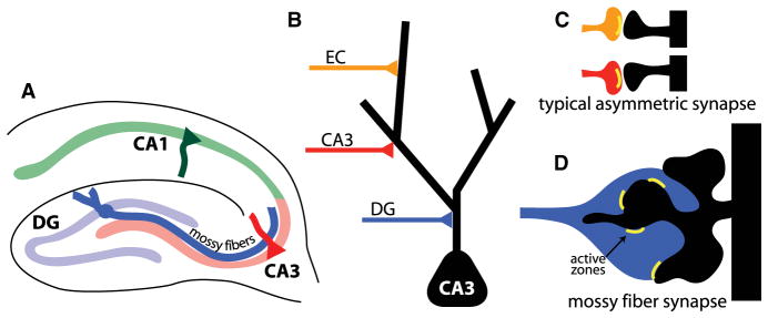
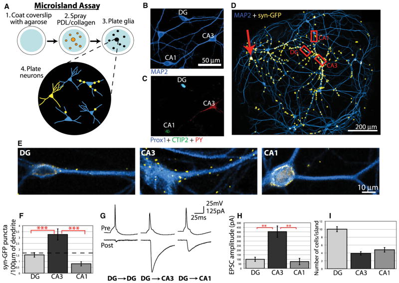
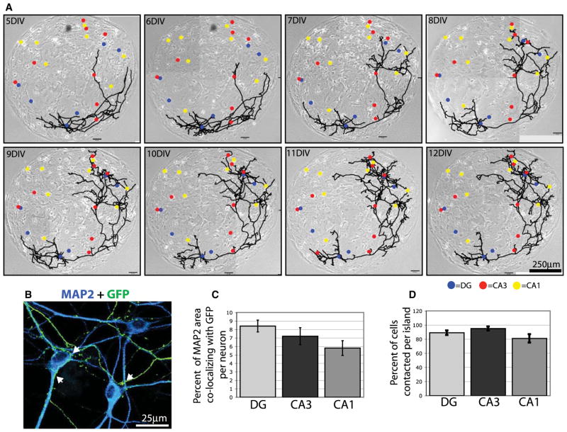
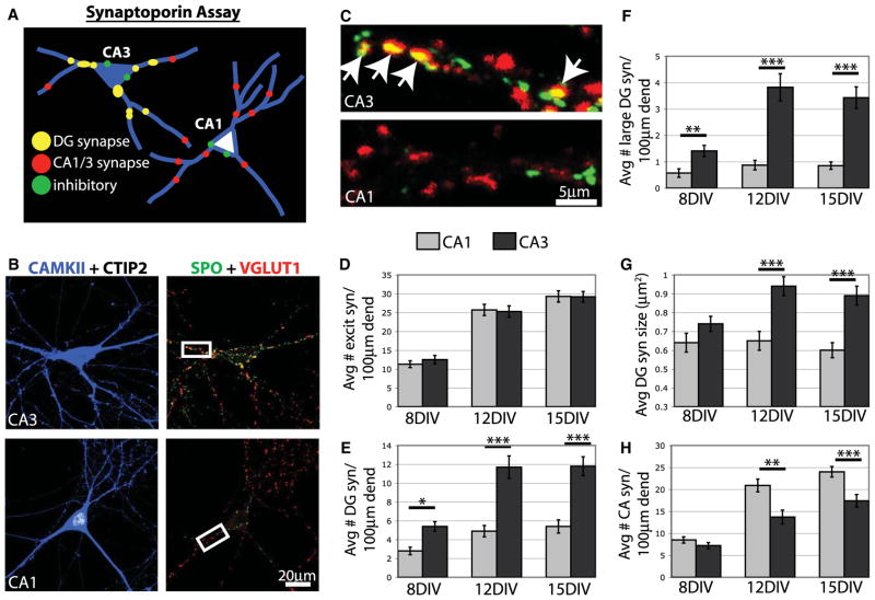
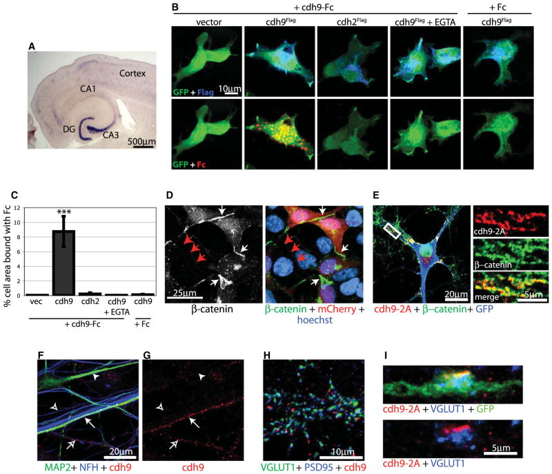
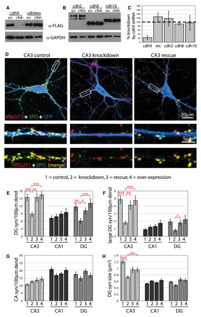
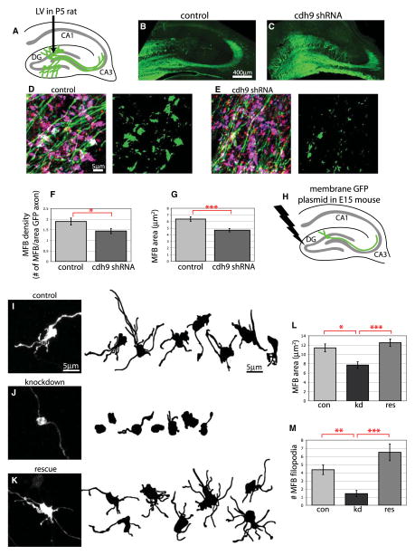
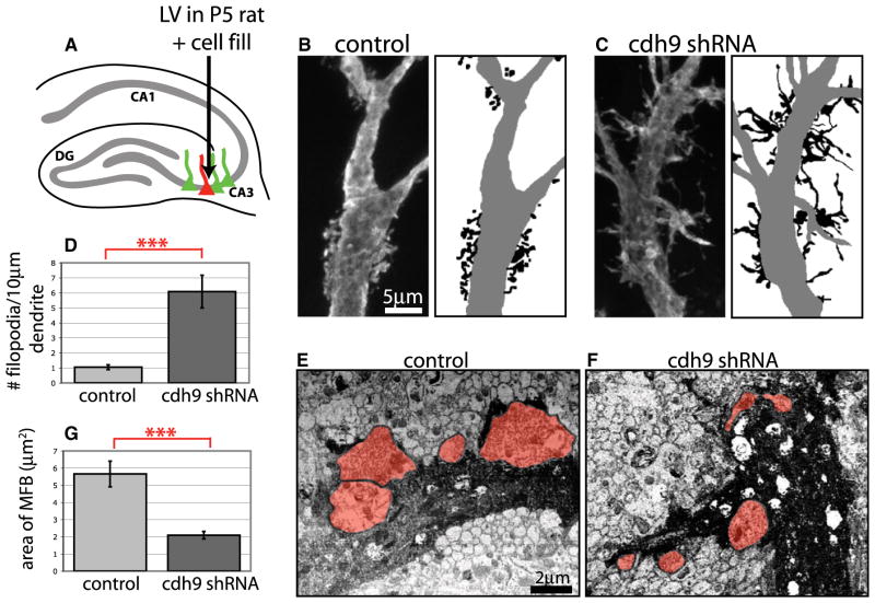
Our understanding of mechanisms that regulate the differentiation of specific classes of synapses is limited. Here, we investigate the formation of synapses between hippocampal dentate gyrus (DG) neurons and their target CA3 neurons and find that DG neurons preferentially form synapses with CA3 rather than DG or CA1 neurons in culture, suggesting that specific interactions between DG and CA3 neurons drive synapse formation. Cadherin-9 is expressed selectively in DG and CA3 neurons, and downregulation of cadherin-9 in CA3 neurons leads to a selective decrease in the number and size of DG synapses onto CA3 neurons. In addition, loss of cadherin-9 from DG or CA3 neurons in vivo leads to striking defects in the formation and differentiation of the DG-CA3 mossy fiber synapse. These observations indicate that cadherin-9 bidirectionally regulates DG-CA3 synapse development and highlight the critical role of differentially expressed molecular cues in establishing specific connections in the mammalian brain.


Figures

Figure 1
Figure 1. Specificity of Synaptic Connections in the Hippocampus
(A) Diagrammatic representation of mossy fiber pathway of the hippocampus. (B–D) Types of synapses on the apical dendrite of CA3 pyramidal neurons.
Figure 2
Figure 2. DG Neurons Preferentially Innervate CA3 Neurons in Microcultures
(A) Diagram of microisland assay. (B) After 12 DIV, hippocampal neurons were immunostained for the neuronal marker MAP2. (C) The same field of view in (B) but immunostained for Prox1, CTIP2, and PY to identify DG, CA1, and CA3 neurons, respectively (also see Figure S1). (D) A 12 DIV microisland with a synaptophysin-GFP transfected DG neuron (red arrow) immunostained for MAP2 and GFP. Multiple fields of view were merged, and the GFP signal was thresholded and enlarged for visibility. (E) Magnified images of the regions outlined in (D) showing preferential formation of DG synapses on CA3 neurons. For simplicity only MAP2 and synaptophysin-GFP are shown, but all cells were immunostained for Prox1 and CTIP2 to identify cell types. GFP fluorescence around the cell soma is the result of nonspecific background staining, and only synaptophysin-GFP puncta on dendrites were analyzed. (F) Quantification of synaptophysin-GFP puncta on DG, CA3, and CA1 neurons in hippocampal microislands. The dotted line represents the expected number of synapses per length of dendrite if all cells were innervated equally (n > 50 cells of each type from 21 islands). (G) Example traces of paired recordings from hippocampal microislands in which single-action potentials were elicited in presynaptic DG neurons (upper traces), and postsynaptic EPSCs were measured by voltage clamp (lower traces). (H) Average evoked EPSC amplitudes for paired recordings. (I) Average number of DG, CA3, and CA1 neurons on each microisland. Statistics were performed using ANOVA followed by posttests where ***p < 0.001 and ** p < 0.01. Error bars represent SEM.
Figure 3
Figure 3. Preferential Synapse Formation Is Independent of Axon Guidance in Microcultures
(A) Phase-contrast images of a microisland merged with line drawings of a GFP-expressing neuron growing on the island. Cell bodies of all neurons on the island are indicated by color-coded dots. Blue, DG; red, CA3; yellow, CA1. (B) A magnified region of a microisland with a DG neuron transfected with GFP (transfected cell soma not shown) and immunostained with antibodies against MAP2 to label all neurons and GFP to label the transfected DG axon. Arrowheads indicate points of contact between the axon and target cell dendrites. (C and D) Quantification of contact between GFP-expressing DG axons and all neurons on microislands. Graph in (C) shows the percentage of MAP2 area that colocalizes with GFP per neuron and, therefore, represents the extent that each potential target neuron comes into contact with the DG axon. Graph in (D) shows the percentage of each cell type that was contacted per island. Error bars represent SEM. No differences among cell types are significant by ANOVA.
Figure 4
Figure 4. Specificity Develops at the Onset of Synapse Formation
(A) Diagram of SPO assay. P0 hippocampal neurons are grown in standard low-density mass cultures and immunostained with cell and synapse markers. (B) After 15 DIV, hippocampal neurons were immunostained for CAMKII (blue), CTIP2 (white), SPO (green), and VGlut1 (red). (C) Magnified images of boxed regions in (B). DG synapses express both SPO and VGlut1 and appear yellow. Arrows indicate DG synapses on the CA3 neuron, but none is visible on a comparable region of the CA1 neuron. (D–H) Graphs showing the total number of excitatory synapses (all VGlut1 puncta), DG synapses (SPO + VGlut1 puncta), large DG synapses (SPO + Vglut1 >1.0 μm2), DG synapse size, and CA synapses (VGlut1 only puncta) on CA1 and CA3 neurons over time. In all cases, n > 30 cells for each cell type from 3 cultures. Statistics were performed using ANOVA followed by posttests where ***p < 0.001, **p < 0.01, and *p < 0.05. Error bars represent SEM. Avg, average; dend, dendrite; excit syn, excitatory synapse.
Figure 5
Figure 5. cdh9 Is a Classic Type II Cadherin
(A) In situ hybridization for cdh9 mRNA in a horizontal section of a P14 mouse brain. (B) 293T cells cotransfected with GFP and Flag-tagged cadherin constructs were treated with Fc or cdh9-Fc as indicated. Top row shows GFP (green) and Flag (blue) expression. Bottom row shows the same cells as above stained for GFP (green) and bound Fc (red). (C) Quantification of Fc binding. The percentage of GFP cell area (green) that colocalized with Fc (red) was determined. The only condition to show significant binding was cdh9-Fc treatment on cdh9 cells. Statistics determined by ANOVA and error bars represent SEM. ***p > 0.001 compared to all other conditions. (D) 293T cells cotransfected with mCherry and cdh9 were immunostained for β-catenin (green), mCherry (red), and Hoechst (blue). Note that β-catenin clusters at junctions between transfected cells expressing cdh9 and mCherry (white arrows), but not between untransfected cells (red arrows). (E) Cultured hippocampal neuron transfected with cdh9-2A-GFP and immunostained for cdh9-2A (red), GFP (blue), and β-catenin (green). The white box indicates a region of higher magnification at right showing that cdh9-2A colocalizes with β-catenin in dendrites, and colocalization appears yellow. (F and G) Hippocampal neurons immunostained for MAP2 (green) to label dendrites, NFH (blue) to label axons, and endogenous cdh9 (red). cdh9 localizes to dendrites (closed arrow) and axons (open arrow) of a subset of neurons in mixed hippocampal cultures. Dendrites (closed arrowhead) and axons (open arrowhead) not expressing cdh9 are also shown. (H) Hippocampal neurons immunostained for VGlut1 (green) to label presynaptic sites, PSD95 (blue) to label postsynaptic sites, and endogenous cdh9 (red). cdh9 is adjacent but not overlapping synaptic release sites. (I) DG axon infected in vivo with a lentivirus expressing cdh9-2A-GFP. In DG axons, cdh9-2A (red) was found adjacent to clusters of synaptic vesicles identified by VGlut1 (blue).
Figure 6
Figure 6. cdh9 Specifically Regulates DG Synapse Formation In Vitro
(A and B) 293T cells were cotransfected with C-terminal FLAG-tagged cadherin constructs and scrambled shRNA (scr) or cdh9 shRNA (c9sh) as indicated, and cell lysates were analyzed by immunoblotting. cdh9 expression is significantly reduced by cdh9 shRNA but not a cdh9 rescue construct containing silent point mutations, or the cadherins cdh2 (N-cadherin), cdh8, or cdh10. (C) Quantification of cdh9 shRNA western blots (n = 3). (D) CA3 neurons expressing GFP and scrambled shRNA (control), cdh9 shRNA (knockdown), or cdh9 shRNA plus cdh9res (rescue) were immunostained for GFP (blue), SPO (green), and VGlut1 (red). White boxes indicate regions of higher magnification shown below each neuron. DG synapses appear yellow and are reduced on CA3 knockdown neurons. (E–H) Graphs show the average number of DG synapses (syn), CA synapses, and DG synapse size on neurons expressing scrambled shRNA (1), cdh9 shRNA3 (2), cdh9 shRNA plus cdh9res (3), or cdh9 overexpression (4) (n > 30 cells for each cell type from at least 3 cultures). Error bars represent SEM. Statistics were performed using ANOVA followed by posttests (***p < 0.001, **p < 0.01, and *p < 0.05). dend, dendrite.
Figure 7
Figure 7. Reduction of cdh9 Expression in DG Neurons In Vivo Disrupts Mossy Fiber Bouton Formation
(A) Diagram of DG lentivirus injection strategy used for (B)–(G). (B and C) Lentivirus infections of the DG with GFP control or GFP plus cdh9 shRNA. Overall development and axon growth appear normal in both control and cdh9 shRNA-expressing cells. (D and E) DG mossy fibers in the stratum lucidum following lentiviral infection. Sections were immunostained with antibodies against GFP (green) to label infected axons and SPO (red) and VGlut1 (blue) to label mossy fiber synapses, which appear purple in the merged images. To the right, the same images have been thresholded to reveal the areas of triple colocalization. Axons infected with cdh9 shRNA have fewer mossy fiber boutons. (F and G) Graphs showing that both the number (F) and size (G) of mossy fiber boutons (MFB) are significantly reduced in axons expressing cdh9 shRNA compared to control (n > 20 fields of view from at least 3 different injections per condition). (H) Diagram of in utero electroporation strategy used for (I)–(M). (I–K) Examples of individual mossy fiber boutons coexpressing membrane GFP with scrambled shRNA (control), cdh9 shRNA (knockdown), or cdh9 shRNA plus cdh9res (rescue). Confocal z projection of a mossy fiber bouton immunostained for GFP is shown at left, and line drawings of several other boutons from each condition are shown at right. (L and M) Graphs showing that mossy fiber bouton size and complexity are reduced upon cdh9 knockdown and are fully rescued by coexpression of cdh9res (n = 14–22 boutons from at least 3 different animals for each condition). For all graphs, error bars represent SEM; ***p < 0.001, **p < 0.01, *p < 0.05 by two-tailed t test (F and G) or ANOVA (L and M).
Figure 8
Figure 8. Reduction of cdh9 in CA3 Neurons In Vivo Causes Pre- and Postsynaptic Defects
(A) Diagram of CA3 lentivirus injection strategy. (B and C) CA3 neurons infected with lentiviruses and filled with LY dye to reveal spine morphology. Line drawings of images illustrating disruption of mossy fiber TEs in cdh9 shRNA-expressing neurons are shown at right. Dendrites are gray, and spines and spines/filopodia are black. (D) Graph showing that the number of filopodia per 10 μm of proximal dendrite is significantly greater in CA3 neurons infected with cdh9 shRNA compared to control (n = 7 neurons for each). (E and F) Scanning electron microscopy images of photoconverted, LY-filled CA3 neurons infected with lentiviruses. Dendrites of filled neurons are dark, and mossy fiber boutons are shaded red. (G) Graph showing that the average size of mossy fiber boutons contacting cdh9 shRNA dendrites is significantly smaller compared to control as determined by electron microscopy (n = 16 boutons from 1 control neuron and n = 35 boutons from 2 cdh9 shRNA neurons). In all cases, error bars represent SEM; ***p < 0.001, two-tailed t test.

Comment in

  • Cadherins as matchmakers.
    Rebsam A, Mason CA. Rebsam A, et al. Neuron. 2011 Aug 25;71(4):566-8. doi: 10.1016/j.neuron.2011.08.005. Neuron. 2011. PMID: 21867873

References

    1. Ackley BD, Jin Y. Genetic analysis of synaptic target recognition and assembly. Trends Neurosci. 2004;27:540–547. - PubMed
    1. Amaral DG, Dent JA. Development of the mossy fibers of the dentate gyrus: I. A light and electron microscopic study of the mossy fibers and their expansions. J Comp Neurol. 1981;195:51–86. - PubMed
    1. Ango F, di Cristo G, Higashiyama H, Bennett V, Wu P, Huang ZJ. Ankyrin-based subcellular gradient of neurofascin, an immunoglobulin family protein, directs GABAergic innervation at purkinje axon initial segment. Cell. 2004;119:257–272. - PubMed
    1. Arikkath J. Regulation of dendrite and spine morphogenesis and plasticity by catenins. Mol Neurobiol. 2009;40:46–54. - PubMed
    1. Arikkath J, Reichardt LF. Cadherins and catenins at synapses: roles in synaptogenesis and synaptic plasticity. Trends Neurosci. 2008;31:487–494. - PMC - PubMed

Publication types