. 2011 Aug 26;35(2):249-59.
doi: 10.1016/j.immuni.2011.08.008.

CD8α(+) dendritic cells are the critical source of interleukin-12 that controls acute infection by Toxoplasma gondii tachyzoites

Affiliations

CD8α(+) dendritic cells are the critical source of interleukin-12 that controls acute infection by Toxoplasma gondii tachyzoites

Mona Mashayekhi et al. Immunity. .

Abstract

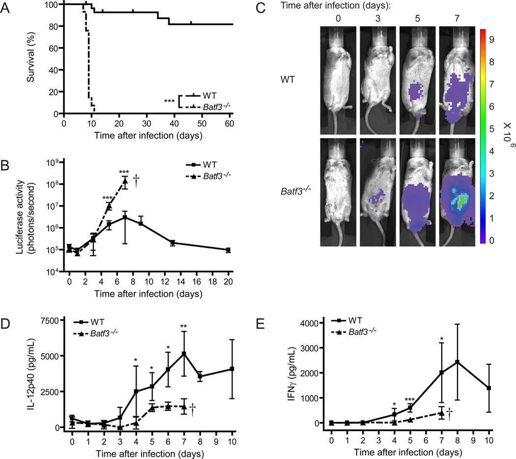
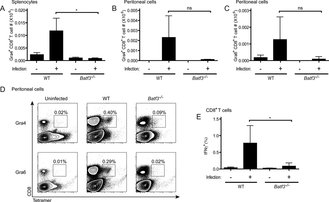
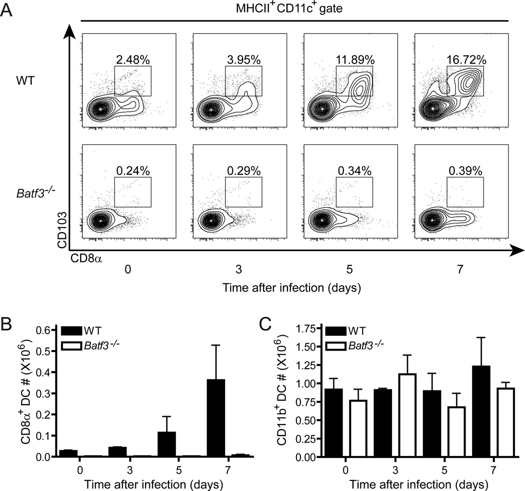
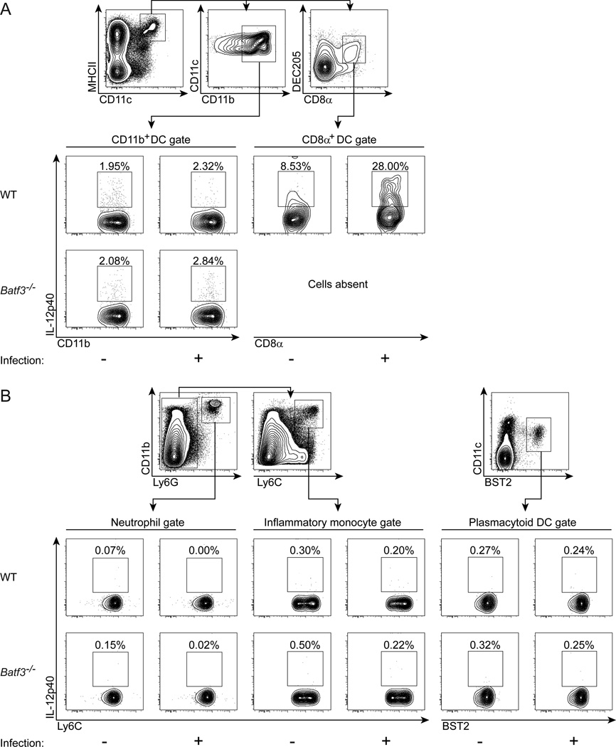
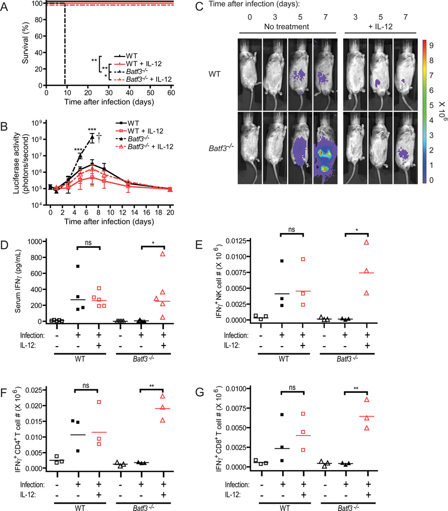
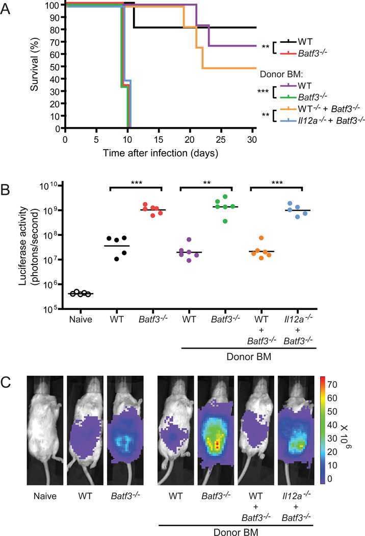
CD8α(+) dendritic cells (DCs) are important in vivo for cross-presentation of antigens derived from intracellular pathogens and tumors. Additionally, secretion of interleukin-12 (IL-12) by CD8α(+) DCs suggests a role for these cells in response to Toxoplasma gondii antigens, although it remains unclear whether these cells are required for protection against T. gondii infection. Toward this goal, we examined T. gondii infection of Batf3(-/-) mice, which selectively lack only lymphoid-resident CD8α(+) DCs and related peripheral CD103(+) DCs. Batf3(-/-) mice were extremely susceptible to T. gondii infection, with decreased production of IL-12 and interferon-γ. IL-12 administration restored resistance in Batf3(-/-) mice, and mice in which IL-12 production was ablated only from CD8α(+) DCs failed to control infection. These results reveal that the function of CD8α(+) DCs extends beyond a role in cross-presentation and includes a critical role for activation of innate immunity through IL-12 production during T. gondii infection.


Conflict of interest statement

The authors have no conflicting financial interests.

Figures

Figure 1
Figure 1. Batf3−/− mice rapidly succumb to infection with an avirulent strain of Toxoplasma gondii
Mice were infected with T. gondii, monitored for survival (A) and parasite burden (B and C), and bled to measure serum cytokines (D and E). (A) Combined survival data from infected C56BL/6, 129S6/SvEV and BALB/c wild-type (solid line, n=30) and Batf3−/− (dashed line, n=29) mice from 8 independent experiments. (B) Infected wild-type (squares) and Batf3−/− (triangles) mice underwent whole body in vivo imaging throughout the course of infection to measure bioluminescence. Data shown is combined parasite burden from infected 129S6/SvEV mice from 2 independent experiments (n=5–8 at each time-point, representative of 6 independent experiments). (C) Representative bioluminescence images of infected 129S6/SvEV mice throughout the course of infection. (D and E) Infected 129S6/SvEV wild-type (squares) and Batf3−/− (triangles) mice were bled at various time-points after infection, and serum analyzed for cytokine concentrations. Data represents combined serum concentrations of IL-12p40 (D) and IFNγ (E) through the course of infection from 2–3 independent experiments (n=3–5 at each time-point). (B, D, E) Data are represented as mean +/− standard deviation. *: 0.01<P<0.05, **: 0.001<P<0.01, ***: P<0.001.
Figure 2
Figure 2. CD8+ T cell priming to T. gondii is defective in Batf3-deficient mice
BALB/c wild-type and Batf3−/− mice were infected with T. gondii, sacrificed on day 8 after infection, and analyzed for CD8+ T cell priming by tetramer staining ex vivo (A–D) and intracellular cytokine staining following peptide re-stimulation in vitro (E). (A) Representative plots of Ld-GRA4 and Ld-GRA6 tetramer staining in the peritoneum, with percentage of total peritoneal cells that are tetramer positive shown. (B–D) Absolute numbers of CD8+ tetramer-positive cells in the peritoneum (B and C) or spleen (D) specific for GRA4 (B and D) or GRA6 (C) on day 8 after infection (n=3, representative of 2 independent experiments). (E) Absolute numbers of IFNγ-positive CD8+ T cells as measured by intracellular cytokine staining after overnight re-stimulation of whole splenocytes with the GRA4 peptide (n=5). (B–E) Data are represented as mean +/− standard deviation. Not significant (ns): P>0.05, *: 0.01<P<0.05.
Figure 3
Figure 3. Splenic CD8α+ dendritic cells expand after T. gondii infection in wild-type mice
129S6/SvEV wild-type and Batf3−/− mice were infected with T. gondii, sacrificed on days 0, 3, 5 and 7 after infection, and analyzed for changes in dendritic cell subsets by flow cytometry. (A) Representative flow cytometry plots gated on Aqua-negative, MHCII+, CD11c+ conventional dendritic cells. (B and C) Absolute numbers of CD8α+ CD103+ DCs (B) and CD11b+ DCs (C) in the spleen of wild-type (black bars) and Batf3−/− (white bars) mice throughout the course of infection (n=3, representative of 2 independent experiments). Data are represented as mean +/− standard deviation.
Figure 4
Figure 4. CD8α+ dendritic cells are the major producers of IL-12 after T. gondii infection in wild-type mice
129S6/SvEV wild-type and Batf3−/− mice were infected with T. gondii, sacrificed on day 3 after infection, and analyzed for the cellular source of IL-12 by intracellular cytokine staining. (A) Representative flow cytometry plots gated on MHCII+ CD11c+ expressing CD11b+ DCs or CD8α+ DEC205+ DCs. (B) Representative flow cytometry plots gated on Ly-6G+ CD11b+ neutrophils, Ly-6G Ly-6C+ CD11b+ inflammatory monocytes, or CD11c+ Bst2+ plasmacytoid DCs. (n=3, representative of 3 independent experiments).
Figure 5
Figure 5. Administration of IL-12 rescues Batf3-deficient mice during T. gondii infection
129S6/SvEV mice were infected with T. gondii and injected with saline or 0.5µg of recombinant murine IL-12 on days 0, 1, 2, 3, and 4 after infection. (A) Survival data from infected mice (WT: solid black line; WT + IL-12: solid red line; Batf3−/−: dashed black line; Batf3−/− + IL-12: dashed red line) (n=3–5, representative of 3 independent experiments). (B) Combined parasite burden from whole body in vivo bioluminescence imaging of infected mice (WT: black squares; WT + IL-12: red squares; Batf3−/−: black triangles; Batf3−/− + IL-12: red triangles) from 2 independent experiments (n=3–8 at each time-point, representative of 4 independent experiments). Data are represented as mean +/− standard deviation. (C) Representative bioluminescence images of infected mice throughout the course of infection. (D) Serum concentrations of IFNγ on day 4 after infection (n=4–5). (E–G) Absolute numbers of IFNγ-positive NK (E), CD4+ T (F), and CD8+ T (G) cells in the spleen directly ex vivo on day 3 after infection as measured by intracellular cytokine staining (n=3). (D–G) Horizontal lines represent the geometric mean. Not significant (ns): P>0.05, *: 0.01<P<0.05, **: 0.001<P<0.01, ***: P<0.001.
Figure 6
Figure 6. CD8α+ dendritic cells are the only cells whose IL-12 production is protective against acute T. gondii infection
BALB/c chimeras and control non-chimeric mice were infected with T. gondii and monitored for survival (A) and parasite burden (B–C). (A) Survival data from non-chimeric wild-type (black line) and Batf3−/− (red line) mice were compared to lethally irradiated recipients which received only wild-type (purple line) or Batf3−/− (green line) BM, or a 1:1 mixture of wild-type with Batf3−/− BM (orange line) or Il12a−/− with Batf3−/− BM (blue line) (n=5–6, representative of 2 independent experiments). Parasite burden (B) and representative images (C) on day 7 after infection from the groups in (A). (B) Horizontal lines represent the geometric mean. **: 0.001<P<0.01, ***: P<0.001.

Comment in

References

    1. Aliberti J, Schulz O, Pennington DJ, Tsujimura H, Sousa Reis E, Ozato K, Sher A. Essential role for ICSBP in the in vivo development of murine CD8alpha + dendritic cells. Blood. 2003;101:305–310. - PubMed
    1. Bar-On L, Birnberg T, Lewis KL, Edelson BT, Bruder D, Hildner K, Buer J, Murphy KM, Reizis B, Jung S. CX3CR1+ CD8alpha+ dendritic cells are a steady-state population related to plasmacytoid dendritic cells. Proc. Natl. Acad. Sci U. S A. 2010;107:14745–14750. - PMC - PubMed
    1. Bliss SK, Butcher BA, Denkers EY. Rapid recruitment of neutrophils containing prestored IL-12 during microbial infection. J Immunol. 2000;165:4515–4521. - PubMed
    1. Bliss SK, Gavrilescu LC, Alcaraz A, Denkers EY. Neutrophil depletion during Toxoplasma gondii infection leads to impaired immunity and lethal systemic pathology. Infect. Immun. 2001;69:4898–4905. - PMC - PubMed
    1. Bliss SK, Zhang Y, Denkers EY. Murine neutrophil stimulation by Toxoplasma gondii antigen drives high level production of IFN-gamma-independent IL-12. J Immunol. 1999;163:2081–2088. - PubMed

Publication types

MeSH terms