. 2011 Oct 21;286(42):36228-37.
doi: 10.1074/jbc.M111.244061. Epub 2011 Aug 25.

Dexamethasone enhances 1alpha,25-dihydroxyvitamin D3 effects by increasing vitamin D receptor transcription

Affiliations

Dexamethasone enhances 1alpha,25-dihydroxyvitamin D3 effects by increasing vitamin D receptor transcription

Alejandro A Hidalgo et al. J Biol Chem. .

Abstract

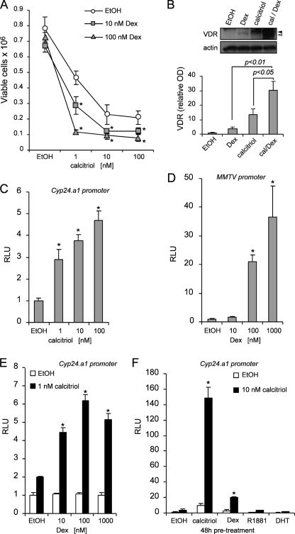
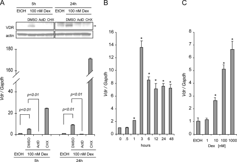
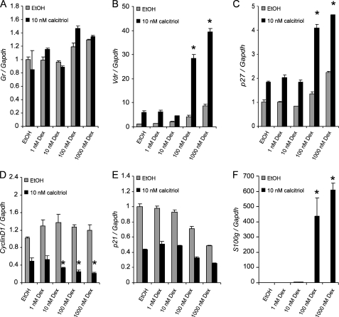
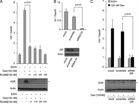
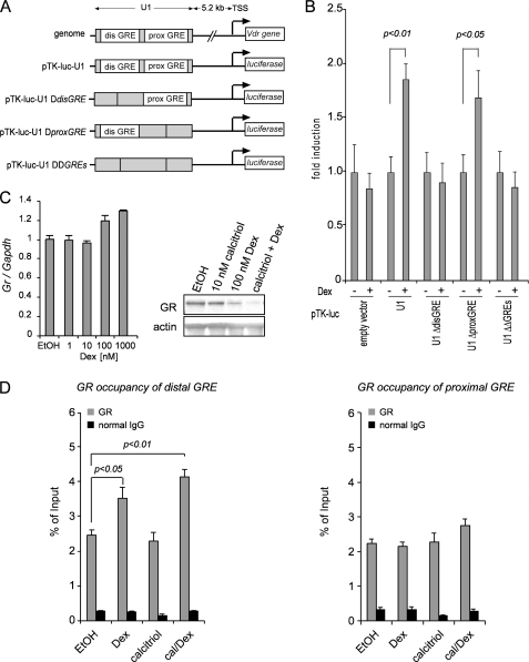
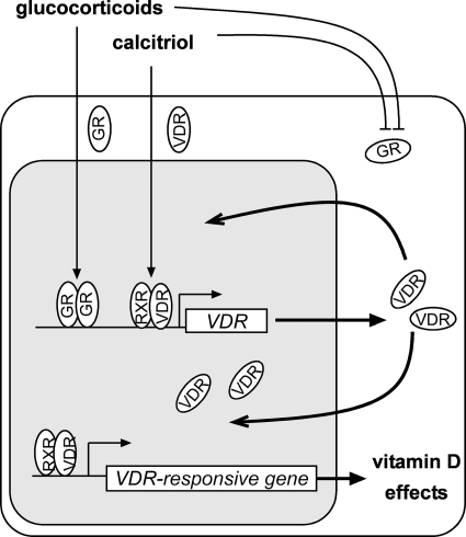
Calcitriol, the active form of vitamin D, in combination with the glucocorticoid dexamethasone (Dex) has been shown to increase the antitumor effects of calcitriol in squamous cell carcinoma. In this study we found that pretreatment with Dex potentiates calcitriol effects by inhibiting cell growth and increasing vitamin D receptor (VDR) and VDR-mediated transcription. Treatment with actinomycin D inhibits Vdr mRNA synthesis, indicating that Dex regulates VDR expression at transcriptional level. Real time PCR shows that treatment with Dex increases Vdr transcripts in a time- and a dose-dependent manner, indicating that Dex directly regulates expression of Vdr. RU486, an inhibitor of glucocorticoids, inhibits Dex-induced Vdr expression. In addition, the silencing of glucocorticoid receptor (GR) abolishes the induction of Vdr by Dex, indicating that Dex increases Vdr transcripts in a GR-dependent manner. A fragment located 5.2 kb upstream of Vdr transcription start site containing two putative glucocorticoid response elements (GREs) was evaluated using a luciferase-based reporter assay. Treatment with 100 nm Dex induces transcription of luciferase driven by the fragment. Deletion of the GRE distal to transcription start site was sufficient to abolish Dex induction of luciferase. Also, chromatin immunoprecipitation reveals recruitment of GR to distal GRE with Dex treatment. We conclude that Dex increases VDR and vitamin D effects by increasing Vdr de novo transcription in a GR-dependent manner.


Figures

FIGURE 1.
FIGURE 1.
Dexamethasone increases the anti-proliferative and VDR-mediated transcriptional effects of calcitriol. A, 48 h of pretreatment with EtOH control (○), 10 (□), and 100 (△) nm Dex sensitizes SCC cells to calcitriol treatment (1–100 nm). Inhibition of cell growth was assessed after 72 h of treatment of calcitriol by trypan blue dye exclusion viability assay. The data represent the means ± S.D. of three independent experiments. *, p < 0.05 compared with corresponding untreated (EtOH) conditions. B, combinatory treatment with calcitriol (cal) and Dex increases VDR protein expression compared with treatments with either calcitriol or Dex alone by Western blot analysis. Calcitriol increased both phosphorylated and unphosphorylated forms of VDR (52), as indicated by closed and open arrowheads, respectively. Dex alone only increased the unphosphorylated form of VDR. Calcitriol/Dex combination treatment further increased both forms of VDR (upper panel). Densitometric analysis was performed using ImageJ software (lower panel). The results are the means ± S.D. of three independent experiments. C, 24 h with calcitriol induces VDR-mediated transcription of the hCYP24.A1 promoter by luciferase reporter assay. D, 24 h with Dex induces GR-mediated transcription of the MMTV promoter by luciferase reporter assay. E, 48 h of pretreatment with Dex potentiates VDR-mediated transcription of the hCYP24.A1 promoter with 1 nm of calcitriol treatment for 24 h. F, 48 h of pretreatment with several steroid hormones demonstrate that the potentiation of VDR-mediated transcription with 10 nm of calcitriol for 24 h is specific to calcitriol and Dex. Other steroid hormones do not affect VDR-mediated transcription. The samples were normalized to Renilla reporter control (C) or total protein (D–F), and the results are expressed relative to EtOH controls. The data (C–F) are representative of at least three independent experiments. Significance (p value) was assessed by Student's t test (unpaired, two-tailed). RLU, relative light unit.
FIGURE 2.
FIGURE 2.
Dexamethasone regulates VDR at the transcriptional level. A, actinomycin D and CHX inhibited transcription or translation of VDR. SCC cells were pretreated with Me2SO, 10 μg/ml cycloheximide, or 10 μg/ml actinomycin D for 1 h followed by 100 nm Dex for 5 or 24 h. VDR was assessed at the transcriptional level (lower panel) and protein (upper panel) by using real time PCR and Western blot analysis, respectively. The results are representative of three independent experiments. B, time-dependent (0.5 to 48 h) induction of Vdr mRNA expression in SCC cells treated with ethanol vehicle control or 100 nm Dex. C, dose-dependent induction of Vdr mRNA. The cells were treated for 6 h with increasing concentrations of Dex or EtOH vehicle control. TaqMan® assay was used for relative quantitation of Vdr gene expression. The expression of the Vdr gene was normalized to mouse glyceraldehyde-3-phosphate dehydrogenase (Gapdh) expression and expressed relative to 0 h (B) or EtOH control (C) for each of the experiments being evaluated. The data represent the means ± S.D. of three technical replicates from one representative experiment. The experiments were repeated four times.
FIGURE 3.
FIGURE 3.
Dose-dependent regulation of VDR target genes by dexamethasone. The cells were pretreated for 48 h with increasing concentrations of Dex or EtOH vehicle control follow by treatment for 24 h with 10 nm of calcitriol or EtOH vehicle control. Expression of VDR target genes Gr (A), Vdr (B), p27 (C), Cyclin D1 (D), p21 (E), and S100g (F) were assessed using TaqMan® gene expression assays for relative quantitative gene expression by real time PCR. The expression levels of the VDR target genes were normalized to mouse glyceraldehyde-3-phosphate dehydrogenase (Gapdh) expression and expressed relative to EtOH control for each of the genes being evaluated. The data represent the means ± S.D. of three independent experiments. The asterisks indicate p < 0.05 compared with EtOH pretreated, calcitriol-induced samples.
FIGURE 4.
FIGURE 4.
Dexamethasone increases de novo Vdr transcription in a GR-dependent manner. A, the anti-glucocorticoid RU486 inhibits Vdr mRNA synthesis. The cells were treated for 24 h with 100 nm Dex alone or in combination with 1, 10, or 50 molar ratios of RU486. Expression of VDR was assessed at the transcriptional level (upper panel) and protein (lower panel) by using real time PCR and Western blot analysis, respectively. B, silencing of GR by using siRNA. The cells were transfected with mock, scramble siRNAs, or siRNA-GR. After 72 h, the cells were harvested, and expression of GR was assessed at the transcriptional level (upper panel) and protein (lower panel) by using real time PCR and Western blot analysis, respectively. C, induction of Vdr by Dex was studied in cells transfected with siRNA targeting Gr. The cells were transfected with mock, scramble siRNAs, or siRNA-GR for 48 h prior to treatment with 100 nm Dex or EtOH vehicle control for an additional 24 h. Expression of VDR was assessed at the transcriptional level (upper panel) and protein (lower panel) by using real time PCR and Western blot analysis, respectively. The expression of Vdr (A and C) and Gr (B) was assessed by real time PCR using TaqMan® gene expression assays. Relative expression of Vdr (A and C) and Gr (B) gene was normalized to mouse glyceraldehyde-3-phosphate dehydrogenase (Gapdh) expression and expressed relative to EtOH vehicle control (A) or compared with the ethanol-treated mock control (B and C). The data represent the means ± S.D. of three independent experiments.
FIGURE 5.
FIGURE 5.
A GRE in the regulatory region upstream of Vdr gene drives transcriptional activation of Vdr that is mediated by dexamethasone. A, schematic representations of Vdr gene containing the distal and proximal GREs in the U1 regulatory region. Deletions introduced on putative GREs are shown. B, the distal GRE in U1 region drives GR-mediated transcription of Vdr by luciferase reporter assay. The cells in each well were transfected with 1 μg of each reporter construct plus 0.2 μg of a plasmid expressing Renilla luciferase (internal control) for 16 h. The cells were induced with 100 nm Dex for 24 h. Cell extracts were collected and used to determine firefly and Renilla luciferase activity. The data represent the means ± S.D. of four independent experiments. C, Dex treatment decreases GR expression at the protein level (right panel) but not at the transcriptional level (left panel). D, recruitment of GR to U1 regulatory region. Dex and combined calcitriol/Dex treatments increase recruitment of GR to the distal GRE. Occupancy of GR is not significantly increased on proximal GRE with hormone treatments. GR occupancy was determined as DNA enrichment by using real time PCR and expressed as percentage of input. The data represent the means ± S.D. of three technical replicates from one representative experiment. The experiments were repeated four times. dis, distal; prox, proximal; TSS, transcriptional start site.
FIGURE 6.
FIGURE 6.
Cross-talk between VDR and GR signaling axes. Proposed model illustrating how calcitriol and glucocorticoids function together to increase VDR and affect the expression of VDR-responsive genes. Based on our data, glucocorticoids increase expression of the Vdr gene to increase VDR protein. In the presence of calcitriol, VDR transcriptionally regulates VDR target genes including Vdr gene itself. This positive feedback between the GR and VDR signaling axes may be inhibited in the presence of high levels of glucocorticoids and calcitriol that reduce GR.

Similar articles

Cited by

References

    1. Dhawan P., Christakos S. (2010) J. Cell. Biochem. 110, 1314–1323 - PubMed
    1. Kallay E., Pietschmann P., Toyokuni S., Bajna E., Hahn P., Mazzucco K., Bieglmayer C., Kato S., Cross H. S. (2001) Carcinogenesis 22, 1429–1435 - PubMed
    1. Trump D. L., Potter D. M., Muindi J., Brufsky A., Johnson C. S. (2006) Cancer 106, 2136–2142 - PubMed
    1. Chung I., Karpf A. R., Muindi J. R., Conroy J. M., Nowak N. J., Johnson C. S., Trump D. L. (2007) J. Biol. Chem. 282, 8704–8714 - PubMed
    1. Chung I., Yu W. D., Karpf A. R., Flynn G., Bernardi R. J., Modzelewski R. A., Johnson C. S., Trump D. L. (2007) J. Steroid Biochem. Mol. Biol. 103, 768–770 - PubMed

Publication types

MeSH terms

LinkOut - more resources