. 2011 Nov;189(3):795-808.
doi: 10.1534/genetics.111.131458. Epub 2011 Aug 25.

Complex mechanisms regulate developmental expression of the matA (HMG) mating type gene in homothallic Aspergillus nidulans

Affiliations

Complex mechanisms regulate developmental expression of the matA (HMG) mating type gene in homothallic Aspergillus nidulans

Wioletta Czaja et al. Genetics. 2011 Nov.

Abstract

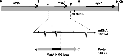
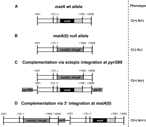
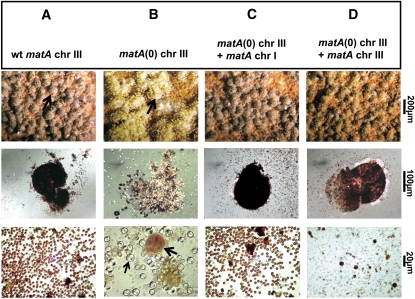
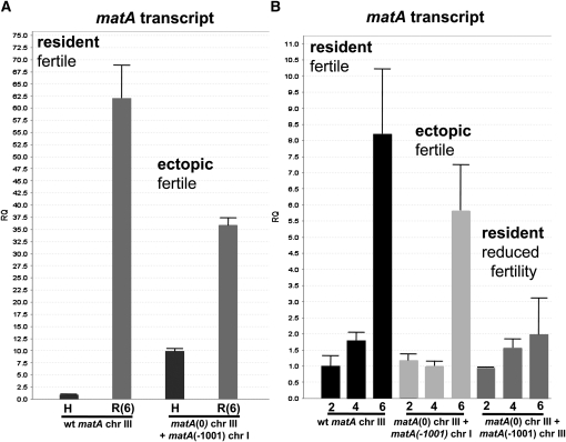
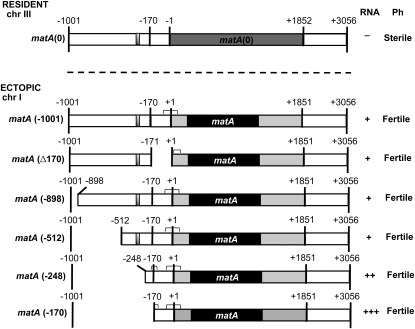
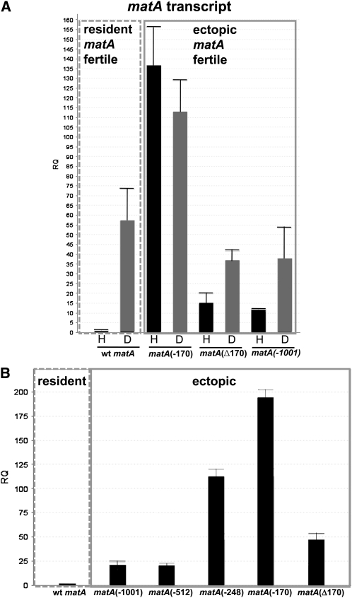
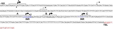
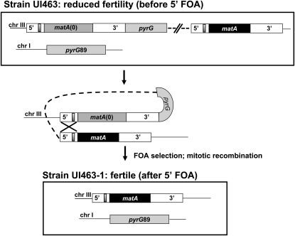
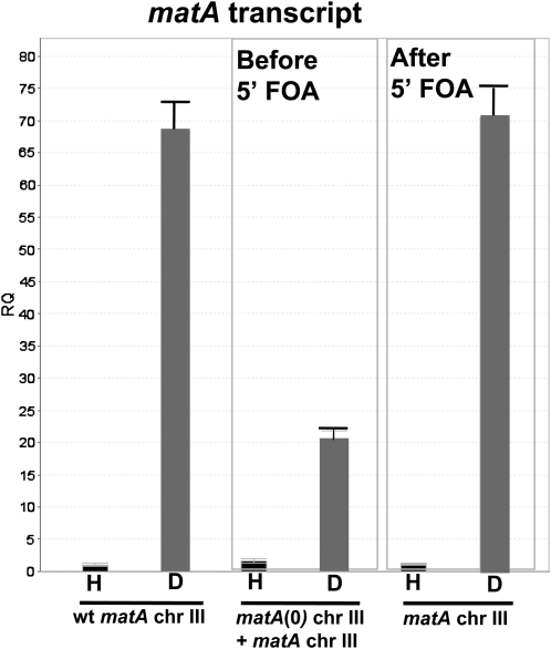
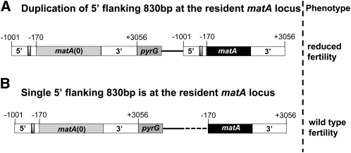
Sexual reproduction is a fundamental developmental process that allows for genetic diversity through the control of zygote formation, recombination, and gametogenesis. The correct regulation of these events is paramount. Sexual reproduction in filamentous fungi, including mating strategy (self-fertilization/homothallism or outcrossing/heterothallism), is determined by the expression of mating type genes at mat loci. Aspergillus nidulans matA encodes a critical regulator that is a fungal ortholog of the hSRY/SOX9 HMG box proteins. In contrast to well-studied outcrossing systems, the molecular basis of homothallism and role of mating type genes during a self-fertile sexual cycle remain largely unknown. In this study the genetic model organism, A. nidulans, has been used to investigate the regulation and molecular functions of the matA mating type gene in a homothallic system. Our data demonstrate that complex regulatory mechanisms underlie functional matA expression during self-fertilization and sexual reproduction in A. nidulans. matA expression is suppressed in vegetative hyphae and is progressively derepressed during the sexual cycle. Elevated levels of matA transcript are required for differentiation of fruiting bodies, karyogamy, meiosis, and efficient formation of meiotic progeny. matA expression is driven from both initiator (Inr) and novel promoter elements that are tightly developmentally regulated by position-dependent and position-independent mechanisms. Deletion of an upstream silencing element, matA SE, results in derepressed expression from wild-type (wt) promoter elements and activation of an additional promoter. These studies provide novel insights into the molecular basis of homothallism in fungi and genetic regulation of sexual reproduction in eukaryotes.


Figures

Figure 1
Figure 1
Genetic context of the matA gene on chromosome III. Black arrows indicate arrangement and orientation of genes (coding regions) flanking the matA locus. The matA transcript and protein structure are shown. Solid triangle indicates the reference transcriptional start site 1 (TSS1), the start of the transcriptional unit. Open triangle indicates the end of the matA transcript.
Figure 2
Figure 2
Schematic representation of the deletion and complementation of matA. (A) Wild type matA allele; solid bar represents coding region (ORF), lightly shaded bars represent matA 5′- and 3′-UTRs, and open bars represent upstream 5′ and downstream 3′ genomic sequences flanking the transcriptional unit. Horizontal solid line represents vector sequence of pWP3. Physical distance is marked (−1001 to +3056 bp). (B) Null matA(0) allele; the matA transcriptional unit was deleted and replaced with the Aspergillus fumigatus argB (AfargB) marker. (C) Complementation via ectopic integration; matA complementing transgene was integrated ectopically at the pyrG89 locus on chromosome I. (D) Complementation via 3′ integration at the matA(0) locus; matA transgene carried on pWP3 was integrated at matA(0) locus via homology between 3′ flanking sequences. The corresponding phenotype is shown: C, cleistothecia; A, ascospores; (+), presence; (−), absence.
Figure 3
Figure 3
Sexual development in wild type (wt), matA(0), and complemented A. nidulans strains. Representative strains (A–D) were induced to undergo sexual development as described in Materials and Methods. Abundance of cleistothecia and internal reproductive tissue were examined under specific levels of magnification as indicated. (A, C, and D, top row) Morphology and abundance of mature cleistothecia on rich medium agar plates in wild-type and complemented strains (arrow in A indicates a single cleistothecium). (B, top row) Cluster of hülle cells in the matA(0) strain. (A, C, and D, middle row) Individual mature cleistothecia in wild-type and complemented strains. (B, middle row) Mass of hülle cells in the matA(0) strain. (A, C, and D, bottom row) Efficiency of ascospore production in wild-type and complemented strains, respectively. (B, bottom row) Sexual development in the matA(0) strain is aborted at the stage of unfertilized protocleistothecia, shown as brown spherical structures (thick black arrow) with individual hülle cells (thin black arrow).
Figure 4
Figure 4
Comparative transcriptional expression of matA at resident and ectopic loci. Phenotype associated with a specific abundance of matA transcript in the reproductive tissue is indicated. (A) Relative quantitation (RQ) of matA expression from the endogenous matA locus on chromosome (chr) III and from the ectopically integrated matA transgene on chr I. The transgene carries 1000 bp of 5′ flanking upstream sequences. H, undifferentiated hyphae; (R6) reproductive tissue, 6 days postinduction. (B) Relative quantitation of matA expression over a developmental time course at 2, 4, and 6 days postinduction of sexual development. Expression profiles of the endogenous matA, ectopically integrated matA transgene and the matA transgene integrated at resident or ectopic loci are shown.
Figure 5
Figure 5
Deletion analysis of the 5′ flanking region of matA. The solid bar indicates the matA coding region, lightly shaded bars represent 5′- and 3′-UTRs. The silencer element is indicated as a small shaded box. Chromosomal positions in the genome are indicated (chr III and chr I). Positions of 5′ deletion end points and internal deletions of upstream flanking sequences are indicated by − and Δ, respectively. Transgene deletions were integrated ectopically at the pyrG locus on chr I. Complementation was observed for all transgene constructs. Transcript presence (+) or absence (−) is indicated. Phenotypes associated with each deletion are indicated. Horizontal brackets indicate positions of mRNA cap sites determined for hyphal and developmental RNAs using RACE (refer to Figure 7).
Figure 6
Figure 6
Comparative expression of matA transgene deletions. (A) Developmental expression of matA in transgene strains lacking either 830 bp of upstream sequence or the internal 170 bp proximal to the start of transcription. The level of matA transcript was analyzed in the undifferentiated hyphae, H, and in the reproductive tissue, D, at 4 days postinduction of sexual development. The dashed line box indicates expression of matA at the resident locus in the wild-type strain. The solid line box indicates expression of matA transgenes at the ectopic locus. (B) Expression in additional 5′ deletion strains used to determine position of upstream repressor and promoter elements. Specific 5′ deletion end points or internal deletions are indicated by − or Δ, respectively.
Figure 7
Figure 7
Identification of both Inr and novel promoter elements that regulate matA transcription. Mapping of mRNA cap sites identified three zones of transcriptional initiation in hyphae and developmental tissue from wild-type and deletion strains (A–C). Each zone is indicated by brackets. An asterisk marks a fourth zone near position −170 observed in hyphal tissue from derepressed strains matA (−248) and matA (−170). The position of the majority of cap sites within a zone is indicated by a thick arrow. Thin arrows mark two minor sites of mRNA start sites. The reference +1 represents the 5′ end of the largest cDNA previously identified from a sexual development library. Potential Inrs are indicated in blue; novel promoter elements are underlined. The translational start site (TSL) and beginning of the ORF are in red text. A black dot below the text marks the −170 position for reference.
Figure 8
Figure 8
Schematic depiction of matA flanking sequence duplication and restoration of wild-type matA gene structure at the endogenous locus. Genetic organization of the matA locus after 3′ homologous integration of pWP3 carrying the matA transgene (solid box). 5′ and 3′ flanking regions are shown with the silencer element (shaded box). The DNA fragment containing pyrG and the matA transgene allows alignment of duplicated 5′ flanking regions and rearrangement during mitotic recombination. The matA(0) allele and pWP3 carrying pyrG are excised and wild-type matA allele is restored at chromosome III. Recombinant events containing wild-type matA and the pyrG89 mutation (chr I) were selected after plating on media supplemented with 5′-FOA.
Figure 9
Figure 9
Developmental expression of matA at the resident locus. Expression of matA was analyzed in undifferentiated hyphae, H, and in reproductive tissue 6 days postinduction of sexual development, D. Expression of the matA transgene at the matA(0) locus is suppressed in the reproductive tissue (before 5′-FOA). Wild-type expression of matA is restored at the resident locus (after 5′-FOA) upon removal of matA(0) and plasmid sequences and the recovery of wild-type genomic structure.
Figure 10
Figure 10
Duplication of 830 bp of matA 5′ flanking sequences at the resident matA locus interferes with the expression and molecular function of the matA transgene. Structure of the complemented endogenous matA(0) locus and associated phenotype are shown. Bars represent matA with flanking regions and pyrG/pyrG89 gene. Solid line represents vector sequence of pWP3; dashed line represents deleted 5′ flanking sequences from −1001 to −171. Shaded box represents silencer element. (A) Integration of the pWP3 plasmid via 3′ homology introduces a duplication of 830 bp of 5′ flanking sequences at the resident matA locus and interferes with fertility. (B) Integration of the pWP3 (∆830 bp), via 3′ homology at the resident matA locus results in wild-type fertility.

Similar articles

Cited by

References

    1. Benjamin C. R., 1955. Ascocarps of Aspergillus and Penicillium, pp. 669–687 in Mycologia. Mycological Society of America, Washington, DC
    1. Boeke J. D., LaCroute F., Fink G. R., 1984. A positive selection for mutants lacking orotidine-5′-phosphate decarboxylase activity in yeast: 5-fluoro-orotic acid resistance. Mol. Gen. Genet. 197: 345–346 - PubMed
    1. Bruggeman J., Debets A. J., Wijngaarden P. J., deVisser J. A., Hoekstra R. F., 2003. Sex slows down the accumulation of deleterious mutations in the homothallic fungus Aspergillus nidulans. Genetics 164: 479–485 - PMC - PubMed
    1. Champe S. P., Nagle D. L., Yager L. N., 1994. Sexual sporulation. Prog. Ind. Microbiol. 29: 429–454 - PubMed
    1. Coppin E., Debuchy R., 2000. Co-expression of the mating-type genes involved in internuclear recognition is lethal in Podospora anserina. Genetics 155: 657–669 - PMC - PubMed

Publication types

MeSH terms