Comparative Study
. 2011 Sep 30;109(8):867-79.
doi: 10.1161/CIRCRESAHA.110.236927. Epub 2011 Aug 25.

Regulatory T cells limit vascular endothelial injury and prevent pulmonary hypertension

Affiliations
Comparative Study

Regulatory T cells limit vascular endothelial injury and prevent pulmonary hypertension

Rasa Tamosiuniene et al. Circ Res. .

Abstract

Rationale: Pulmonary arterial hypertension (PAH) is an incurable disease associated with viral infections and connective tissue diseases. The relationship between inflammation and disease pathogenesis in these disorders remains poorly understood.

Objective: To determine whether immune dysregulation due to absent T-cell populations directly contributes to the development of PAH.

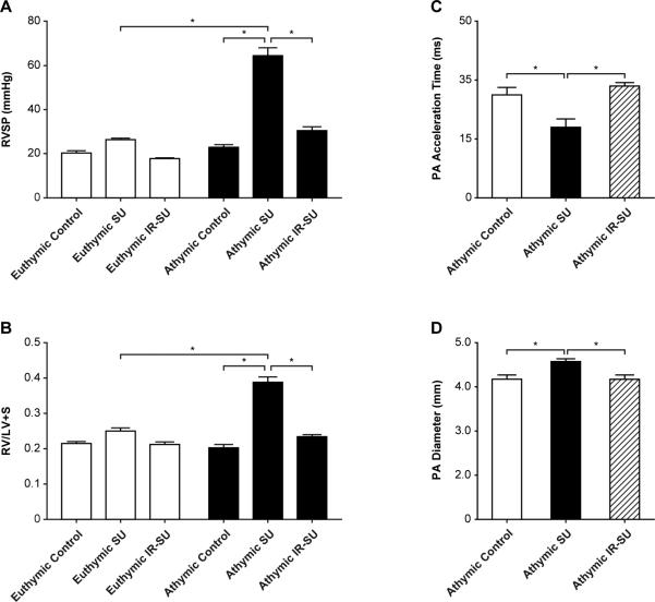
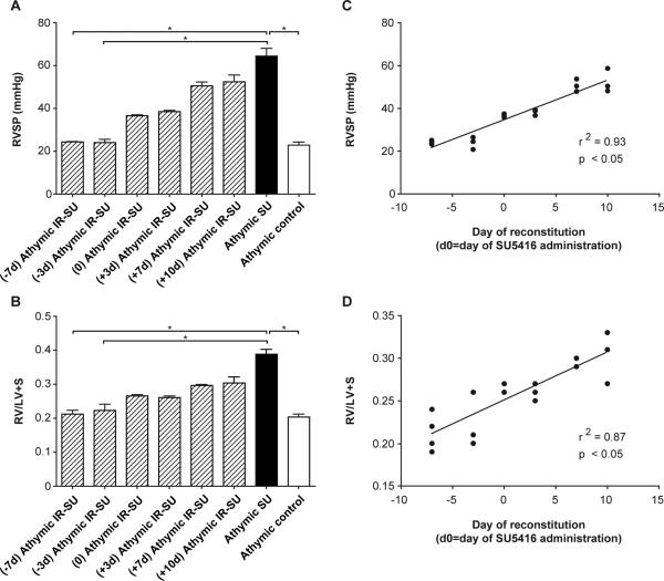
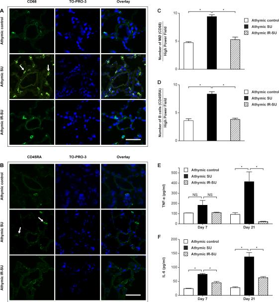
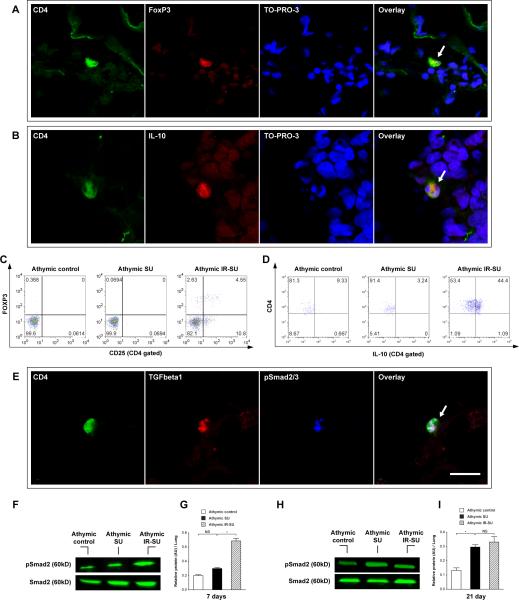
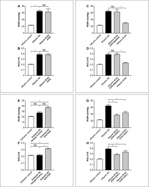
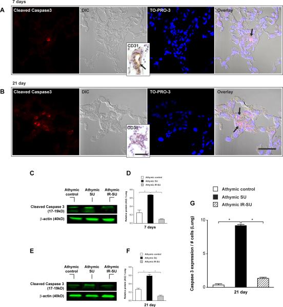
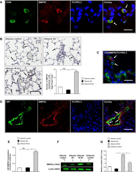
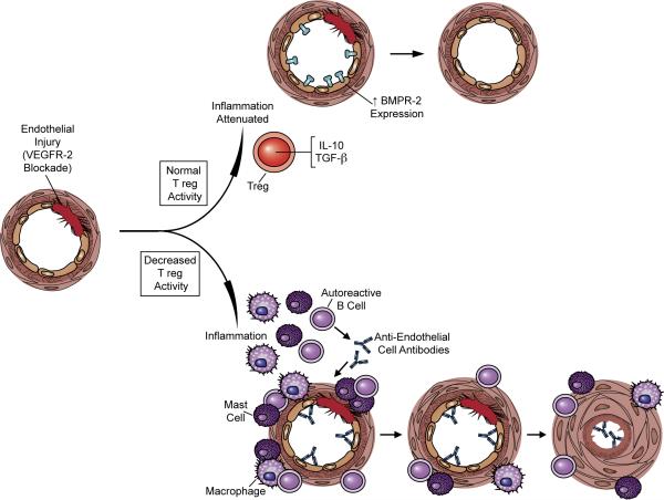
Methods and results: Vascular endothelial growth factor receptor 2 (VEGFR2) blockade induced significant pulmonary endothelial apoptosis in T-cell-deficient rats but not in immune-reconstituted (IR) rats. T cell-lymphopenia in association with VEGFR2 blockade resulted in periarteriolar inflammation with macrophages, and B cells even prior to vascular remodeling and elevated pulmonary pressures. IR prevented early inflammation and attenuated PAH development. IR with either CD8 T cells alone or with CD4-depleted spleen cells was ineffective in preventing PAH, whereas CD4-depleting immunocompetent euthymic animals increased PAH susceptibility. IR with either CD4(+)CD25(hi) or CD4(+)CD25(-) T cell subsets prior to vascular injury attenuated the development of PAH. IR limited perivascular inflammation and endothelial apoptosis in rat lungs in association with increased FoxP3(+), IL-10- and TGF-β-expressing CD4 cells, and upregulation of pulmonary bone morphogenetic protein receptor type 2 (BMPR2)-expressing cells, a receptor that activates endothelial cell survival pathways.

Conclusions: PAH may arise when regulatory T-cell (Treg) activity fails to control endothelial injury. These studies suggest that regulatory T cells normally function to limit vascular injury and may protect against the development of PAH.


Figures

Figure 1
Figure 1
Immune reconstitution of athymic rats prevents PAH development. (A) RVSP measurements in athymic and euthymic rats treated with SU5416 at d21 and vehicle-treated rats (athymic control, euthymic control). (B) RVH measurements as assessed by RV/(LV+S) ratio in athymic and euthymic rats treated with SU5416 at d21 and vehicle-treated control rats (n=10-16/group). (C-D) Sequential echocardiography of athymic controls, athymic SU, and athymic IR-SU at d21 (n=4/group). Data are shown as means with error bars representing SEM. * p<0.05.
Figure 2
Figure 2
Immune reconstitution is most effective if administered prior to vascular injury. The hemodynamic analysis of IR was evaluated at multiple time points relative to SU5416 administration (d0). (A) RVSP measurements evaluated at multiple time points. The X-axis represents the day of IR relative to SU5416 administration (e.g. (-7d) athymic IR-SU indicates athymic rats undergoing IR d7 prior to SU5416 administration). All hemodynamic evaluations performed d21 after vehicle or SU5416 administration (n=19 (d-7 through d+10)). (B) RVH as assessed by RV/(LV+S) ratio evaluation at multiple time points. All evaluations performed d21 after vehicle or SU5416 administration (n=19 (d-7 through d+10)). (C,D) Correlations between RVSP and RVH at multiple time points. Data are shown as means with error bars representing SEM. * p<0.05.
Figure 3
Figure 3
Evidence of anti-inflammatory effect of IR at d7 after SU5416 administration. (A, B) Immunofluorescent images of lung sections from athymic SU animals stained with CD68 (arrows) for macrophages, and with CD45RA (arrows) for B cells at d7 after SU5416 administration (n=8/group). (C,D) Morphometric analysis of macrophages (CD68) and B cells (CD45RA) in lung sections at d7 after SU5416 administration (n=4/group). (E, F) Serum TNF-α and IL-6 evaluated by ELISA at d7 and d21 (n=6-8/group). Data are shown as means with error bars representing SEM. * p<0.05. Scale bars: (A,B)=50 μm.
Figure 4
Figure 4
Seven days after SU5416 administration, IR results in migration of TGFβ+ and IL-10+ expressing T cells into the lungs. (A) Immunofluorescent images of lung sections for Tregs cells (CD4+FoxP3+) (arrow) in athymic IR-SU group at d7 (n=7/group). (B) Immunofluorescent images of lung sections for CD4+IL-10+cells (arrow) in athymic IR-SU group at d7 (n=7/group). (C,D) Flow cytometry data of peripheral blood for FoxP3+and IL-10+ detection on d7 in athymic IR-SU group. (E) Immunofluorescent images of lung sections for CD4+TGFβ+ pSmad2/3+ cells (arrow) in athymic IR-SU group at d7 (n=7/group). (F,G,H,I) Western blot analysis from whole lung lysate and protein levels of pSmad2 and Smad2 on d7 and d21(n=8/group). Data are shown as means with error bars representing SEM. * p<0.05. Scale bar = 6.25 μm.
Figure 5
Figure 5
Localizing regulatory activity in the CD4 T cell subset. (A, B) RVSP and RVH measurements in athymic rats after SU5416 administration and IR with fractionated CD8+ T cells (n=7-14/group). (C,D) RVSP and RVH measurements in athymic rats after SU5416 administration undergoing IR with CD4-depleted spleen cells (n=8-11/group). (E,F) RVSP and RVH measurements in euthymic rats after SU5416 administration and undergoing CD4 depletion (n=8-13/group). (G,H) RVSP and RVH measurements in athymic rats after SU5416 administration and undergoing reconstitution with CD4+CD25hi cells and CD4+CD25- cells (n=14-16/group). Data are shown as means with error bars representing SEM. * p<0.05.
Figure 6
Figure 6
IR of athymic rats leads to reduced pulmonary vascular endothelial cell apoptosis. (A) Immunofluorescent images of serial lung tissue sections for cleaved caspase 3 expression (arrow) at d7 after SU5416 administration. Differential interference contrast (DIC) represents small lung vessel histology and inset shows CD31 expression in small lung vessel (n=8/group). (B) Immunofluorescent images of serial lung tissue sections for cleaved caspase 3 expression (arrow) at d21 after SU5416 administration. DIC image shows an almost occluded vessel and inset shows absent CD31 staining inside the lung vessel wall (n=8/group). (C,D,E,F) Western blot analysis from whole lung lysate for cleaved caspase 3 protein levels at d7 and d21 (n=8/group). (G) Quantitation of activated caspase 3 positive endothelial cells in precapillary pulmonary arteries in lung tissue sections at d21 (n=4/group). Data are shown as means with error bars representing SEM. * p<0.05. Scale bar: (A,B)=50μm; insets=25μm.
Figure 7
Figure 7
IR of athymic rats leads to increased BMPR2 expression in the lung. (A) Immunofluorescent images show colocalization of BMPR2 expressing cells and CD68 in all three animal groups. (B) Immunohistochemistry and quantitation of BMPR2 expressing cells in lungs sections of athymic control, athymic SU and athymic IR-SU rats. (C) Immunofluorescent image of lung tissue shows BMPR2 expressing cells (red) are adjacent to CD4+ cells (green) in athymic IR-SU group. (D) Immunofluorescent images of lung section shows that vWF colocalizes with BMPR2 (arrow) in vascular endothelial cells at d21 in athymic IR-SU rats (n=4/group). (E) The vWF-BMPR2 colocalization coefficient at d21. (F,G) Western blot analysis of BMPR2 protein levels at d21 from whole lung lysates (n=8/group). Data are shown as means with error bars representing SEM. * p<0.05. Scale bar: (A,C)=25 μm; (B,D)=50 μm.
Figure 8
Figure 8
Model of immune regulation contributing to vascular health and immune dysregulation favoring vascular disease following endothelial injury. In this model, endothelial injury causes a local immune response. With normal immune regulation, vascular injury does not culminated in pulmonary vascular remodeling and PAH. The absence of normal immune regulation results in an inappropriately exuberant inflammatory response, accelerated endothelial cell apoptosis, smooth muscle hypertrophy, and increased pulmonary vascular resistance.

References

    1. Nicolls MR, Taraseviciene-Stewart L, Rai PR, Badesch DB, Voelkel NF. Autoimmunity and pulmonary hypertension: a perspective. Eur Respir J. 2005;26(6):1110–1118. - PubMed
    1. Tuder RM, Voelkel NF. Pulmonary hypertension and inflammation. J Lab Clin Med. 1998;132(1):16–24. - PubMed
    1. Dorfmuller P, Perros F, Balabanian K, Humbert M. Inflammation in pulmonary arterial hypertension. Eur Respir J. 2003;22(2):358–363. - PubMed
    1. Rai PR, Cool CD, King JA, Stevens T, Burns N, Winn RA, Kasper M, Voelkel NF. The cancer paradigm of severe pulmonary arterial hypertension. Am J Respir Crit Care Med. 2008;178(6):558–564. - PMC - PubMed
    1. Hassoun PM, Mouthon L, Barbera JA, Eddahibi S, Flores SC, Grimminger F, Jones PL, Maitland ML, Michelakis ED, Morrell NW, Newman JH, Rabinovitch M, Schermuly R, Stenmark KR, Voelkel NF, Yuan JX, Humbert M. Inflammation, growth factors, and pulmonary vascular remodeling. J Am Coll Cardiol. 2009;54(1 Suppl):S10–19. - PubMed

Publication types

MeSH terms