. 2011 Oct 15;92(7):745-51.
doi: 10.1097/TP.0b013e31822bc95a.

Glial cell line-derived neurotrophic factor enhances human islet posttransplantation survival

Affiliations

Glial cell line-derived neurotrophic factor enhances human islet posttransplantation survival

Simon M Mwangi et al. Transplantation. .

Abstract

Background: Development of pretransplantation islet culture strategies that preserve or enhance β-cell viability would eliminate the requirement for the large numbers of islets needed to restore insulin independence in type 1 diabetes patients. We investigated whether glial cell line-derived neurotrophic factor (GDNF) could improve human islet survival and posttransplantation function in diabetic mice.

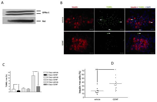
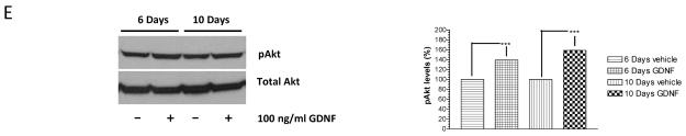
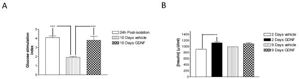
Methods: Human islets were cultured in medium supplemented with or without GDNF (100 ng/mL) and in vitro islet survival and function assessed by analyzing β-cell apoptosis and glucose stimulated insulin release. In vivo effects of GDNF were assessed in streptozotocin-induced diabetic nude mice transplanted under the kidney capsule with 2000 islet equivalents of human islets precultured in medium supplemented with or without GDNF.

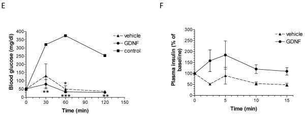
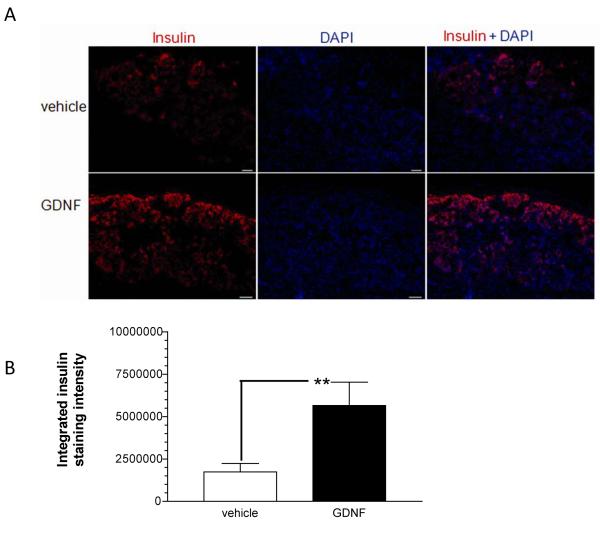
Results: In vitro, human islets cultured for 2 to 10 days in medium supplemented with GDNF showed lower β-cell death, increased Akt phosphorylation, and higher glucose-induced insulin secretion than islets cultured in vehicle. Human islets precultured in medium supplemented with GDNF restored more diabetic mice to normoglycemia and for a longer period after transplantation than islets cultured in vehicle.

Conclusions: Our study shows that GDNF has beneficial effects on human islet survival and could be used to improve islet posttransplantation survival.


Figures

FIGURE 1
FIGURE 1. GDNF enhances human islet survival and function in vitro
(A) Western blot analysis of Ret and GFRα-1 receptor expression in human islets. Data is representative of three separate experiments. (B) Representative images showing insulin (red) and TUNEL (green) staining with DAPI nuclear counter-staining (blue) in human islets cultured for 2 days in vehicle or medium containing GDNF. Scale 20 μm. (C) Score of TUNEL-positive cells in islets cultured for 2–9 days in CIT culture medium alone (vehicle) or CIT culture medium supplemented with GDNF (100 ng/ml). Plotted are means + SEM. ***, P<0.001, **, P<0.01, *, P<0.05, N≥5. (D) Score of insulin-positive cells in islets cultured for 2 days in vehicle or medium containing GDNF. Horizontal lines show the means for each group. **, P<0.01; GDNF, N=13 replicates; vehicle, N=11 replicates. (E) Western blot analysis of phospho-Akt and total Akt levels in human islets cultured for 6 and 10 days in vehicle or medium supplemented with GDNF. Histogram showing phospho-Akt band densities equalized to total Akt levels expressed as a percentage of vehicle. Plotted are means + SEM. ***, P<0.001.
FIGURE 2
FIGURE 2. GDNF preserves human islet function in vitro
(A) Glucose stimulation indices of islets cultured in CIT culture medium for 24 h immediately post-isolation and islets cultured for 10 days in vehicle or medium supplemented with GDNF. Plotted are means + SEM. ***, P<0.001; N=6 replicates each from 2 isolations. (B) Insulin content of human islets cultured for 2 and 9 days in vehicle or GDNF. Plotted are means + SEM. *, P≤0.05. N=2.
FIGURE 3
FIGURE 3. Diabetic mice transplanted with islets cultured in GDNF show better glycemic control than mice transplanted with islets cultured in vehicle
(A) Weekly non-fasting blood glucose levels for study 1–3 control, vehicle and GDNF transplant mice. Plotted are means ± SEM. Group means that are significantly different from control means at each time point are shown by asterisks either above (vehicle) or below (GDNF) the mean value. ***, P<0.001; **, P<0.01; *, P<0.05; control N=5, vehicle N=9, GDNF N=8. (B) Linear regression fit of blood glucose data. ***, P<0.001. (C) Non-fasting blood glucose distribution (quartiles and range) for control mice and mice transplanted with human islets pre-cultured for 14 days in vehicle or medium supplemented with GDNF. (D) Fixed effects and P-values of log linear mixed regression model fit tests of the significance of the difference in blood sugar levels between the treatment groups. (E) Blood glucose levels during intraperitoneal glucose (3 mg/kg) tolerance testing in control, vehicle and GDNF transplant mice performed 65 days post-transplantation. Plotted are means + SEM. N=3 mice. Group means that are significantly different from control mice means at each time point are shown by asterisks either above (vehicle) or below (GDNF) the mean value. ***, P<0.001; **, P<0.01; *, P<0.05. (F) Insulin release in response to intraperitoneal arginine (300 mg/kg) administration performed 63 days post-transplantation. Plotted are means + SEM of plasma insulin levels expressed as a percentage of baseline. N=3 mice in each group.
FIGURE 4
FIGURE 4. GDNF improves islet post-transplantation survival
(A) Representative images showing insulin (red) staining with DAPI (blue) nuclear counter-staining in kidney sections collected 65 days post-transplantation from vehicle (vehicle) and GDNF (GDNF) transplant mice. Scale, 20 μm. (B) Comparison of intensity of insulin staining in kidneys from vehicle (vehicle) and GDNF (GDNF) transplant mice. Plotted are means + SEM. (**, P<0.01; vehicle N=14 replicates, GDNF N=10 replicates from 4 mice each).

Similar articles

Cited by

References

    1. Shapiro AMJ, Lakey JRT, Ryan EA, et al. Islet Transplantation in Seven Patients with Type 1 Diabetes Mellitus Using a Glucocorticoid-Free Immunosuppressive Regimen. N Engl J Med. 2000;343(4):230. - PubMed
    1. Shapiro AM, Ricordi C, Hering BJ, et al. International trial of the Edmonton protocol for islet transplantation. N Engl J Med. 2006;355(13):1318. - PubMed
    1. Hering BJ, Kandaswamy R, Ansite JD, et al. Single-Donor, Marginal-Dose Islet Transplantation in Patients With Type 1 Diabetes. JAMA. 2005;293(7):830. - PubMed
    1. Froud T, Ricordi C, Baidal DA, et al. Islet Transplantation in Type 1 Diabetes Mellitus Using Cultured Islets and Steroid-Free Immunosuppression: Miami Experience. American Journal of Transplantation. 2005;5(8):2037. - PubMed
    1. O'Connell PJ, Hawthorne WJ, Holmes-Walker DJ, et al. Clinical islet transplantation in type 1 diabetes mellitus: results of Australia's first trial. Med J Aust. 2006;184(5):221. - PubMed

Publication types

MeSH terms