

Human monocytes accelerate proliferation and blunt differentiation of preadipocytes in association with suppression of C/EBPΑ mRNA
- PMID: 21869759
- PMCID: PMC4364279
- DOI: 10.1038/oby.2011.275
Human monocytes accelerate proliferation and blunt differentiation of preadipocytes in association with suppression of C/EBPΑ mRNA
Abstract
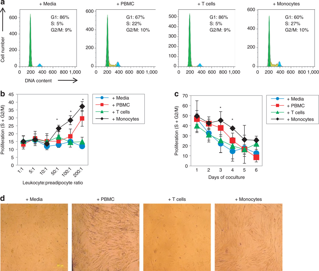
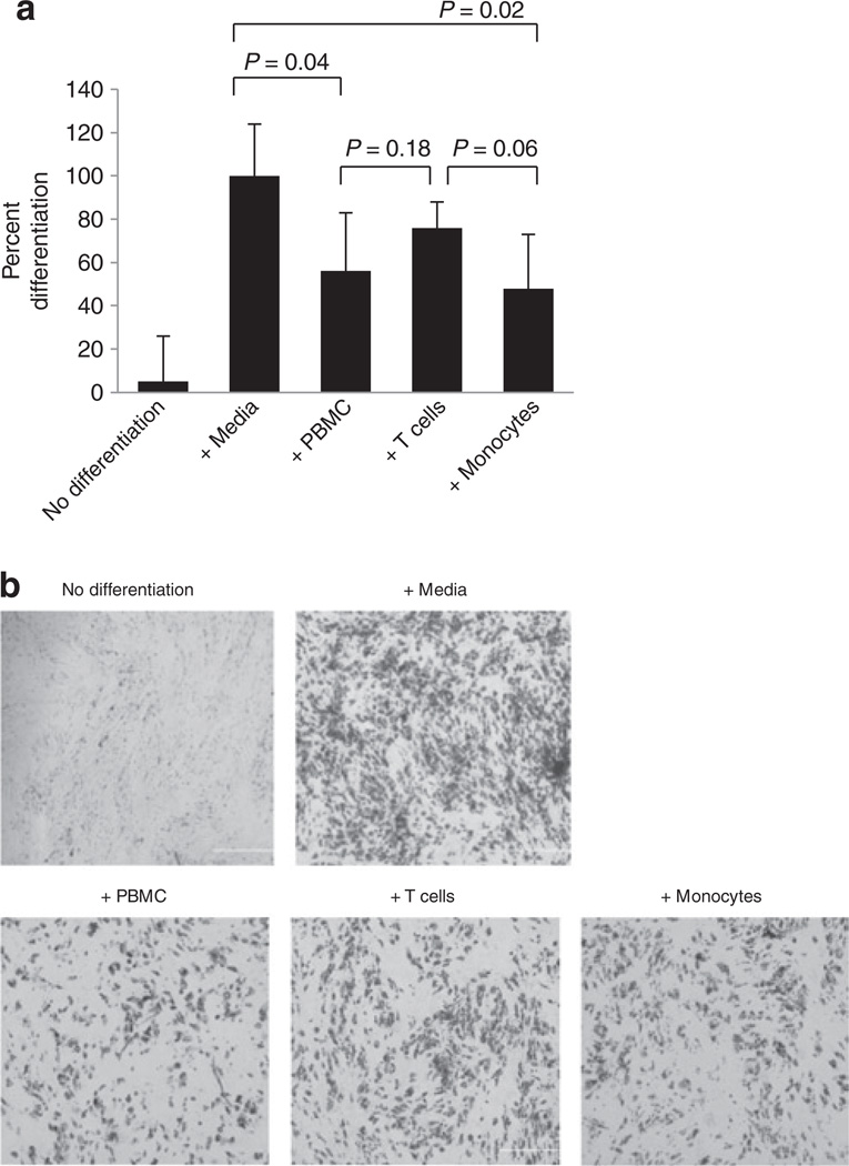
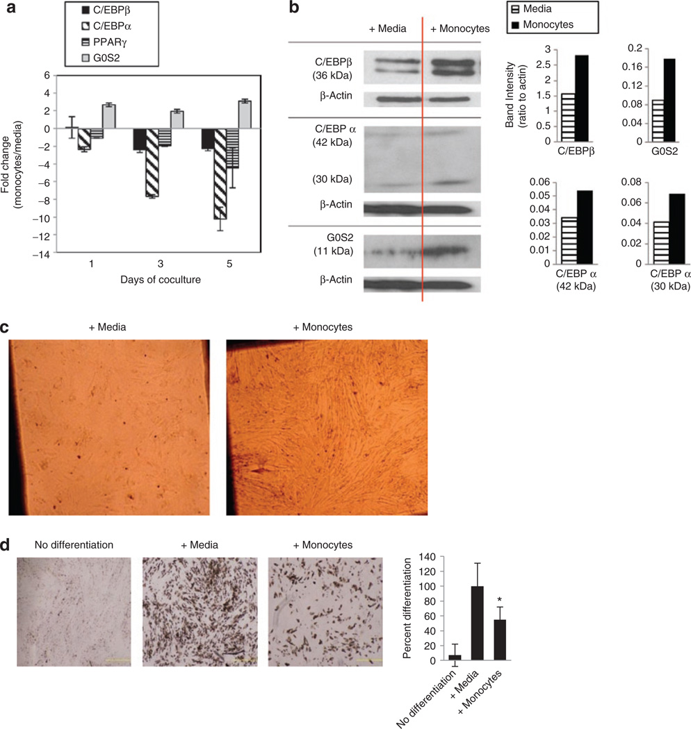
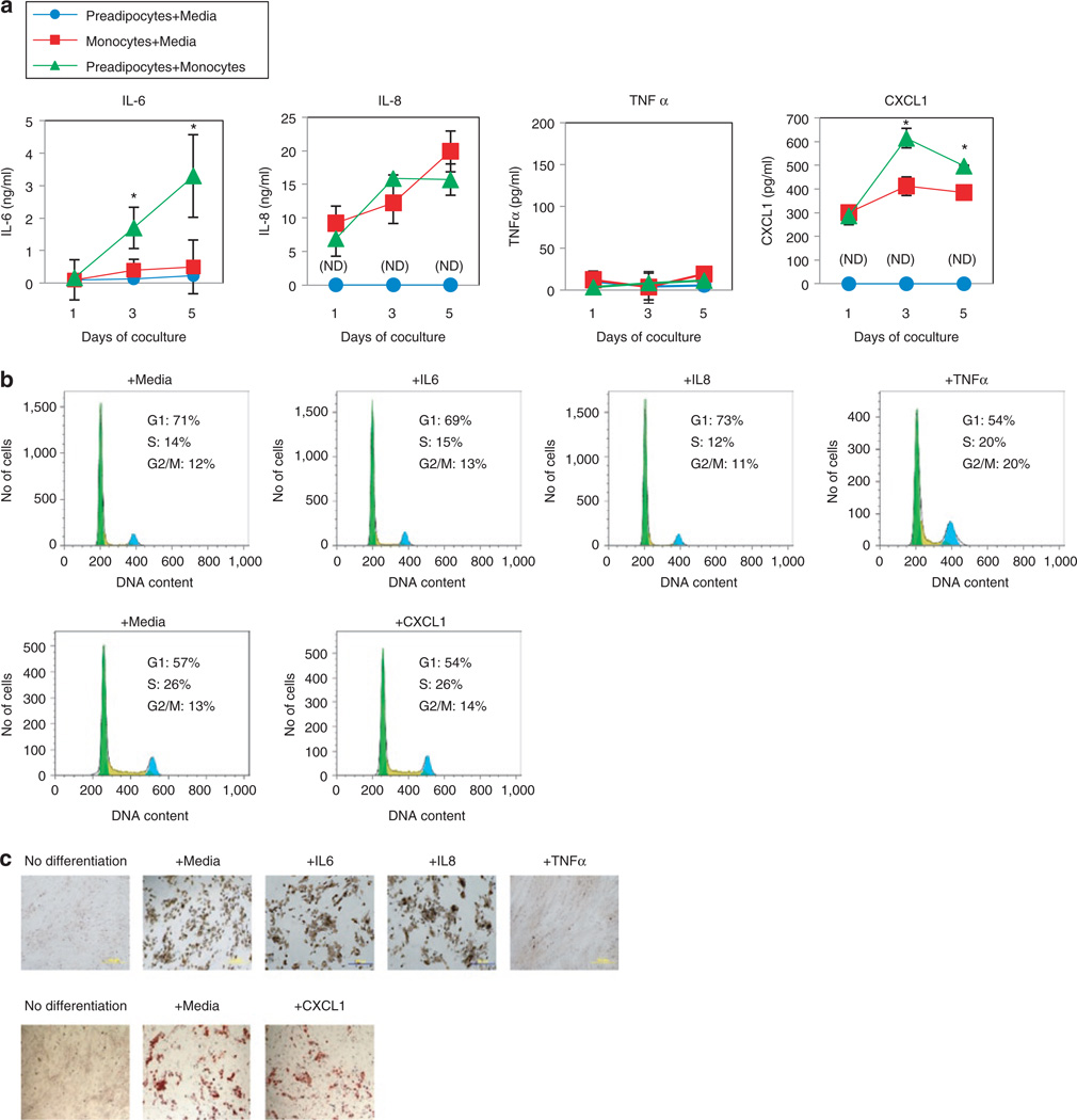
Obesity, type 2 diabetes, and HIV-associated lipodystrophy are associated with abnormalities in adipocyte growth and differentiation. In persons with these conditions, adipose depots contain increased numbers of macrophages, but the origins of these cells and their specific effects are uncertain. Peripheral blood mononuclear cells (PBMC)-derived monocytes, but not T cells, cocultured via transwells with primary subcutaneous preadipocytes, increased proliferation (approximately twofold) and reduced differentiation (~50%) of preadipocytes. Gene expression analyses in proliferating preadipocytes (i.e., prior to hormonal induction of terminal differentiation) revealed that monocytes down-regulated mRNA levels of CCAAT/enhancer binding protein, alpha (C/EBPα) and up-regulated mRNA levels of G0/G1 switch 2 (G0S2) message, genes important for the regulation of adipogenesis and the cell cycle. These data indicate that circulating peripheral blood monocytes can disrupt adipogenesis by interfering with a critical step in C/EBPα and G0S2 transcription required for preadipocytes to make the transition from proliferation to differentiation. Interactions between preadipocytes and monocytes also increased the inflammatory cytokines IL-6 and IL-8, as well as a novel chemotactic cytokine, CXCL1. Additionally, the levels of both IL-6 and CXCL1 were highest when preadipocytes and monocytes were cultured together, compared to each cell in culture alone. Such cross-talk amplifies the production of mediators of tissue inflammation.
Conflict of interest statement
The authors declared no conflict of interest.
Figures
Similar articles
-
Altered fat differentiation and adipocytokine expression are inter-related and linked to morphological changes and insulin resistance in HIV-1-infected lipodystrophic patients.Antivir Ther. 2004 Aug;9(4):555-64. Antivir Ther. 2004. PMID: 15456087
-
Effects of ghrelin on the proliferation and differentiation of 3T3-L1 preadipocytes.J Huazhong Univ Sci Technolog Med Sci. 2009 Apr;29(2):227-30. doi: 10.1007/s11596-009-0218-x. Epub 2009 Apr 28. J Huazhong Univ Sci Technolog Med Sci. 2009. PMID: 19399410
-
Tumor necrosis factor alpha and interleukin 11 secreted by malignant breast epithelial cells inhibit adipocyte differentiation by selectively down-regulating CCAAT/enhancer binding protein alpha and peroxisome proliferator-activated receptor gamma: mechanism of desmoplastic reaction.Cancer Res. 2001 Mar 1;61(5):2250-5. Cancer Res. 2001. PMID: 11280794
-
Lipin expression preceding peroxisome proliferator-activated receptor-gamma is critical for adipogenesis in vivo and in vitro.J Biol Chem. 2004 Jul 9;279(28):29558-64. doi: 10.1074/jbc.M403506200. Epub 2004 Apr 29. J Biol Chem. 2004. PMID: 15123608
-
Impact of inflammatory cytokine and adipokine gene variations in the development of HIV-associated lipodystrophy.J Gene Med. 2023 Aug;25(8):e3512. doi: 10.1002/jgm.3512. Epub 2023 Apr 26. J Gene Med. 2023. PMID: 37186064 Review.
Cited by
-
CTNNB1/β-catenin dysfunction contributes to adiposity by regulating the cross-talk of mature adipocytes and preadipocytes.Sci Adv. 2020 Jan 8;6(2):eaax9605. doi: 10.1126/sciadv.aax9605. eCollection 2020 Jan. Sci Adv. 2020. PMID: 31934629 Free PMC article.
-
G0S2 ameliorates oxidized low-density lipoprotein-induced vascular endothelial cell injury by regulating mitochondrial apoptosis.Ann Transl Med. 2022 Dec;10(24):1383. doi: 10.21037/atm-22-5618. Ann Transl Med. 2022. PMID: 36660674 Free PMC article.
-
Adipose tissue macrophages impair preadipocyte differentiation in humans.PLoS One. 2017 Feb 2;12(2):e0170728. doi: 10.1371/journal.pone.0170728. eCollection 2017. PLoS One. 2017. PMID: 28151993 Free PMC article.
References
-
- Wu H, Ghosh S, Perrard XD, et al. T-cell accumulation and regulated on activation, normal T cell expressed and secreted upregulation in adipose tissue in obesity. Circulation. 2007;115:1029–1038. - PubMed
-
- Jan V, Cervera P, Maachi M, et al. Altered fat differentiation and adipocytokine expression are inter-related and linked to morphological changes and insulin resistance in HIV-1-infected lipodystrophic patients. Antivir Ther (Lond) 2004;9:555–564. - PubMed
-
- Bedford PA, Todorovic V, Westcott ED, et al. Adipose tissue of human omentum is a major source of dendritic cells, which lose MHC Class II and stimulatory function in Crohn’s disease. J Leukoc Biol. 2006;80:546–554. - PubMed
-
- Kappert K, Meyborg H, Clemenz M, et al. Insulin facilitates monocyte migration: a possible link to tissue inflammation in insulin-resistance. Biochem Biophys Res Commun. 2008;365:503–508. - PubMed
Publication types
MeSH terms
Substances
Grants and funding
LinkOut - more resources
Full Text Sources
Medical
Molecular Biology Databases
