. 2012 Feb;19(2):345-55.
doi: 10.1038/cdd.2011.110. Epub 2011 Aug 26.

L-selectin and SDF-1 enhance the migration of mouse and human cardiac mesoangioblasts

Affiliations

L-selectin and SDF-1 enhance the migration of mouse and human cardiac mesoangioblasts

A Bernal et al. Cell Death Differ. 2012 Feb.

Abstract

Efficient delivery of stem cells to heart regions is still a major problem for cell therapy. Here, we report experiments aimed to improve migration of mouse and human cardiac mesoangioblasts to the damaged heart. Cardiac mesoangioblasts were induced to transmigrate through the endothelium by factors released by cardiomyocytes or cytokines, among which stromal-derived factor 1 (SDF-1) was the most potent. Cardiac mesoangioblasts were also delivered into the left ventricular (LV) chamber of mice after coronary artery ligation (CAL), and their in vivo homing to the damaged heart was found to be quite modest. Pretreatment of cardiac mesoangioblasts with SDF-1 or transient expression of L-selectin induced a two- to three-fold increase in their transmigration and homing to the damaged heart. Therefore, combined pretreatment with SDF-1 and L-selectin generated modified cardiac mesoangioblasts, 50% of which, after injection into the LV chamber of mice early after CAL, home directly to the damaged free wall of the heart. Finally, modified mouse cardiac mesoangioblasts, injected into the LV chamber regenerate a larger surface of the ventricle in long-term experiments in comparison with their control counterparts. This study defines the requirements for efficient homing of cardiac mesoangioblasts to the damaged heart and offers a new potent tool to optimize efficiency of future cell therapy protocols for cardiovascular diseases.


Figures
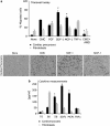
Figure 1
Figure 1
Induction of cardiac mesoangioblast transmigration by cytokines in vitro. (a) Mouse cardiac mesoangioblasts or fibroblasts were plated on endothelium-coated Transwell filters and induced to migrate for 6 h in presence of cardiomyocytes (CMCs) or different cytokines (30 ng/ml FGF; 50 ng/ml SDF-1; 80 ng/ml MCP-1; 30 ng/ml TNF-α; 10 ng/ml AMD3100, CXCR4 antagonist). A representative out of five independent experiments run in duplicate is shown (left; *α<0.02). A representative image of the transmigrated cardiac mesoangioblasts in the presence of CMCs, SDF-1 or MCP-1 is also shown. Bar, 30 μm. (b) Supernatants from fibroblasts or CMCs were collected after 4 days, and different cytokines were detected using the mouse multi-cytokine detection system. One out of three experiments is shown (*α<0.025)
Figure 2
Figure 2
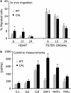
Homing of cardiac mesoangioblasts in vivo. (a) Cardiac mesoangioblasts were injected into the LV chamber of mice subjected to coronary artery ligation (CAL) or control mice. After 6, 12 or 24 h, the heart and filter organs were collected and the number of migrated cells was calculated by real-time PCR for GFP. A mean of three independent experiments run in triplicate is shown (*α<0.03). (b) Myocardium free walls of the hearts from 2-month-old WT or CAL mice were homogenized and analyzed for cytokine expression using the mouse multi-cytokine detection system. Concentrations have been normalized to protein load. One out of three experiments is shown (*α<0.025)
Figure 3
Figure 3
SDF-1 improves cardiac mesoangioblast migration through CD44, MMPs and cav-1. (a) Cardiac mesoangioblasts pretreated with different cytokines (50 ng/ml IL8 and SDF-1; 80 ng/ml MCP-1; 30 ng/ml TNF-α; 30 ng/ml FGF) were plated on endothelium-coated filters and induced to migrate for 6 h in the presence of mature cardiomyocytes in the lower chamber. FGF was used as a positive control. One representative out of five independent experiments run in duplicate is shown (left; *α<0.01). A representative image of the transmigrated SDF-1-pretreated cardiac mesoangioblasts is also shown (right). Bar, 30 μm. (b) GFP-cardiac mesoangioblasts pretreated with 50 ng/ml SDF-1 were injected into the LV chamber of control or CAL mice, and after 6 h, the heart and filter organs were analyzed by real-time PCR for the presence of migrated cells (*α<0.03, +α<0.02). (c) A gene array containing oligos corresponding to different mouse adhesion molecules was hybridized with cDNAs probes retrotranscribed from RNA of cardiac mesoangioblasts pretreated with SDF-1. Relative RNA levels of selected mRNAs normalized to β-actin expression are shown. Data presented are the mean of two independent experiments (*α<0.01). (d) SDF-1-pretreated cardiac mesoangioblasts were incubated with the MMP inhibitor, GM1489 or siRNA cav-1 inhibitor or with Abs against CD44 and αv integrin and induced to migrate for 6 h through endothelium-coated filters and in the presence of mature cardiomyocytes in the lower chamber. A representative out of three independent experiments run in duplicate is shown (*α<0.02,+α<0.005)
Figure 4
Figure 4
-selectin increases cardiac mesoangioblast migration and has synergistic effects with SDF-1 pretreatment. (a) Cardiac mesoangioblasts transfected with one or two vectors expressing different surface molecules and EGFP were plated on endothelium-coated filters and induced to migrate for 6 h in the presence of mature cardiomyocytes in the lower chamber. A representative out of five independent experiments run in duplicate is shown (left panel) (left; *α<0.01). A representative image of the transmigrated cardiac mesoangioblasts transfected with -selectin is also shown (right). Bar, 30 μm. (b) Cardiac mesoangioblasts transfected with vectors expressing -selectin and/or β2-integrin and GFP were injected into the LV chamber of CAL mice, and after 6 h, the heart and filter organs were collected. Percentage of migrated cells was calculated after performing real-time PCR for GFP. A mean of three independent experiments run in triplicate is shown (*α<0.02). (c) SDF-1-pretreated cardiac mesoangioblasts, transfected or not with vectors expressing -selectin and GFP, were induced to migrate for 6 h through endothelium-coated filters and in the presence of mature cardiomyocytes in the lower chamber. A mean of three independent experiments run in duplicate is shown (left; *α<0.01). A representative image of the transmigrated SDF-1-pretreated cardiac mesoangioblasts transfected with -selectin is also shown (right). Bar, 30 μm. (d) SDF-1-pretreated cardiac mesoangioblasts, transfected or not with vectors expressing -selectin-EGFP, as well as control cardiac mesoangioblasts were injected into the LV chamber of CAL mice, and after 6 h, the heart and filter organs were collected and analyzed by real-time PCR for the presence of GFP. A mean of six independent experiments run in triplicate is shown (*α<0.01)
Figure 5
Figure 5
Pretreatment of mouse cardiac mesoangioblasts increases myocardium regeneration. (a) Infarcted hearts, injected with cardiac mesoangioblasts pretreated with SDF-1 and transfected with the -selectin-EGFP vector or with control cardiac mesoangioblasts, were collected after 6 weeks and subjected to real-time PCR for the presence of GFP. A mean of seven independent experiments run in triplicate is shown (*α<0.02). (b) Infarcted hearts, injected with cardiac mesoangioblasts pretreated with SDF-1 and transfected with the -selectin-EGFP vector or with control cardiac mesoangioblasts, were collected after 6 weeks and sorted for the GFP population after digestion. A representative plot of four independent experiments is shown. (c) Immunostaining of the free wall collected 6 weeks after injection with control cardiac mesoangioblasts or with cardiac mesoangioblasts pretreated with SDF-1 and transfected with the -selectin-EGFP vector. Laminin is shown in red. Nuclei are stained with Hoechst (blue). Magnification: × 450. Scale bar, 100 μm. (d) Immunofluorescence for GFP and sarcomeric actin (red) from a heart region of CAL mice 6 weeks after injection with cardiac mesoangioblasts pretreated with SDF-1 and transfected with -selectin-EGFP. Scale bar, 100 μm. (e) Higher magnification immunofluorescence for GFP and sarcomeric actin (red) from the hearts of CAL mice 6 weeks after injection with cardiac mesoangioblasts pretreated with SDF-1 and transfected with -selectin-EGFP. Nuclei are stained with Hoechst (blue). Scale bar, 20 μm. (f) Western blot for GFP and sarcomeric actin of total GFP-sorted fractions recovered from the hearts of CAL mice 6 weeks after injection with control GFP-cardiac mesoangioblasts or with cardiac mesoangioblasts pretreated with SDF-1 and transfected with the -selectin-EGFP vector. One representative out of five independent experiments is shown. (g) Real-time PCR for late cardiac differentiation genes on mature cardiomyocytes and GFP-sorted fraction coming from the hearts of CAL mice 6 weeks after injection with cardiac mesoangioblasts, both control or pretreated with SDF-1 and transfected with the -selectin-EGFP vector. Quantities have been normalized. A mean of four independent experiments run in triplicate is shown
Figure 6
Figure 6
Pretreatment of human cardiac mesoangioblasts also improves homing to the heart. (a) Human cardiac mesoangioblasts pretreated with SDF-1 or transfected with -selectin construction were plated on endothelium-coated filters and induced to migrate for 6 h in the presence of cardiomyocytes. One representative out of three independent experiments run in duplicate is shown. (left; *α<0.02, +α<0.01). A representative image of the transmigrated SDF-1-pretreated cardiac mesoangioblasts transfected or not with -selectin is also shown (right). Bar, 30 μm. (b) Human cardiac mesoangioblasts transfected with vectors expressing -selectin and GFP and pretreated with SDF-1 were injected into the LV chamber of CAL-SCID mice, and after 6 h, the heart and filter organs were collected. The percentage of migrated cells is calculated after performing real-time PCR for GFP with the different samples. A mean of five independent experiments run in triplicate is shown (*α<0.025, +α<0.015)

Similar articles

Cited by

References

    1. Butcher E. Leukocyte-endothelial cell recognition: three (or more) steps to specificity and diversity. Cell. 1991;67:1033–1036. - PubMed
    1. Fu S, Liesveld J. Mobilization of hematopoietic stem cells. Blood Rev. 2000;14:205–218. - PubMed
    1. Grabovsky V, Feigelson S, Chen C, Bleijs D, Peled A, Cinamon G, et al. Subsecond induction of alpha4 integrin clustering by immobilized chemokines stimulates leukocyte tethering and rolling on endothelial vascular cell adhesion molecule 1 under flow conditions. J Exp Med. 2000;192:495–506. - PMC - PubMed
    1. Peled A, Kollet O, Ponomaryov T, Petit I, Franitza S, Grabovsky V, et al. The chemokine SDF-1 activates the integrins LFA-1, VLA-4, and VLA-5 on immature human CD34(+) cells: role in transendothelial/stromal migration and engraftment of NOD/SCID mice. Blood. 2000;95:3289–3296. - PubMed
    1. Springer T. Traffic signals for lymphocyte recirculation and leukocyte emigration: the multistep paradigm. Cell. 1994;76:301–314. - PubMed

Publication types