Review
. 2011:2011:472460.
doi: 10.1155/2011/472460. Epub 2011 Aug 21.

The tonsils revisited: review of the anatomical localization and histological characteristics of the tonsils of domestic and laboratory animals

Affiliations
Review

The tonsils revisited: review of the anatomical localization and histological characteristics of the tonsils of domestic and laboratory animals

Christophe Casteleyn et al. Clin Dev Immunol. 2011.

Abstract

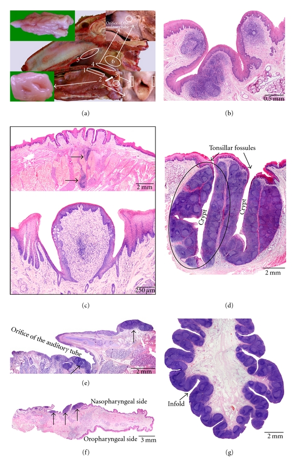
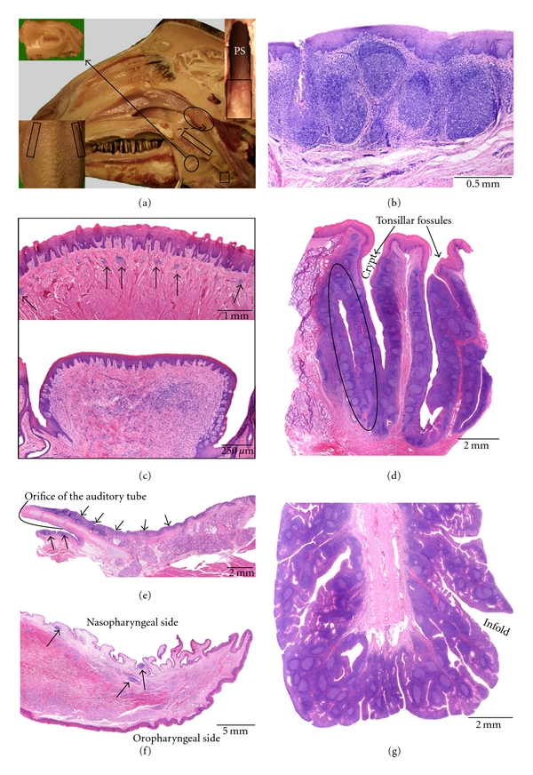
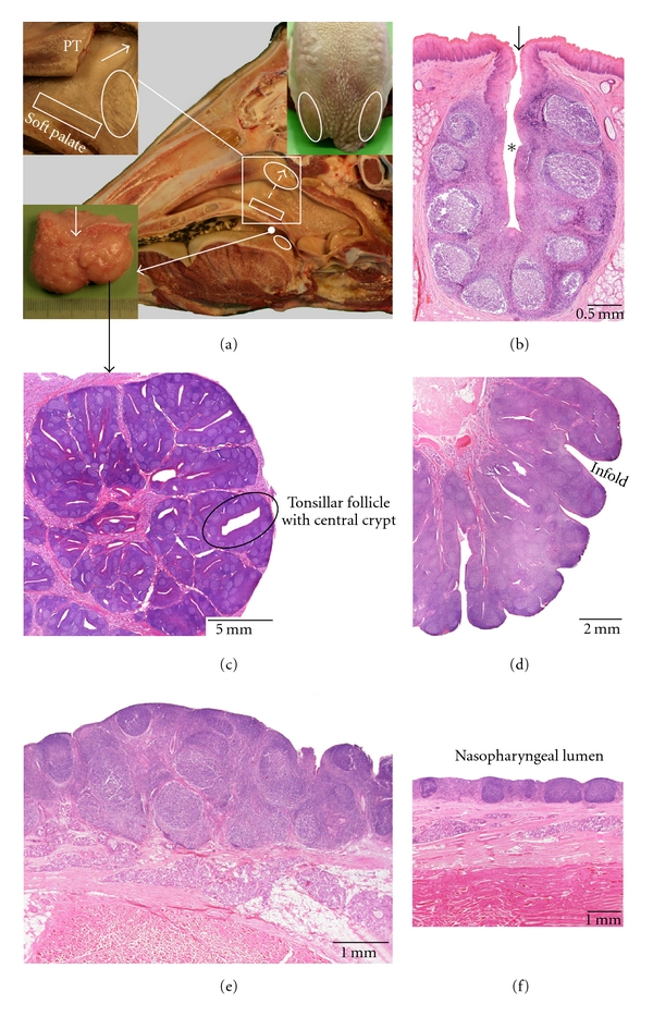
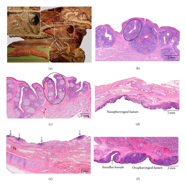
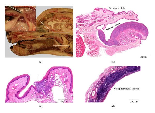
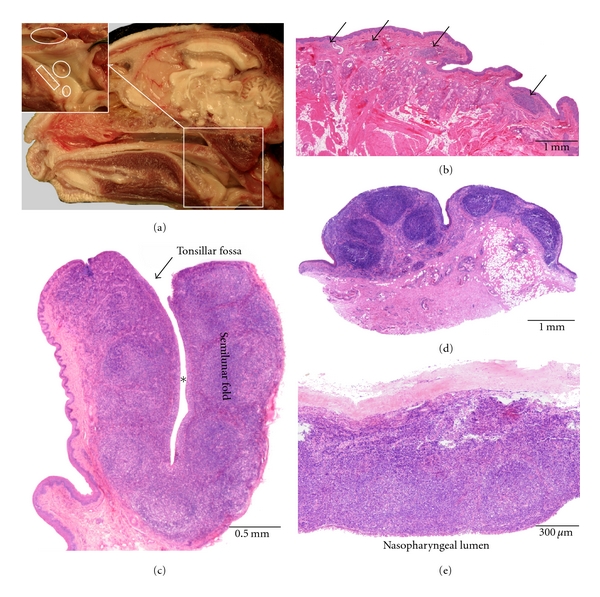
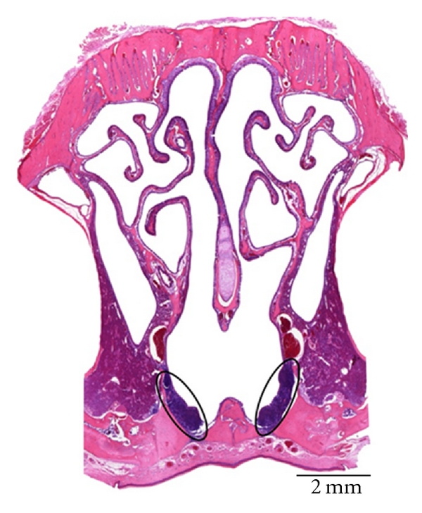
This paper gives an overview of the anatomical localization and histological characteristics of the tonsils that are present in ten conventional domestic animal species, including the sheep, goat, ox, pig, horse, dog, cat, rabbit, rat, and pigeon. Anatomical macrographs and histological images of the tonsils are shown. Six tonsils can be present in domestic animals, that is, the lingual, palatine, paraepiglottic, pharyngeal, and tubal tonsils and the tonsil of the soft palate. Only in the sheep and goat, all six tonsils are present. Proper tonsils are absent in the rat, and pigeon. In the rabbit, only the palatine tonsils can be noticed, whereas the pig does not present palatine tonsils. The paraepiglottic tonsils lack in the ox, horse, and dog. In addition, the dog and cat are devoid of the tubal tonsil and the tonsil of the soft palate.


Figures

Figure 1
Figure 1
Anatomical localization and histological characteristics of the ovine tonsils. (a) Median section through an ovine head showing the location of the lingual tonsil (1), the palatine tonsil (2) of which a larger magnification is shown in the lower left insert, the paraepiglottic tonsil (3) that is located at the lateral basis of the epiglottis (lower right insert), the tonsil of the soft palate (4) consisting of scattered lymphoid nodules at the nasopharyngeal side of the soft palate (higher magnification after 2% acetic acid fixation for 4 h shown in the upper right insert), the pharyngeal tonsil (5) of which a larger magnification is presented in the upper left insert, and the tubal tonsil (6) consisting of scattered lymphoid nodules located caudoventral to the opening of the auditory tube (higher magnification after 2% acetic acid fixation for 4 h also shown in the upper right insert). (b) Histological section through the paraepiglottic tonsil. (c) Histological images of the lingual tonsil. The upper view shows aggregations of lymphoid cells (arrows) in between the lingual muscles and salivary glands. The lower image demonstrates the presence of an aggregation of lymphoid cells in the connective tissue core of a vallate papilla. (d–g) Histological sections through the palatine (d) and tubal (e) tonsils, the tonsil of the soft palate (f), and the pharyngeal tonsil (g), respectively. A tonsillar follicle is encircled in (d); the lymphoid tissue in (e) and (f) is indicated by the arrows.
Figure 2
Figure 2
Anatomical localization and histological characteristics of the caprine tonsils. (a) Median section through a caprine head. A detail of the palatine tonsil is presented in the upper left insert. The location of the lingual tonsil, in the region of the vallate papillae, is shown in the lower left insert (dorsal view). The paraepiglottic tonsil that is located lateral to the basis of the epiglottis (small square box) is only present in a minority of goats. The position of the tonsil of the soft palate at the nasopharyngeal side of the soft palate is indicated by the rectangular box. At the caudal end of the pharyngeal septum (PS in the upper right insert), the pharyngeal tonsil (encircled on the overview, delineated by the rectangular box in the upper right insert) can be recognized by the numerous mucosal folds. The tubal tonsil is located caudoventral to the opening of the auditory tube, but is not visible on this view. (b) Histological section through the paraepiglottic tonsil. (c) Histological images of the lingual tonsil. The upper view shows aggregations of lymphoid cells (arrows) in between the lingual muscles and salivary glands. The lower image demonstrates the presence of an aggregation of lymphoid cells in the connective tissue core of a vallate papilla. (d–g) Histological sections through the palatine (d) and tubal (e) tonsils, the tonsil of the soft palate (f), and the pharyngeal tonsil (g), respectively. A tonsillar follicle is encircled in (d); the lymphoid tissue in (e) and (f) is indicated by the arrows.
Figure 3
Figure 3
Anatomical localization and histological characteristics of the bovine tonsils. (a) Median section through a bovine head. In the upper left insert, the caudal part of the pharyngeal tonsil (PT, encircled in the central image) has been partly removed to demonstrate the tubal tonsil (encircled in the insert, indicated by the interrupted arrow in the central image) caudoventral to the opening of the auditory tube (arrow in the insert). The tonsil of the soft palate (boxed area) is located at the nasopharyngeal side of the soft palate. The bilobated palatine tonsil is shown in the lower left insert. Its sinus is indicated by the arrow. The position of the lingual tonsil at the root of the tongue is presented in the upper right insert. (b) Histological section through a tonsillar follicle belonging to the lingual tonsil. The tonsillar fossula and crypt are indicated by the arrow and the asterisk, respectively. (c–f) Histological sections through parts of the palatine (c), pharyngeal (d) and tubal (e) tonsils, and the tonsil of the soft palate (f), respectively.
Figure 4
Figure 4
Anatomical localization and histological characteristics of the porcine tonsils. (a) Median section through a porcine head. The pharyngeal tonsil (large oval area) and tubal tonsil (small oval area) in the nasopharynx, the tonsil of the soft palate at the oral side of the soft palate (larger rectangular area, detail shown in upper right insert), and the lingual tonsil (smaller rectangular area) and the paraepiglottic tonsil at the lateral base of the epiglottis (encircled area, detail shown in the lower right insert) are indicated. (b–f) Histological sections through parts of the lingual tonsil (b), the tonsil of the soft palate (c), the pharyngeal tonsil (d), the tubal tonsil (e), and the paraepiglottic tonsil (f), respectively. The encircled areas in (c), (d), and (f) delineate a tonsillar follicle with a central crypt.
Figure 5
Figure 5
Anatomical localization and histological characteristics of the equine tonsils. (a) Median section through an equine head. The upper left insert shows the pharyngeal and tubal tonsils that are only visible after 4 h fixation in 2% acetic acid. The encircled area in this insert represents the area of the pharyngeal tonsil. Caudoventral to the slit-like opening of the auditory tube (white arrows), numerous white nodules belonging the tubal tonsil (black arrows) can be observed (R: rostral, C: caudal, GP: guttural pouch). The tonsil of the soft palate at the oropharyngeal side of the soft palate (small encircled area), the lingual tonsil (boxed area), and the palatine tonsil (larger encircled area, detail shown in the lower left insert) are additionally illustrated. (b) Histological section through the lingual tonsil. (c–f) Histological sections through parts of the palatine (c), pharyngeal (d), and tubal (e) tonsils and the tonsil of the soft palate (f), respectively. Primary lymphoid follicles belonging to the tubal tonsil are indicated by arrows. Tonsillar follicles are encircled in (b) and (c).
Figure 6
Figure 6
Anatomical localization and histological characteristics of the canine tonsils. (a) Median section through a canine head. The insert shows a higher magnification of the region in which the tonsils are located. In the dog, the lingual (lower boxed area of the insert), palatine (asterisk), and pharyngeal tonsils (upper boxed area of the insert caudodorsal to the opening of the auditory tube (arrow)) are present. (b) Histological section through the palatine tonsil that is partly covered by the semilunar fold. (c) Histological view of a part of the lingual tonsil that is composed of scattered aggregations of lymphoid cells (arrow). (d) Histological section through a part of the pharyngeal tonsil.
Figure 7
Figure 7
Anatomical localization and histological characteristics of the feline tonsils. (a) Median section through a feline head. The insert shows a larger magnification of the region in which the tonsils are located. The lingual (rectangular box), palatine (larger circle), paraepiglottic (smaller circle), and pharyngeal tonsils (oval) are indicated. (b) Histological section through a part of the lingual tonsil that is composed of scattered aggregations of lymphoid cells (arrows). (c) Histological section through the palatine tonsil which contains a central tonsillar fossa (asterisk). (d) and (e) Histological sections through the paraepiglottic tonsil (d) and a part of the pharyngeal tonsil (e), respectively.
Figure 8
Figure 8
Anatomical localization and histological characteristics of the rabbit tonsils. (a) Rostral view on the paired palatine tonsil (arrows). An excised palatine tonsil is illustrated at higher magnification in the insert. (b) Histological section through a palatine tonsil. The tonsillar fossa is indicated by the asterisk. (c) Histological cross-section through the rabbit nose showing the locations of NALT. Organized lymphoid tissue is present at the bottom of the ventral nasal meatus (boxed areas). Diffuse lymphoid tissue is located along the mucosal linings of the lateral walls of the nasal cavity (arrows).
Figure 9
Figure 9
Histological cross section through the nose of a rat. The NALT at the bottom of the ventral nasal meatus is encircled.
Figure 10
Figure 10
(a) Ventral view of the palate of a pigeon showing the location of the lymphoid tissue around the choanal and infundibular clefts (boxed area). (b) and (c) Histological sections of the palate showing organized lymphoid tissue that is present underneath the respiratory epithelium lining the nasal cavity (b) and underneath the keratinized stratified squamous epithelium lining the oral cavity (c).

Similar articles

Cited by

References

    1. Kuper CF, Koornstra PJ, Hameleers DMH, et al. The role of nasopharyngeal lymphoid tissue. Immunology Today. 1992;13(6):219–224. - PubMed
    1. Strid J, Hourihane J, Kimber I, Callard R, Strobel S. Disruption of the stratum corneum allows potent epicutaneous immunization with protein antigens resulting in a dominant systemic Th2 response. European Journal of Immunology. 2004;34(8):2100–2109. - PubMed
    1. Liebler-Tenorio EM, Pabst R. MALT structure and function in farm animals. Veterinary Research. 2006;37(3):257–280. - PubMed
    1. Casteleyn C. Morphological characteristics of the ovine tonsils—a study of their volume and surface area, lymphoid tissue organization, lining epithelia and vascular architecture. Faculty of Veterinary Medicine, Ghent University; 2010. p. p. 9. Ph.D. thesis.
    1. Vaerman JP, Férin J. Local immunological response in the vagina, cervix and endometrium. Acta Endocrinologica. 1975;78(194):281–305. - PubMed

Publication types

LinkOut - more resources