. 2011 Feb 1;4(1):63-76.
doi: 10.1007/s12410-010-9058-0.

Molecular Imaging of Healing After Myocardial Infarction

Affiliations

Molecular Imaging of Healing After Myocardial Infarction

Nivedita K Naresh et al. Curr Cardiovasc Imaging Rep. .

Abstract

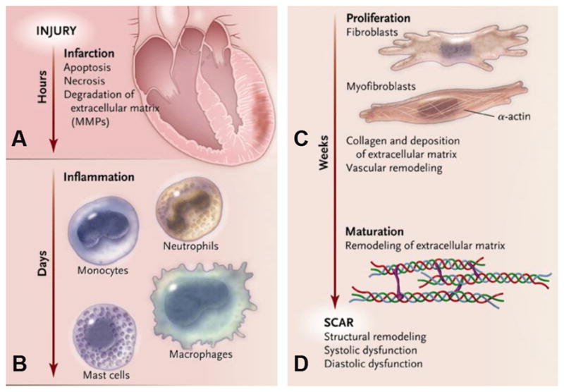
The progression from acute myocardial infarction (MI) to heart failure continues to be a major cause of morbidity and mortality. Potential new therapies for improved infarct healing such as stem cells, gene therapy, and tissue engineering are being investigated. Noninvasive imaging plays a central role in the evaluation of MI and infarct healing, both clinically and in preclinical research. Traditionally, imaging has been used to assess cardiac structure, function, perfusion, and viability. However, new imaging methods can be used to assess biological processes at the cellular and molecular level. We review molecular imaging techniques for evaluating the biology of infarct healing and repair. Specifically, we cover recent advances in imaging the various phases of MI and infarct healing such as apoptosis, inflammation, angiogenesis, extracellular matrix deposition, and scar formation. Significant progress has been made in preclinical molecular imaging, and future challenges include translation of these methods to clinical practice.


Figures

Figure 1
Figure 1
Molecular MRI (echo time, 4 ms) of CM apoptosis in myocardium exposed to mild-moderate injury. A, Mouse injected with AnxCLIO-Cy5.5. B, mouse with a similar degree of injury but injected with the control (annexin-inactivated) agent Inact_CLIOCy5.5. Significant midmyocardial uptake of the active probe (signal hypointensity) is seen in the region of injury (yellow arrows). No significant uptake of the control probe is seen (B). (Reprinted with permission from Sosnovik DE, Garanger E, Aikawa E et al. Molecular MRI of cardiomyocyte apoptosis with simultaneous delayed-enhancement MRI distinguishes apoptotic and necrotic myocytes in vivo: potential for midmyocardial salvage in acute ischemia. Circulation Cardiovascular Imaging 2009 November; 2(6):460-7)
Figure 2
Figure 2
In vivo and ex vivo imaging of monocytes at day 3 post-MI labeled using iron oxide nanoparticles 3 days before MI and its comparison with all groups at Day 3. SPION_MI clearly showed a hypointense signal by magnetic resonance imaging (in vivo and ex vivo) in the myocardial infarction. The fibrosis of this group was calculated to 17.67+3.43% (%vol). There were numerous fluorescent-loaded cells in the myocardial infarction, corresponding to CD68-positive cells. Ø_MI did not show hypointense signal by magnetic resonance imaging, or iron-loaded cells by histology. The fibrosis was 19.56+8.21%. The SPION_Sham group did not show an inflammatory infiltrate or myocardial infarction. The SPION_MI_AntiCCL5 showed a hypointense signal by magnetic resonance imaging, smaller fibrosis (10.67+5.51%) and less iron-loaded CD68-positive cells in the myocardial infarction. *P < 0.05. (Reprinted from Montet-Abou K, Daire JL, Hyacinthe JN et al. In vivo labelling of resting monocytes in the reticuloendothelial system with fluorescent iron oxide nanoparticles prior to injury reveals that they are mobilized to infarcted myocardium. European Heart Journal 2010 June; 31(11):1410-20, by permission of Oxford University Press.)
Figure 3
Figure 3
Tracking post-MI macrophage infiltration using Gd-liposomes. Gd-DTPA-enhanced inversion-recovery (IR) MRI demonstrates the region of infarction 2 days after MI (A, arrows). No myocardial enhancement was seen at day 2 post-MI before administering Gd-DTPA and after administering Gd-liposomes. (B). However, on day 4 post-MI (C), macrophages labeled with Gd-liposomes have infiltrated the infarct zone and show enhancement on IR images (arrows).
Figure 4
Figure 4
SPECT imaging of αv integrins. (A) In vivo microSPECT-CT images of 201Tl perfusion (top row, green) and 99mTc-NC100692 (middle row, red) in IGF-1 rat at 4 weeks post-MI were reconstructed in short and horizontal and vertical long axes and fused (bottom row) with a reference contrast CT image (grayscale). All post-MI rats had an anterolateral 201Tl perfusion defect (yellow solid arrows) and focal uptake of 99mTc-NC100692 in defect area. The contrast agent permitted better definition of myocardium allowing differentiation of focal myocardial uptake of targeted radiotracer from uptake within chest wall at the thoracotomy site (dashed yellow arrows). (B) Representative circumferential count profile of middle myocardial section of IGF-1 rat at 4 weeks post-MI. Count profiles for both 99mTc-NC100692 (solid circles) and 201Tl perfusion (open circles) are demonstrated. (Reprinted from Journal of Molecular Cell Cardiology 2010 June; 48(6), Dobrucki LW, Tsutsumi Y, Kalinowski L et al. Analysis of angiogenesis induced by local IGF-1 expression after myocardial infarction using microSPECT-CT imaging.:1071–1079, Copyright(2009), with permission from Elsevier.)
Figure 5
Figure 5
Imaging of fibrosis and scar post-MI using a collagen targeted contrast agent. Gradient-echo IR MR images (b,e) and corresponding picrosirius red–stained histologic sections of the LV (c, f). Arrows point to area of scarring. Standard anatomic MR images acquired by using a double IR gradient-echo sequence (a, d). Regions of contrast enhancement on midventricular short-axis MR images of the LV at two section locations obtained 40 minutes after EP-3533 injection correlate closely with photomicrographs of picrosirius red–stained tissue sections shown at nine times their original size. (Reprinted with permission from Helm PA, Caravan P, French BA et al. Postinfarction myocardial scarring in mice: molecular MR imaging with use of a collagen-targeting contrast agent. Radiology 2008 June; 247(3):788-96.)
Figure 6
Figure 6
In vivo SPECT imaging of tenascin-C. Comparison of SPECT imaging between 111In-anti-TNC-Fab and 99mTc MIBI. Transverse dual-isotope SPECT images (AC) and autoradiographies of the same rats (D, E). The uptake of 111In-anti-TNC-Fab (red in A, C, D) and 99mTc-MIBI (green in B, C, E) in acute MI heart (upper panels), in sham-operated heart (middle panels), and in normal rat heart (lower panels). A indicates anterior left ventricular wall; L, lateral left ventricular wall; P, posterior left ventricular wall; and S, septal wall. Red color indicates the uptake of 111In-anti-TNC-Fab and green color, the uptake of 99mTc-MIBI. Yellow broken lines circle myocardium. White arrows indicate sutured incision of the left intercostal space just below the myocardium. (Reprinted with permission from Odaka K, Uehara T, Arano Y et al. Noninvasive detection of cardiac repair after acute myocardial infarction in rats by 111In Fab fragment of monoclonal antibody specific for tenascin-C. International Heart Journal 2008 July; 49(4):481-92.)
Figure 7
Figure 7
Different phases of infarct healing. (Reprinted with permission from Jugdutt [49])

Similar articles

Cited by

References

    1. Lloyd-Jones D, Adams RJ, Brown TM, et al. Heart disease and stroke statistics--2010 update: a report from the American Heart Association. Circulation. 2010;121(7):e46–e215. - PubMed
    1. Leor J, Amsalem Y, Cohen S. Cells, scaffolds, and molecules for myocardial tissue engineering. Pharmacol Ther. 2005;105(2):151–63. - PubMed
    1. Frangogiannis NG. The immune system and cardiac repair. Pharmacol Res. 2008;58(2):88–111. - PMC - PubMed
    1. Nahrendorf M, Pittet MJ, Swirski FK. Monocytes: protagonists of infarct inflammation and repair after myocardial infarction. Circulation. 2010;121(22):2437–45. - PMC - PubMed
    1. Frantz S, Bauersachs J, Ertl G. Post-infarct remodelling: contribution of wound healing and inflammation. Cardiovasc Res. 2009;81(3):474–81. - PMC - PubMed

LinkOut - more resources