

Behçet's Syndrome and Thrombosis
- PMID: 21869912
- PMCID: PMC3152448
- DOI: 10.4084/MJHID.2011.026
Behçet's Syndrome and Thrombosis
Abstract
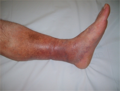
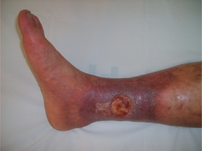
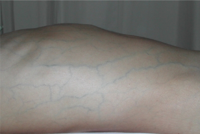
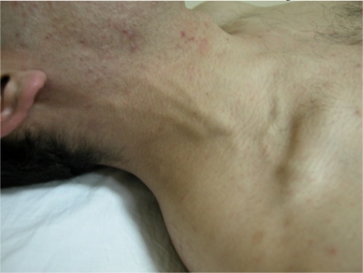
Behçet syndrome (BS) is a multisystem vasculitis with unknown etiology and a unique geographic distribution. The disease course is characterized by exacerbations and remissions while abating as the years pass. The usual onset is in the third decade. Recurrent skin mucosa lesions and sight threatening panuveitis are the hallmark of the disease. Males are more severely affected than females. Vascular involvement can occur in up to 40% of cases. BS is unique among the vasculitides in that it may involve all sizes and types of vessels. It affects the veins more than the arteries. Lower extremity vein thrombosis is the most frequent manifestation of vascular involvement, followed by vena cava thrombosis, pulmonary artery aneurysms, Budd-Chiari syndrome, peripheral artery aneurysms, dural sinus thrombosis and abdominal aorta aneurysms. Vascular involvement is frequently associated with constitut onal symptoms and increased acute phase response and is the major cause of increased mortality. A predominantly neutrophilic vasculitis around the vaso vasorum is typical of BS. The thrombus is tightly adherent to the vessel wall which probably explains why thromboembolism is so rare despite the high frequency of venous disease. Thrombophilic factors do not seem to explain thrombotic tendency in BS. Immunosuppressive treatment is essential in suppression and preventing the attacks.
Figures
References
-
- Behcet H. Uber rezidivierende, aphthose, dürch ein Virus verursachte Geshwure am Munde, am Auge und an den Genitalien. Dematologische Wochenschrift. 1937;36:1152–1157.
-
- Yurdakul S, Hamuryudan V, Fresko I, Yazıcı H. Behçet’s syndrome. In: Hochberg MC, Silman AJ, Smolen YS, Weinblatt ME, Weisman MH, editors. Rheumatology. 5th edn. Philadelphia: Mosby Elsevier; 2011. pp. 1575–1581.
-
- Direskeneli H, Saruhan-Direskeneli S. Disease Mechanisms. In: Yazici Y, Yazici H, editors. Behçet’s Syndrome. 1th edn. New York: Springer; 2010. pp. 243–264.
-
- Gul A, Ohno S. Genetics of Behçet’s disease. In: Yazici Y, Yazici H, editors. Behçet’s Syndrome. 1th edn. New York: Springer; 2010. pp. 265–276.