. 2012 Mar;7(1):187-201.
doi: 10.1007/s11481-011-9309-0. Epub 2011 Aug 26.

Clozapine protects dopaminergic neurons from inflammation-induced damage by inhibiting microglial overactivation

Affiliations

Clozapine protects dopaminergic neurons from inflammation-induced damage by inhibiting microglial overactivation

Xiaoming Hu et al. J Neuroimmune Pharmacol. 2012 Mar.

Abstract

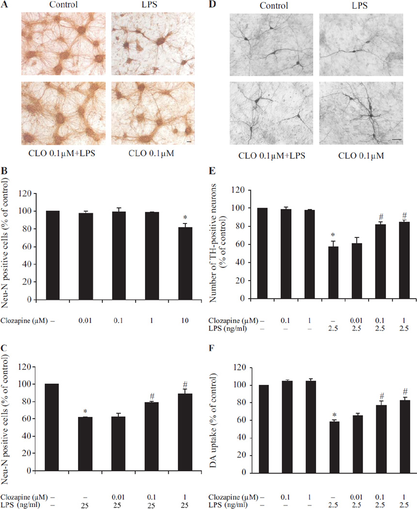
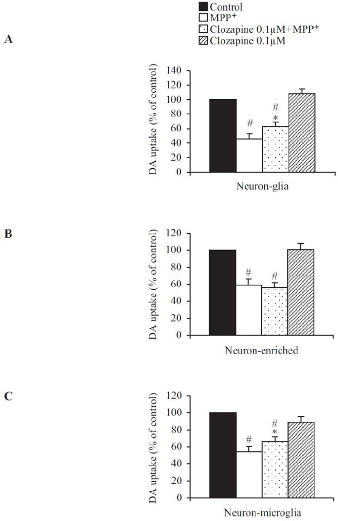
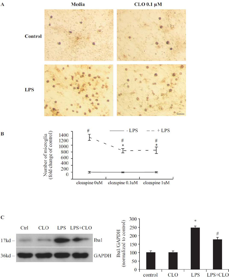
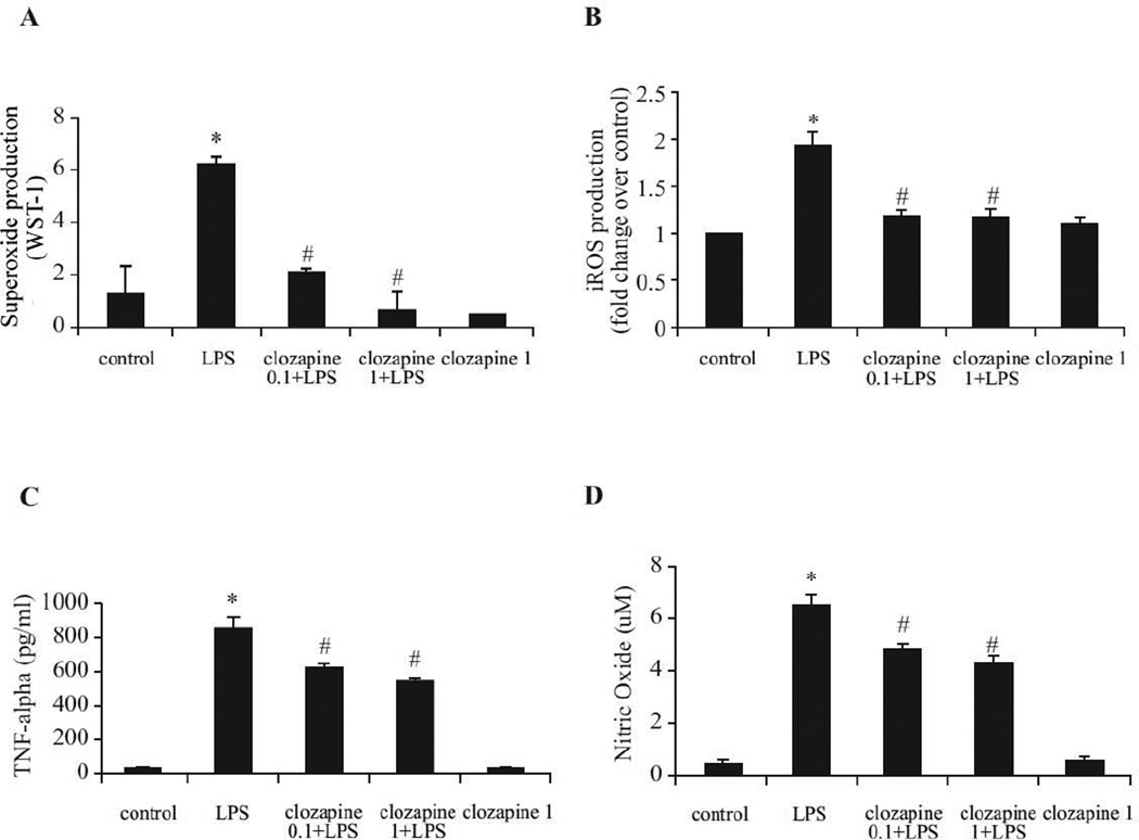
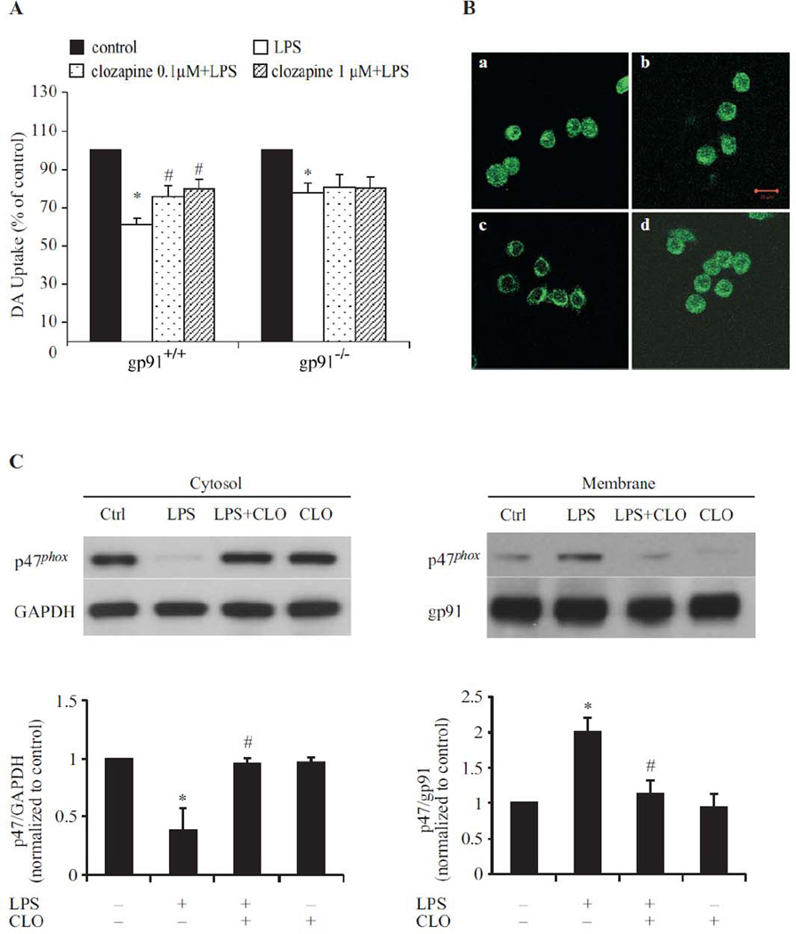
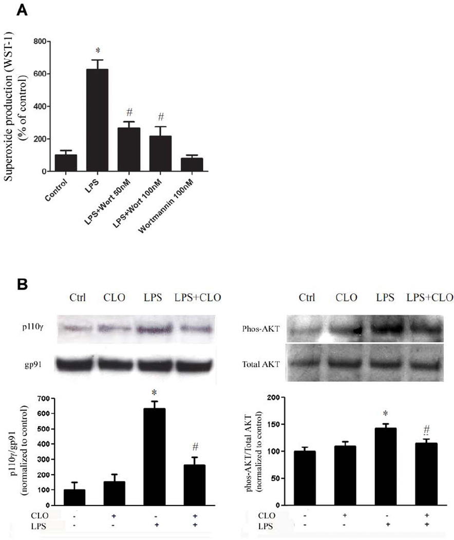
Increasing evidence suggests a possible involvement of neuroinflammation in some psychiatric disorders, and also pharmacological reports indicate that anti-inflammatory effects are associated with therapeutic actions of psychoactive drugs, such as anti-depressants and antipsychotics. The purpose of this study was to explore whether clozapine, a widely used antipsychotic drugs, displays anti-inflammatory and neuroprotective effects. Using primary cortical and mesencephalic neuron-glia cultures, we found that clozapine was protective against inflammation-related neurodegeneration induced by lipopolysaccharide (LPS). Pretreatment of cortical or mesencephalic neuron-glia cultures with clozapine (0.1 or 1 μM) for 24 h attenuated LPS-induced neurotoxicity. Clozapine also protected neurons against 1-methyl-4-phenylpyridinium(+) (MPP(+))-induced neurotoxicity, but only in cultures containing microglia, indicating an indispensable role of microglia in clozapine-afforded neuroprotection. Further observation revealed attenuated LPS-induced microglial activation in primary neuron-glia cultures and in HAPI microglial cell line with clozapine pretreatment. Clozapine ameliorated the production of microglia-derived superoxide and intracellular reactive oxygen species (ROS), as well as the production of nitric oxide and TNF-α following LPS. In addition, the protective effect of clozapine was not observed in neuron-glia cultures from mice lacking functional NADPH oxidase (PHOX), a key enzyme for superoxide production in immune cells. Further mechanistic studies demonstrated that clozapine pretreatment inhibited LPS-induced translocation of cytosolic subunit p47(phox) to the membrane in microglia, which was most likely through inhibiting the phosphoinositide 3-kinase (PI3K) pathway. Taken together, this study demonstrates that clozapine exerts neuroprotective effect via the attenuation of microglia activation through inhibition of PHOX-generated ROS production and suggests potential use of antipsychotic drugs for neuroprotection.


Conflict of interest statement

Conflict of interest:

The authors declare that they have nocompeting interests or conflicts of interest.

Figures

Fig. 1
Fig. 1. Clozapine was neuroprotective in a LPS-induced neurotoxcity model
Cortical or mesencephalic neuron–glia cultures were seeded in 24-well plates at 5 × 10⁵ cells/well. (A) Representative images of control, LPS (25 ng/ml) without or with clozapine-treated cortical neuron-glia cultures stained by MAP2 antibody. (B) Bar graph showing the quantification of NeuN-positive neurons in cultures treated with different concentration of clozapine (0.01– 10 µM). (C) Quantification of NeuN-positive neurons in the cultures treated with clozapine (0.01–1 µM) for 24hrs prior to LPS administration. (D) Representative images of control, LPS (2.5 ng/ml) without or with clozapine-treated mesencephalic neuron-glia cultures stained by TH antibody. (E) Bar graph showing the quantification of TH-positive neurons in the mesencephalic neuron-glia cultures treated with clozapine (0.01–1 µM) and/or LPS. (F) Mesencephalic neuron-glia cultures were treated with clozapine (0.01–1 µM) for 24hrs before LPS (2.5 ng/ml) administration. The functional status of DA neurons was quantified by the [3H] DA uptake assay seven days after LPS stimulation. Results were expressed as a percentage of the vehicle-treated control cultures and were the mean ± SEM from three independent experiments in triplicate. *, P<0.05 compared with the vehicle-treated control cultures; #, P<0.05, versus LPS-treated cultures. Scale bar: 50 µm.
Fig. 2
Fig. 2. The neuroprotective effect of clozapine was microglia-dependent
Clozapine (0.1 µM) or vehicle was added to the following different cell cultures: (A) neuron-glia cultures, (B) neuron-enriched cultures, or (C) neuron-microglia co-cultures by adding 1×10⁵/well of enriched microglia to the neuron-enriched cultures. MPP+ (0.25 µM) was added 24hrs after clozapine pretreatment. Neurotoxicity was quantified by the [3H] DA uptake assay 7 days after MPP+ treatment. Results were expressed as a percentage of corresponding control cultures and were the mean ± SEM from three to four independent experiments in triplicate. *, P < 0.05 compared with vehicle-treated control culture cultures; #, P<0.05, versus LPS-treated cultures.
Fig. 3
Fig. 3. Clozapine inhibited LPS-induced microglial activation without affecting microglial viability
(A) Mesencephalic neuron-glia cultures were treated with LPS and/or clozapine (0.1 and 1 µM). Cultures were fixed at 7 days after treatments. Activation of microglia was visualized by immunostaining of the Iba1 antigen, a microglia marker. The images presented are representative of three independent experiments. Scale bar: 50 µm. (B) Quantification of Iba1-positive microglia in panel A. Results were the mean ± SEM from three independent experiments in triplicate. (C) Western blot analysis of microglial activation. Cell lysates of mesencephalic neuron-glia cultures were prepared 7 days after LPS and/or clozapine (0.1 µM) treatment. Immunoblot analysis was performed to assess Iba1 antigen. GAPDH was used as loading control. The ratio of densitometry values of Iba1 and GAPDH was analyzed and normalized to control. The experiment has been performed three times. Results were presented as the mean ± SEM. (D, E) HAPI cells were pretreated with vehicle or clozapine (0.1 or 1µM) for 24 hrs followed by stimulation with LPS (2.5 ng/ml) for 24 hrs. Expression of OX6 (MHCII) was monitored by flow cytometry. Percentage of OX-6 positive cells was analyzed on a FACSCalibur. Values are mean ± SEM from three experiments. (F) Microglia-enriched culture was treated with clozapine (0.1 and 1 µM) for 24h or 48h. Cell viability was measured by PMS/MTS assay. Results were expressed as a percentage of control and were the mean ± SEM. *, P < 0.05 compared with vehicle-treated control culture cultures; #, P<0.05, versus LPS-treated cultures.
Fig. 4
Fig. 4. Clozapine inhibited LPS-induced production of reactive oxygen species (ROS) and pro-inflammatory factors
(A, B) Microglia-enriched cultures seeded at 5 × 10⁴/well were pretreated with clozapine (0.1 and 1 µM) for 24hrs before LPS (2.5 ng/ml) administration. (A) Production of extracellular superoxide was measured as SOD-inhibitable reduction of WST-1. (B) Intracellular ROS (iROS) production was determined by a fluorescence probe DCFH-DA. Results were normalized to fold change of control. (C, D) Rat primary midbrain neuron-glia cultures were pretreated with vehicle or clozapine (0.1 and 1 µM) for 24 hrs before the LPS (2.5 ng/ml) stimulation. (C) The level of TNF-α in medium was determined at 3 hrs after LPS challenge. (D) Nitric oxide was measured at 24 hrs after LPS treatment.. Results were expressed as mean ± SEM. from three to four independent experiments in triplicate. *, P<0.05 compared with the vehicle-treated control cultures; #, P<0.05, versus LPS-treated cultures.
Fig. 5
Fig. 5. Microglia PHOX was the target of clozapine (CLO)-induced neuroprotection in LPS-induced neurotoxicity
(A) Gp91phox+/+ and gp91phox–/– mice neuron-glia cultures were pretreated with vehicle or clozapine for 24hrs followed by 2.5 ng/ml LPS treatment. Neurotoxicity was assessed by the [3H] DA uptake assay. Results were expressed as a percentage of the control culture, and were the means ± SEM of six individual experiments in triplicate in each experiment. *, P < 0.05 compared with vehicle-treated control culture cultures; #, P<0.05, versus LPS-treated cultures. (B) HAPI cells seeded in a dish at 5 × 10⁴ cells/well were treated with LPS for 10 min in the absence or presence of clozapine pretreatment for 24 hrs. Cells were incubated with a rabbit polyclonal antibody against p47phox and then with a FITC-conjugated goat anti-rabbit antibody. Focal planes spaced at 0.4 µm intervals were imaged. The signal of p47phox is shown. Scale bar: 20 µm. (C) Western blot assays for p47phox levels in membrane and cytoslic fractions of HAPI cells 10 min after treatment. Densitometry analysis was performed with values of p47phox normalized to each respective loading control (GAPDH for cytosolic fraction, gp91 for membrane fraction) and further normalized to vehicle-treated controls. Experiments were performed at least three times.
Fig. 6
Fig. 6. Clozapine impairs activation of PHOX through inhibiting PI3K pathway
(A) Microglia-enriched cultures seeded at 5 × 10⁴/well were pretreated with wortmannin (50nM, 100 nM) for 10 min before LPS (10 ng/ml) administration. Production of extracellular superoxide was measured as SOD-inhibitable reduction of WST-1. Results were expressed as a percentage of the control. *, P < 0.05 compared with vehicle-treated control. (B) Microglia-enriched cultures were pretreated with clozapine (1 µM) for 30 min before LPS (10 ng/ml) administration. After 10 min LPS administration, cells were collected for western blot for the determination of levels of membrane p110γ and cytosolic phosphorylated AKT. Densitometry analysis was performed with values normalized to each respective loading control (gp91phox as a membrane marker, total AKT for its phosphorylated level) and further normalized to vehicle-treated controls. Experiments were performed at least three times. *, P < 0.05 compared with vehicle-treated control; #, P<0.05, versus LPS-treated cultures.

References

    1. Adams RA, Bauer J, Flick MJ, Sikorski SL, Nuriel T, Lassmann H, Degen JL, Akassoglou K. The fibrin-derived gamma377-395 peptide inhibits microglia activation and suppresses relapsing paralysis in central nervous system autoimmune disease. J Exp Med. 2007;204:571–582. - PMC - PubMed
    1. Altshuler LL, Casanova MF, Goldberg TE, Kleinman JE. The hippocampus and parahippocampus in schizophrenia, suicide, and control brains. Arch Gen Psychiatry. 1990;47:1029–1034. - PubMed
    1. Anderson KE, Boyle KB, Davidson K, Chessa TA, Kulkarni S, Jarvis GE, Sindrilaru A, Scharffetter-Kochanek K, Rausch O, Stephens LR, Hawkins PT. CD18-dependent activation of the neutrophil NADPH oxidase during phagocytosis of Escherichia coli or Staphylococcus aureus is regulated by class III but not class I or II PI3Ks. Blood. 2008;112:5202–5211. - PubMed
    1. Araki N, Johnson MT, Swanson JA. A role for phosphoinositide 3-kinase in the completion of macropinocytosis and phagocytosis by macrophages. J Cell Biol. 1996;135:1249–1260. - PMC - PubMed
    1. Babior BM. NADPH oxidase: an update. Blood. 1999;93:1464–1476. - PubMed

Publication types

MeSH terms

LinkOut - more resources