. 2011 Oct;32(4):367-74.
doi: 10.1007/s10059-011-0121-8. Epub 2011 Aug 23.

Dramatic increase in the signal and sensitivity of detection via self-assembly of branched DNA

Affiliations

Dramatic increase in the signal and sensitivity of detection via self-assembly of branched DNA

Kyung-Tae Kim et al. Mol Cells. 2011 Oct.

Abstract

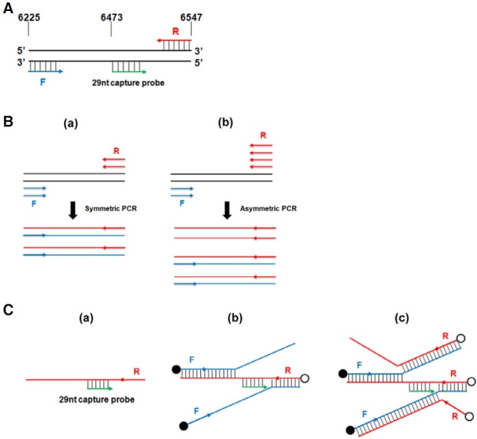
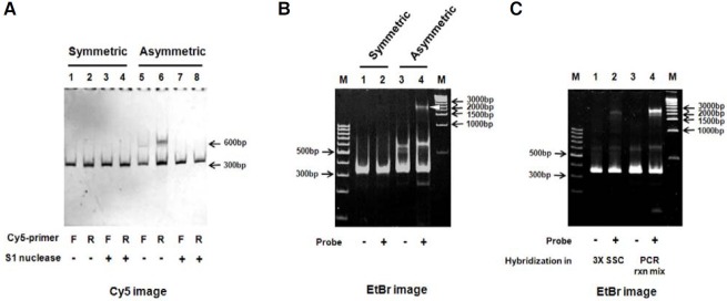
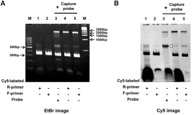
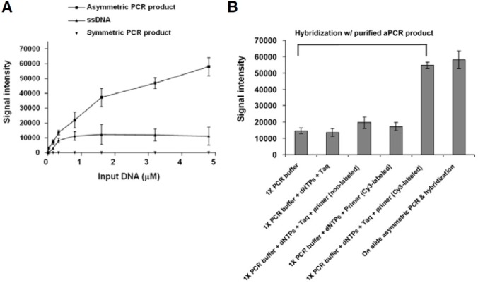
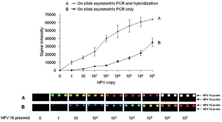
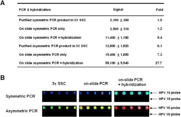
In molecular testing using PCR, the target DNA is amplified via PCR and the sequence of interest is investigated via hybridization with short oligonucleotide capture probes that are either in a solution or immobilized on solid supports such as beads or glass slides. In this report, we report the discovery of assembly of DNA complex(es) between a capture probe and multiple strands of the PCR product. The DNA complex most likely has branched structure. The assembly of branched DNA was facilitated by the product of asymmetric PCR. The amount of branched DNA assembled was increased five fold when the asymmetric PCR product was denatured and hybridized with a capture probe all in the same PCR reaction mixture. The major branched DNA species appeared to contain three reverse strands (the strand complementary to the capture probe) and two forward strands. The DNA was sensitive to S1 nuclease suggesting that it had single-stranded gaps. Branched DNA also appeared to be assembled with the capture probes immobilized on the surface of solid support when the product of asymmetric PCR was hybridized. Assembly of the branched DNA was also increased when hybridization was performed in complete PCR reaction mixture suggesting the requirement of DNA synthesis. Integration of asymmetric PCR, heat denaturation and hybridization in the same PCR reaction mixture with the capture probes immobilized on the surface of solid support achieved dramatic increase in the signal and sensitivity of detection of DNA. Such a system should be advantageously applied for development of automated process for detection of DNA.


Figures

Fig. 1.
Fig. 1.. Model of the bDNA formed between capture probes and the strands of the PCR product. (A) The L1 region of the HPV16 DNA as a model system. Shown are the regions of L1, the PCR primers, and the capture probes (29 nt) that were used in this study. F: forward primer; and R: reverse primer. Also shown are the coordinates of the L1 region in the HPV16 DNA (GenBank accession number K02718). (B) Generation of excess single stranded DNA by aPCR. (a) sPCR in the presence of equimolar amount of forward and reverse primers generates complete double-stranded PCR product. (b) aPCR in the presence of an excess amount of a primer (for example, reverse primer) generates an excess amount of single-stranded DNA (for example, the strand extended by reverse primer) in addition to double stranded DNA. (C) Model of the assembly of bDNA. In this model, the strands extended by forward (F) and reverse (R) PCR primers are shown. (a) The single-stranded reverse strand produced by aPCR will bind to the capture probe. (b) Once the reverse strand binds to a capture probe, this leaves two open spaces for the strand extended by forward primers will bind to the strand bound to a capture probe. (c) The DNA structure shown in (b) has two open regions in the bound forward strands for binding of two DNA strands extended by reverse primers. When PCR is carried out in the presence of the primers labeled with fluorescent molecule, the structure shown in (c) will have 5 labels (indicated by open and closed circles).
Fig. 2.
Fig. 2.. Synthesis of single-stranded DNA by aPCR and assembly of bDNA. (A) sPCR and aPCR products were labeled by either Cy5-forward (F) or Cy5-reverse (R) primers, and the product was digested with S1 nuclease after purification of DNA. After 4% polyacrylamide gel electrophoresis, the gel was scanned for Cy5 image by LAS-4000 Biomolecular Imager. sPCR: lane 1, +Cy5-forward primer; lane 2, +Cy5-reverse primer; lane 3, DNA in lane 1 digested with S1; lane 4, DNA in lane 2 digested with S1. aPCR: lane 5, +Cy5-forward primer; lane 6, +Cy5- reverse primer; lane 7, DNA in lane 5 digested with S1; lane 8, DNA in lane 6 digested with S1. (B) Assembly of bDNA. sPCR and aPCR were carried out in the presence of 107 copies of HPV16 plasmid. After the PCR, 29 nt HPV16 capture probe (10 pmol) was added to some of the PCR reaction mixtures, and all of the mixtures were heated at 94℃ for 5 min for the denaturation of the PCR product and incubated at 50℃ for 1 h for the hybridization. The DNA was analyzed via 4% polyacrylamide gel electrophoresis. Hybridization of sPCR product in the absence (lane 1) and presence (lane 2) of capture probe; hybridization of aPCR product in the absence (lane 3) and presence (lane 4) of capture probe. M: size markers. The arrow in lane 4 indicates the bDNA of 2,500 bp. (C) Purified Cy3-labeled aPCR product was hybridized with HPV16 capture probe either in 3x SSC (lane 2) or in complete PCR reaction mixture (lane 4).
Fig. 3.
Fig. 3.. Characterization of bDNA. aPCR was carried out, and the mixture was heated at 94℃ and incubated at 50℃ for 1 h in the absence (lanes 1 and 2) or presence (lanes 3-5) of capture probe as described in “Materials and Methods”. The process was carried out in the presence of either a forward primer, a reverse primer, or a capture probe that was labeled with Cy5. After the gel electrophoresis, the gel was scanned for the Cy5 image, and the fluorescent intensity of the branched DNA was determined with the LAS-4000 Imager, as described in “Materials and Methods”. The gel was also stained with ethidium bromide. (A) Ethidium bromide image. (B) Cy5 image. Lane 1: + Cy5-reverse primer; lane 2, + Cy5-forward primer; lane 3, + Cy5-capture probe; lane 4, + Cy5-reverse primer; lane 5, + Cy5-forward primer.
Fig. 4.
Fig. 4.. Assembly of bDNA on solid support. (A) Comparison of the signal obtained from the hybridization of different preparations of amplified HPV16 DNA with the capture probes immobilized on a glass slide. Increasing amounts of Cy3-labeled single- stranded DNA (extended by Cy3-labeled reverse primer), and products of aPCR or sPCR, were added to the hybridization buffer (30 μl of 5 mM Tris-HCl [pH 7.5], 0.5 mM EDTA, and 1 M NaCl). The mixtures were transferred to the chambers that were assembled on the surface of a glass slide that contained immobilized HPV16 and 18 capture probes. The slides were heated at 94℃ for 5 min and incubated at 50℃ for 1 h. The DNA was labeled during the PCR with the addition of Cy3-labeled PCR primers. Note that the labeled PCR primers were diluted 1:10 with non-labeled primers, which made the signal fall within the dynamic range of the used laser scanner. The experiments were carried out several times, and similar results were obtained. (B) Increased signal of detection by hybridization of aPCR product with immobilized capture probes by hybridization in complete PCR reaction mixture. Cy3-labeled aPCR product (0.3 μM) was hybridized with the immobilized capture probes in PCR buffer in the presence of various combination of dNTP, PCR primers (non-labeled or Cy3-labeled), and Taq DNA polymerase.
Fig. 5.
Fig. 5.. Sensitivity of detection of HPV16 DNA via integrated aPCR and hybridization on a glass slide. Top: The signal intensity that was obtained with different numbers of HPV16 plasmid. The values were corrected for the background signal (1,500 units). Line A, aPCR was carried out with serially diluted HPV16 plasmid in the presence of Cy3-labeled primers (the labeled primers were diluted 5 fold with non-labeled primers), heated at 94℃ (for the denaturation), and incubated at 50℃ (for the hybridization), all in the same PCR reaction mixture, on glass slides that contained immobilized HPV16 and 18 capture probes. After the hybridization, the slides were washed and scanned as described in “Materials and Methods”. Line B, aPCR was carried out; but at the end of the reaction, the slides were washed and scanned without hybridization. Bottom: The fluorescent images of the capture probe spots. HPV18 capture probes served as non-specific probes. This experiment was carried out several times and similar results were obtained.
Fig. 6.
Fig. 6.. Comparison of the signal intensities obtained via sPCR and aPCR and hybridization. aPCR and sPCR were carried out in the presence of 105 molecules of HPV16 plasmid and Cy3-labeled PCR primers (diluted 5 fold with non-labeled primers), and the purified product was heated at 94℃ for 5 min and hybridized with the immobilized capture probes in 3x SSC for 1 h on glass slides. aPCR and sPCR were also carried out on the surface of immobilized probes, and the slides were scanned after PCR alone or after heating and hybridization. (A) The signal values were corrected for the background signal. (B) The fluorescent images of the capture probe spots are shown. The HPV18 capture probe served as a specificity control.

Similar articles

Cited by

References

    1. Albrecht V., Chevallier A., Magnone V., Barbry P., Vandenbos F., Bongain A., Lefebvre J.C., Giordanengo V. Easy and fast detection and genotyping of high-risk human papillomaviruse by dedicated DNA microarrays. J. Virol. Methods. (2006);137:236–244. - PubMed
    1. Brewer B.J., Fangman W.L. The localization of replication origins on ARS plasmids in S. cerevisiae. Cell. (1987);56:463–471. - PubMed
    1. Chung I.H., Yoo H.S., Eah J.Y., Yoon H.K., Jung J.W., Hwang S.Y., Kim C.B. A DNA microarray for idenficiation of seleceted Korean birds based on mitochondrial cytochrome c oxidase I gene sequences. Mol. Cells. (2010);30:295–301. - PubMed
    1. Collins M.L., Irvine B., Tyner D., Fine E., Zayati C., Chang C., Horn T., Ahle D., Detmer J., Shen L.P., et al. A branched DNA signal amplification assay for quantification of nucleic acid targets below 100 molecules/ml. Nucleic Acids Res. (1997);25:2979–2984. - PMC - PubMed
    1. Dunderdale H.J., Benson F.E., Parson C.A., Sharples G.J., Lloyd R.G., West S.G. Formation and resolution of recombination intermediates by E.coli RecA and RuvC proteins. Nature. (1991);354:506–510. - PubMed

Publication types

LinkOut - more resources