Comparative Study
. 2011 Sep 20;50(37):7919-32.
doi: 10.1021/bi200873u. Epub 2011 Aug 26.

What determines the activity of antimicrobial and cytolytic peptides in model membranes

Affiliations
Comparative Study

What determines the activity of antimicrobial and cytolytic peptides in model membranes

Kim S Clark et al. Biochemistry. .

Abstract

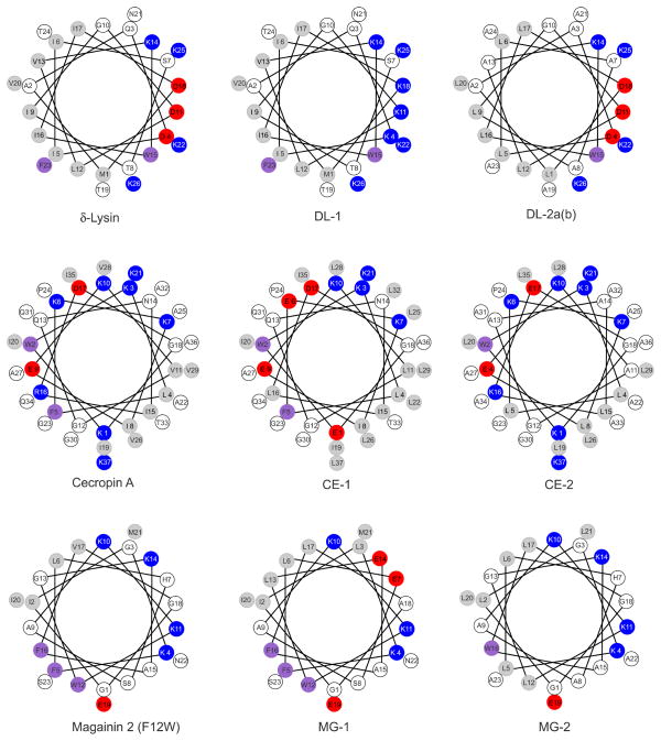
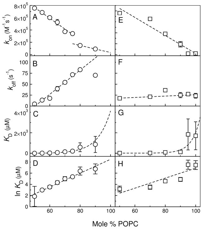
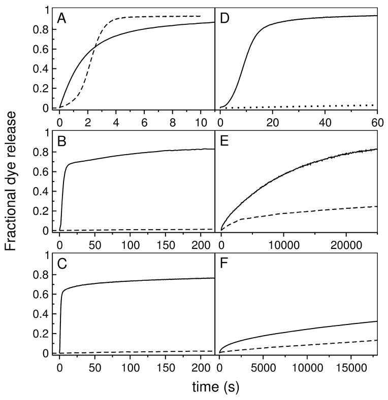
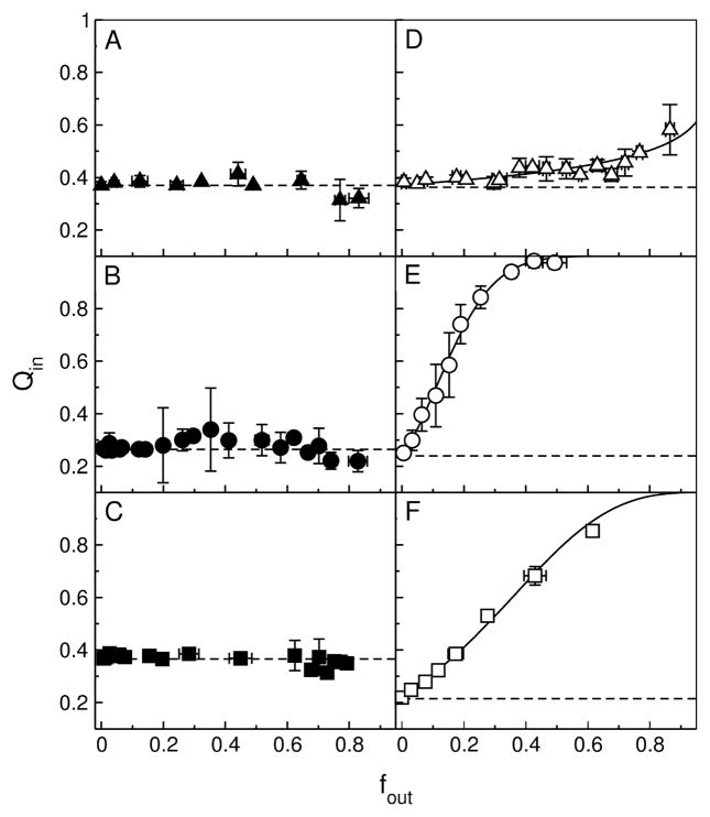
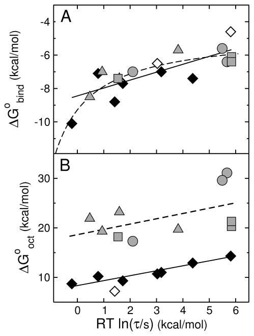
We previously proposed three hypotheses relating the mechanism of antimicrobial and cytolytic peptides in model membranes to the Gibbs free energies of binding and insertion into the membrane [Almeida, P. F., and Pokorny, A. (2009) Biochemistry 48, 8083-8093]. Two sets of peptides were designed to test those hypotheses, by mutating of the sequences of δ-lysin, cecropin A, and magainin 2. Peptide binding and activity were measured on phosphatidylcholine membranes. In the first set, the peptide charge was changed by mutating basic to acidic residues or vice versa, but the amino acid sequence was not altered much otherwise. The type of dye release changed from graded to all-or-none according to prediction. However, location of charged residues in the sequence with the correct spacing to form salt bridges failed to improve binding. In the second set, the charged and other key residues were kept in the same positions, whereas most of the sequence was significantly but conservatively simplified, maintaining the same hydrophobicity and amphipathicity. This set behaved completely different from predicted. The type of release, which was expected to be maintained, changed dramatically from all-or-none to graded in the mutants of cecropin and magainin. Finally, contrary to the hypotheses, the results indicate that the Gibbs energy of binding to the membrane, not the Gibbs energy of insertion, is the primary determinant of peptide activity.


Figures

FIGURE 1
FIGURE 1
Thermodynamic cycle for peptide binding to the membrane interface and insertion into the bilayer. The folding equilibrium in water lies toward the unstructured state and is determined by ΔGfo, which is typically small in comparison with the other terms. The Gibbs energy of binding to the interface ( ΔGifo) includes contributions from the hydrophobic effect and secondary structure formation. Transfer, as an α-helix, from water to the bilayer interior is approximated by transfer to octanol ( ΔGocto). The Gibbs energy of transfer from the surface to the interior of the bilayer is approximately ΔGoctoΔGifo=ΔGoctifo. Modified with permission from ref (38). Copyright 2007 Elsevier.
FIGURE 2
FIGURE 2
Helical wheel projection of the original and variant peptides studied. The colors reflect the hydrophobicities according to the Wimley-White interfacial scale (18). White, hydrophilic or neutral; light gray, hydrophobic; magenta, aromatic; red, negatively charged; and blue, positively charged, at pH 7.5. The only difference between DL-2a and DL-2b is the replacement of Ala 13 and 23 by Leu in DL-2b.
FIGURE 3
FIGURE 3
Example of a curve of binding kinetics, acquired for DL-1 binding to 100 μM POPC LUV. The data correspond to fluorescence emission from the lipid fluorophore 7MC-POPE incorporated in the membrane, through FRET, upon excitation of the Trp residue on the peptide. The line is a single exponential fit to the data.
FIGURE 4
FIGURE 4
Representative CD spectra of the mutant peptides (20 μM) in 5 mM LUV of POPC, except for CE-2 and MG-2, which were in POPC/POPG 1:1. (A) DL-1, (B) CE-1, (C) MG-1, (D) DL-2a, (E) CE-2, and (F) MG-2.
FIGURE 5
FIGURE 5
Kinetics of peptide binding to LUV of POPC (Dl-1, CE-1, MG-1, and DL-2a) or POPC/POPG 1:1 (CE-2 and MG-2). The apparent rate constant (kapp), obtained from a single exponential fit to the traces of binding kinetics, is plotted against the lipid concentration. (A) DL-1, (B) CE-1, (C) MG-1, and (D) DL-2a. The error bars are standard deviations from 2–4 independent experiments and the lines are linear regressions, which yield kon from the slope and koff from the y-intercept. In the case of MG-1 the data are averages from POPC/POPG LUV 50:50, 70:30, 80:20, and 90:10, since there was no detectable dependence on membrane composition. The values of kon, koff, and KD obtained are listed in Table 2.
FIGURE 6
FIGURE 6
Kinetics of binding of CE-2 (left panels) and MG-2 (right) to LUV as a function of membrane composition, from POPC/POPG 1:1 to pure POPC. The data show the dependence of kon (A, E), koff (B, F), KD (C, D), and ln KD on the lipid composition. Extrapolation to pure POPC, to which binding is weak, provides the best estimates of the rate and equilibrium constants for CE-2 and MG-2 in POPC, listed in Table 2.
FIGURE 7
FIGURE 7
Kinetics of CF release induced by the mutant peptides (solid lines) from 50 μM POPC LUVs. The curves for the original peptides are shown for comparison (dashed lines) under the same conditions. (A) DL-1 (solid) and δ-lysin (dashed) from POPC; (B) CE-1 (solid) and cecropin A (dashed); (C) MG-1 (solid) and magainin 2 (dashed); (D) DL-2a (solid) and DL-2b (dotted) from POPC; (E) CE-2 (solid) and cecropin A (dashed); and (F) MG-2 and magainin 2 (dashed). Some curves were acquired for longer times, but are shown in the time frames that allow best comparison. In (D), only the beginning of the curve for DL-2b is shown; the curve has the same shape as for Dl-2a, but on a timescale an order of magnitude larger.
FIGURE 8
FIGURE 8
ANTS/DPX requenching assay for the mutant peptides. (A) DL-1 in POPC, (B) CE-1 in POPC, (C) MG-1 in POPC, (D) DL-2a in POPC, (E) CE-2 in POPC/POPG 1:1, and (F) MG-2 in POPC/POPG 1:1. The solid lines in panels D–F (DL-2a, CE-2, and MG-2) represent the best fits to the equation for graded release (Eq. 5). The corresponding fit parameters are, for DL-2a, α = 0.28 and Ksta = 80 M−1; for CE-2, α = 8.5 and Ksta = 140 M−1; and for MG-2, α = 3.0 and Ksta = 140 M−1. For comparison with DL-2a, for δ-Lysin in POPC, the parameters were α = 0.22 and Ksta = 220 M−1 (15). The dashed horizontal lines represent the behavior expected for all-or-none release.
FIGURE 9
FIGURE 9
(A) Gibbs energy of binding to the membrane interface determined experimentally ( ΔGbindo) as a function of the mean characteristic time τ for CF release from POPC LUVs. Each gray symbol corresponds to a peptide examined here: δ-lysin, DL-1, DL-2a, and DL-2b are shown by gray triangles; cecropin A, CE-1, and CE-2, by gray circles; and magainin 2, MG-1, and MG-2, by gray squares. The data for TP10 variants previously published (16) are shown here for comparison (diamonds): TP10W, TPW-1, TPW-2, and TPW-3; TP10, TP10-COO, TP10W-COO, and TP10-7MC (38, 58). The black symbols correspond to experimental data; the open symbols correspond to TP10 and TP10-COO, for which τ is experimental but the binding affinity is calculated with the Wimley-White scale (neither contains Trp). The straight line is a fit, with a slope of 0.5. The dashed line represents qualitatively the expected behavior limited by diffusion or bilayer response. (B) Gibbs energy of transfer to octanol calculated with the Wimley-White scale ( ΔGocto) as a function of the mean characteristic time τ for CF release from POPC LUV. The points correspond to the same peptides as in (A) and the same symbols were used. Again the TP10 variant data are from our previous paper (16). The open circle corresponds to TP10-7MC for which ΔGoctowas estimated assuming Tyr for the Lys-MC residue (16). The lines are fits, which have slopes of 1.

References

    1. Zasloff M. Antimicrobial peptides of multicellular organisms. Nature. 2002;415:389–395. - PubMed
    1. Giangaspero A, Sandri L, Tossi A. Amphipathic α-helical antimicrobial peptides. Eur J Biochem. 2001;268:5589–5600. - PubMed
    1. Tossi A, Sandri L, Giangaspero A. Amphipathic, α-helical antimicrobial peptides. Biopolymers. 2000;55:4–30. - PubMed
    1. Nickel W. The mystery of nonclassical protein secretion. A current view on cargo proteinsand potential export routes. Eur J Biochem. 2003;270:2109–2119. - PubMed
    1. Lindgren M, Langel U. Clasees and prediction of cell-penetrating peptides. Methods Mol Biol. 2011;683:3–19. - PubMed

Publication types

MeSH terms