

Review of juxtaglomerular cell tumor with focus on pathobiological aspect
- PMID: 21871063
- PMCID: PMC3173291
- DOI: 10.1186/1746-1596-6-80
Review of juxtaglomerular cell tumor with focus on pathobiological aspect
Abstract
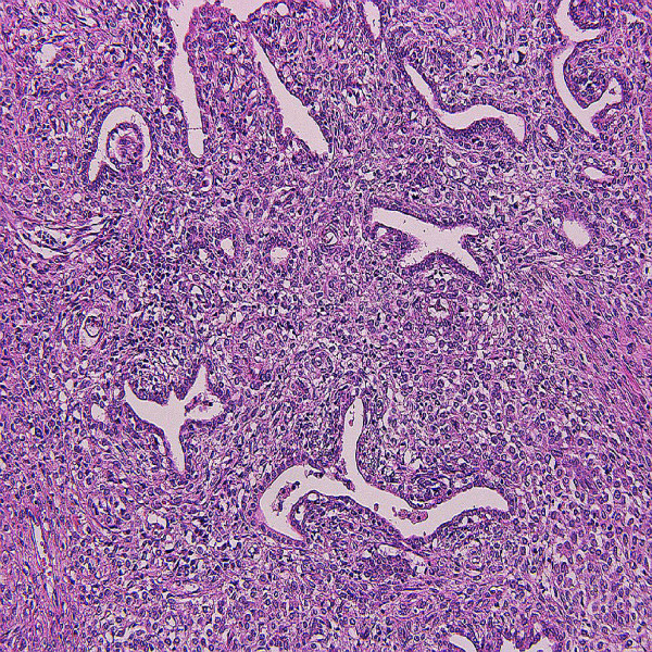
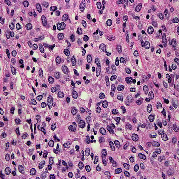
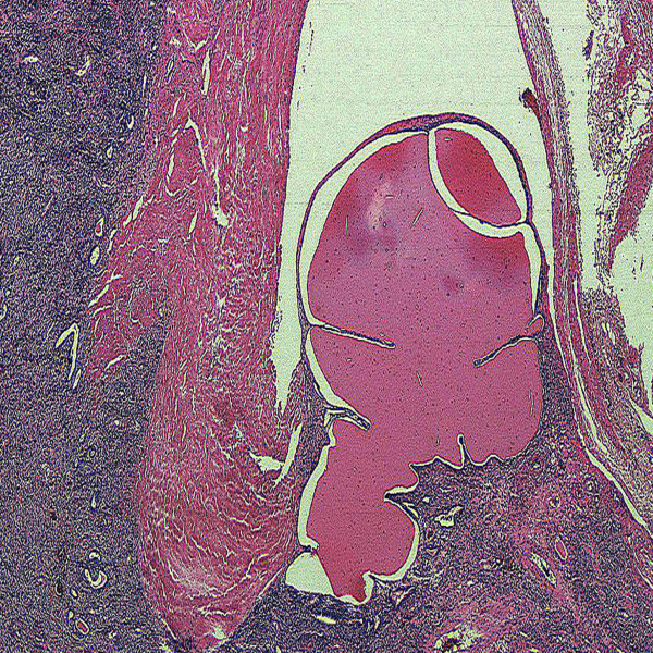
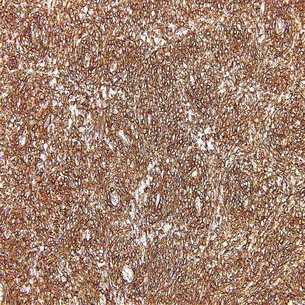
Juxtaglomerular cell tumor (JGCT) generally affects adolescents and young adults. The patients experience symptoms related to hypertension and hypokalemia due to renin-secretion by the tumor. Grossly, the tumor is well circumscribed with fibrous capsule and the cut surface shows yellow or gray-tan color with frequent hemorrhage. Histologically, the tumor is composed of monotonous polygonal cells with entrapped normal tubules. Immunohistochemically, tumor cells exhibit a positive reactivity for renin, vimentin and CD34. Ultrastructurally, neoplastic cells contain rhomboid-shaped renin protogranules. Genetically, losses of chromosomes 9 and 11 were frequently observed. Clinically, the majority of tumors showed a benign course, but rare tumors with vascular invasion or metastasis were reported. JGCT is a curable cause of hypertensive disease if it is discovered early and surgically removed, but may cause a fatal outcome usually by a cerebrovascular attack or may cause fetal demise in pregnancy. Additionally, pathologists and urologists need to recognize that this neoplasm in most cases pursues a benign course, but aggressive forms may develop in some cases.
Figures
References
-
- Kihara I, Kitamura S, Hoshino T, Sieda H, Watanabe T. A hitherto unreported vascular tumor of the kidney: A proposal of "juxtaglomerular cell tumor". Acta Path Jap. 1968;18(2):197–206. - PubMed
-
- Eddy RL, Sanchez SA. Renin-secreting renal neoplasm and hypertension with hypokalemia. Ann Int Med. 1971;75(5):725–729. - PubMed
-
- Valdes G, Lopez JM, Maritinez P, Rosenberg H, Barriga P, Rodriguez JA, Otipka N. Renin-secreting tumor. Case report. Hypertension. 1980;2(5):714–718. - PubMed
Publication types
MeSH terms
LinkOut - more resources
Full Text Sources
Medical
