. 2011 Oct;179(4):1623-9.
doi: 10.1016/j.ajpath.2011.06.039. Epub 2011 Aug 24.

Brain cell reservoirs of latent virus in presymptomatic HIV-infected individuals

Affiliations

Brain cell reservoirs of latent virus in presymptomatic HIV-infected individuals

Katherine A Thompson et al. Am J Pathol. 2011 Oct.

Abstract

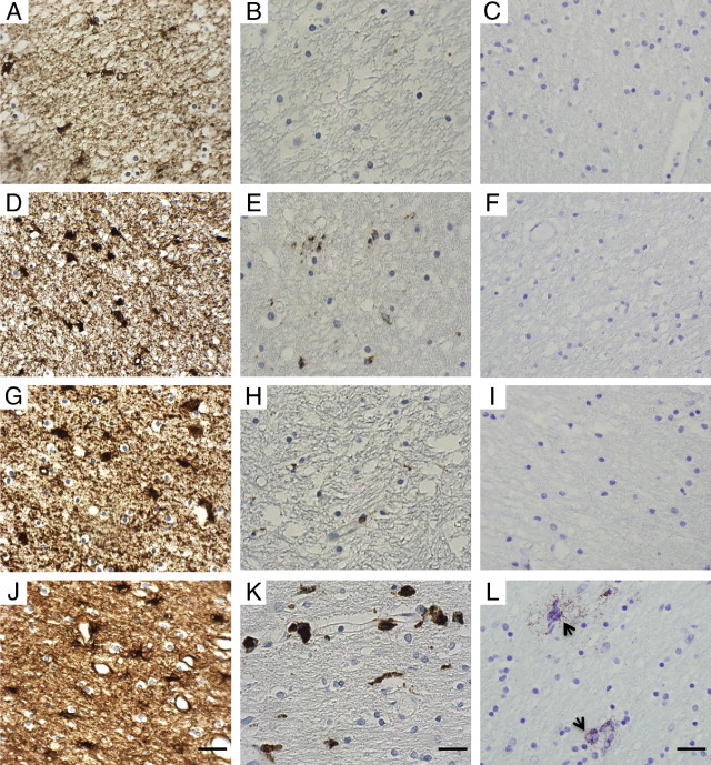
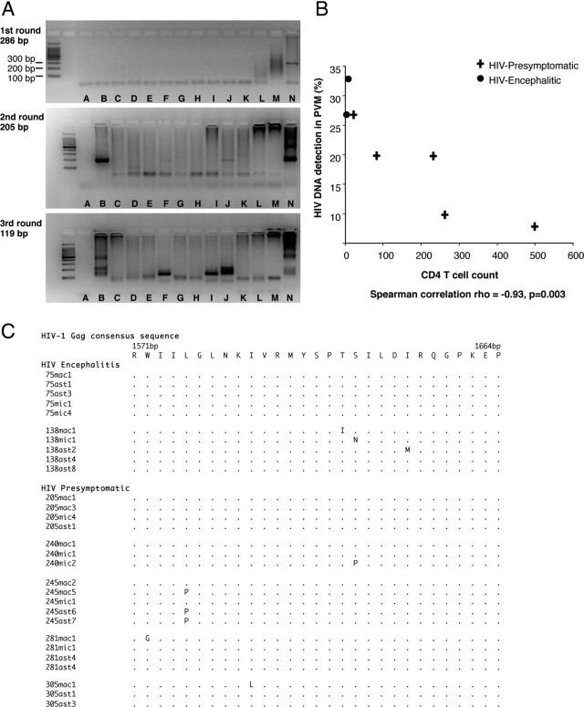
We detected HIV-1 DNA in pure populations of perivascular macrophages, parenchymal microglia, and astrocytes, isolated using laser microdissection from brain tissue of five untreated individuals who died in the presymptomatic stage of infection from non-HIV causes. HIV-1 DNA was detected in the three cell populations, most consistently in perivascular macrophages, without evidence of productive infection. The percentage of PCR reactions detecting HIV-1 DNA in perivascular macrophages correlated inversely with peripheral blood CD4 counts. These findings demonstrate that brain cell reservoirs of latent HIV-1 exist before pathological HIV encephalitis and suggest that perivascular macrophage trafficking of latent virus into the brain increases with immunosuppression.


Figures

Figure 1
Figure 1
Representative photomicrographs of immunohistochemically stained sections of the white matter from the occipital region of the cerebral cortex. A–C: HIV-negative, subject code no. 310. D–F: HIV-positive presymptomatic, code no. 240. G–I: HIV-positive presymptomatic, code no. 245. J–L: HIV-positive encephalitic, code no. 138. A, D, G, and J: GFAP immunoreaction, showing baseline astrocyte numbers in controls and increased astrocyte reactivity in both HIV-positive presymptomatic and HIV-positive encephalitic cases. B, E, H, and K: CD68 immunoreaction, showing baseline microglia in controls with a minor increase in macrophage/microglial reactivity in HIV-positive presymptomatic cases, and prominent reactivity in HIV-positive encephalitic cases. C, F, I, and L:In situ proximity ligation assay for detection of individual p24 protein, showing no evidence of p24 protein (C, F, and I) or showing p24 positivity (L), indicating productive infection with HIV protein (arrows). Hematoxylin counterstain. Scale bars = 40 μm.
Figure 2
Figure 2
Analysis of HIV-1 gag DNA isolated from laser microdissected brain cell populations; astrocytes, perivascular macrophages (PVM), and parenchymal microglia (PM) from HIV-presymptomatic (PS) and HIV-encephalitic (HIVE) cases. A: Representative images of HIV-1 gag DNA PCR resulting in three rounds of PCR products (286, 205, and 119 bp). HIV-1 gag DNA was not consistently detected in PCR amplification from samples in which there were low levels of virus. Cell populations were therefore collected in duplicate (two collections of astrocytes per case), and the triple-nested PCR was performed on each cell population at least 10 times, to increase the frequency of detection. Not every PCR amplification from DNA of cell populations is illustrated (results of PCR amplifications of HIV-1 gag DNA from all cell populations are given in Table 1 and sequencing results are shown here in panel B). Lane A, negative PCR control (water as DNA template) carried through three rounds of PCR. Lane B, code no. 281 (PS), PVM. Lane C, code no. 305 (PS), PVM. Lane D, code no. 281, astrocytes. Lane E, code no. 305, astrocytes. Lane F, code no. 75 (HIVE), astrocytes. Lane G, code no. 75, PVM. Lane H, code no. 138 (HIVE), astrocytes. Lane I, code no. 138, PVM. Lane J, code no. 75, PM. Lane K, code no. 138, PM. Lane L, code no. 281, PM. Lane M, code no. 305, PM. Lane N, Lymph node of HIV-positive patient, positive PCR control (overamplification results in PCR product not running through the gel). B: Association between CD4 T-cell count and the frequency with which HIV-1 DNA was detected by triple-nested PCR in perivascular macrophages (PVM, expressed as a percentage) in the occipital cortex for each of the seven HIV-positive individuals. C: Amino acid sequence alignment of HIV-1 gag DNA from PCR products of laser microdissected brain cell populations. Sequences are aligned and numbered according to the HIV-1 gag consensus sequence. ast, astrocyte; mac, perivascular macrophages; mic, parenchymal microglia.

References

    1. Centers for Disease Control and Prevention 1993 revised classification system for HIV infection and expanded surveillance case definition for AIDS among adolescents and adults. MMWR Recomm Rep. 1992;41(RR-17):1–19. - PubMed
    1. McCrossan M., Marsden M., Carnie F.W., Minnis S., Hansoti B., Anthony I.C., Brettle R.P., Bell J.E., Simmonds P. An immune control model for viral replication in the CNS during presymptomatic HIV infection. Brain. 2006;129:503–516. - PubMed
    1. Thompson K.A., Churchill M.J., Gorry P.R., Sterjovski J., Oelrichs R.B., Wesselingh S.L., McLean C.A. Astrocyte specific viral strains in HIV dementia. Ann Neurol. 2004;56:873–877. - PubMed
    1. Trillo-Pazos G., Diamanturos A., Rislove L., Menza T., Chao W., Belem P., Sadiq S., Morgello S., Sharer L., Volsky D.J. Detection of HIV-1 DNA in microglia/macrophages, astrocytes and neurons isolated from brain tissue with HIV-1 encephalitis by laser capture microdissection. Brain Pathol. 2003;13:144–154. - PMC - PubMed
    1. Thompson K.A., Varrone J.J., Jankovic-Karasoulos T., Wesselingh S.L., McLean C.A. Cell specific temporal infection of the central nervous system in a simian immunodeficiency virus model of human immunodeficiency virus encephalitis. J Neurovirol. 2009;15:300–311. - PubMed

Publication types

Substances