Review
. 2012 Aug;1818(8):2030-47.
doi: 10.1016/j.bbamem.2011.08.015. Epub 2011 Aug 16.

Gap junctions in inherited human disorders of the central nervous system

Affiliations
Review

Gap junctions in inherited human disorders of the central nervous system

Charles K Abrams et al. Biochim Biophys Acta. 2012 Aug.

Abstract

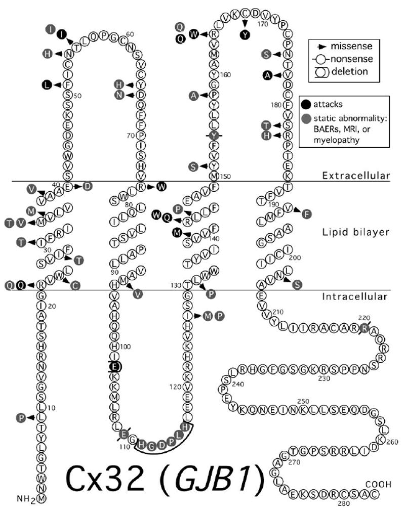
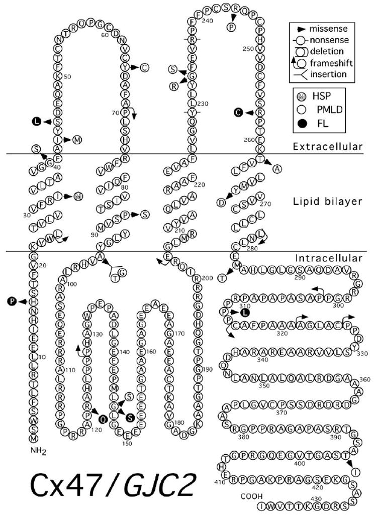
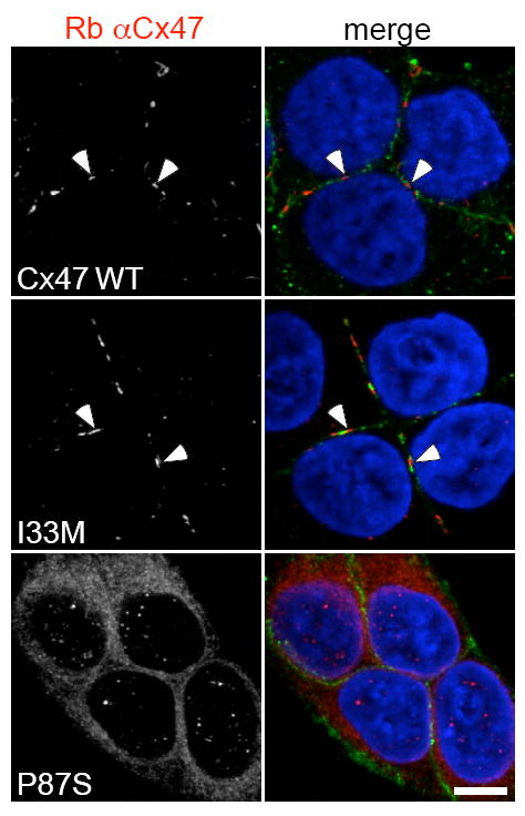
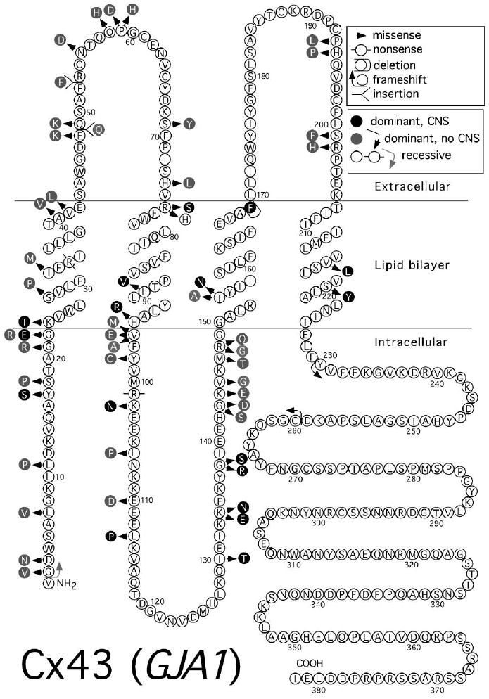
CNS glia and neurons express connexins, the proteins that form gap junctions in vertebrates. We review the connexins expressed by oligodendrocytes and astrocytes, and discuss their proposed physiologic roles. Of the 21 members of the human connexin family, mutations in three are associated with significant central nervous system manifestations. For each, we review the phenotype and discuss possible mechanisms of disease. Mutations in GJB1, the gene for connexin 32 (Cx32) cause the second most common form of Charcot-Marie-Tooth disease (CMT1X). Though the only consistent phenotype in CMT1X patients is a peripheral demyelinating neuropathy, CNS signs and symptoms have been found in some patients. Recessive mutations in GJC2, the gene for Cx47, are one cause of Pelizaeus-Merzbacher-like disease (PMLD), which is characterized by nystagmus within the first 6 months of life, cerebellar ataxia by 4 years, and spasticity by 6 years of age. MRI imaging shows abnormal myelination. A different recessive GJC2 mutation causes a form of hereditary spastic paraparesis, which is a milder phenotype than PMLD. Dominant mutations in GJA1, the gene for Cx43, cause oculodentodigital dysplasia (ODDD), a pleitropic disorder characterized by oculo-facial abnormalities including micropthalmia, microcornia and hypoplastic nares, syndactyly of the fourth to fifth fingers and dental abnormalities. Neurologic manifestations, including spasticity and gait difficulties, are often but not universally seen. Recessive GJA1 mutations cause Hallermann-Streiff syndrome, a disorder showing substantial overlap with ODDD. This article is part of a Special Issue entitled: The Communicating junctions, composition, structure and functions.


Figures

Figure 1
Figure 1
Connexin nomenclature A. Connexins are integral membrane proteins with four transmembrane domains and intracellular N and C termini. B. Each connexon or hemichannel is composed of six subunits called connexins. C. Cell-cell channels are formed by docking of two apposed connexons from apposed cells. D. If all six subunits are identical the connexon is termed homomeric. In cells expressing more than one type of connexin, different connexins may aggregate to form heteromeric connexons. E. Many members of the connexin family can form functional GJs by associating with a like connexon (a homotypic junction) or a different one (a heterotypic junction).
Figure 2
Figure 2
Ultrastructural findings in the CNS myelin of mice lacking expression of Cx32 and Cx47. All are electron micrographs of the ventral funiculus from P31 mice. A. An axon (a) is surrounded by a periaxonal cytoplasmic collar thin myelin sheath (arrow). B. A demyelinated axon. Note that neurofilament packing is tighter than in A. C. An axon still partially surrounded by the adaxonal membrane (arrowheads), but separated from its myelin sheath (arrow) by- extracellular space (asterisks). D. An apoptotic oligodendrocyte nucleus (n). Scale bar, 1μm. From Menichella et al. 2003, [16] used with permission of Society for Neuroscience.
Figure 3
Figure 3
Pathologic findings in the CNS myelin of mice lacking expression of Cx30 globally and Cx43 in astrocytes (Cx30 Cx43astro dKO). A–C. Toluidine blue stained 1μm epoxy sections of the dorsal cuneate of the lumbar spinal cord of non-Cre (Cx43fl/fl Cx30-/- (A) and Cx30 Cx43astro dKO (B, C) mice. D, Electron micrograph showing enlarged oligodendrocyte cytoplasm interpolating between an apparently normal axon and its myelin sheath. Inset depicts ectopic expression of myelin in one of several locations within the oligodendrocyte cytoplasm. E. Myelin whorls, likely representing Wallerian axonal degeneration. F and G. Edema and splitting of myelin sheaths. Scale bar: (A, B) 100μm; (C) 10μm. Magnification: (D) 4000X, inset: 15,000X; (E) 9000X; (F) 8000X; (G) 13,000X, inset: 4000X. From Lutz et al. 2009 [44] used with permission of Society for Neuroscience.
Figure 4
Figure 4
Mutations in Cx32 associated with CNS manifestations. Topology is as depicted in Yeager and Nicholson (1996) [198]
Figure 5
Figure 5
MRI imaging of a CMT1X patient with an acute florid CNS presentation. T1-weighted, T2-weighted, fluid-attenuated inversion recovery (FLAIR), and diffusion-weighted magnetic resonance imaging (DWI) images during onset of an acute CNS syndrome and 1 and 2 months later show splenium of corpus callosum (arrow in the T2, FLAIR and DWI images at onset) and posterior limb of the internal capsule abnormalities at all three time points; signal intensities have almost returned to normal after 2 months. Modified from Paulson et al. (2003) [113], used with permission of Wiley-Davis.
Figure 6
Figure 6
Mutations in Cx47 associated with human disease. Topology is as depicted in Yeager and Nicholson (1996) [198]
Figure 7
Figure 7
MRI imaging of patients with PMLD1. Axial T2-weighted magnetic resonance images of the brain at the level of the basal ganglia. A. Patient with PMLD at 6 years of age. B. Patient with PMD at 7 years of age. The patients in panels A and B show nearly identical patterns consistent with hypomyelination of central white matter, as indicated by diffusely enhanced signal intensity. C. Low signal of normal myelination in an unaffected child. From Uhlenberg et al. (2004) [134] used with permission of Elsevier.
Figure 8
Figure 8
MRI imaging of a patient with SPG44. A. Sagittal T1-weighted imaging shows diffuse thinning of the corpus callosum. B -D. Axial T2-weighted imaging shows symmetric hyperintensity in the region of the corticospinal/corticobulbar tracts at the level of the pons (B, arrows), and the posterior limb of internal capsule (C, arrowhead). In addition, there is diffuse hyperintensity in the subcortical, lobar and periventricular white matter (C and D), and enlarged ventricles (C) Modified from Orthmann-Murphy et al. (2009) [140] Used with Permission of Oxford University Press.
Figure 9
Figure 9
Immunostaining for the I33M and P87S mutants. The SPG44 associated I33M mutant forms GJ plaques while the PMLD1 associated P87S mutant does not. These are confocal images of bulk-selected HeLa cells that express WT Cx47 or the indicated mutants, immunostained with a rabbit antiserum against human Cx47 (red) and a mouse monoclonal antibody against pan-cadherin (green), and counterstained with DAPI. The pan-cadherin staining at cell borders interdigitates with the cell surface staining of Cx47 in cells that express WT Cx47 (arrowheads) or I33M (arrowheads), but surrounds the staining of cells expressing the mutant P87S, which is localized in the endoplasmic reticulum. Scale bar: 10 μm. From Orthmann-Murphy et al. [140] Used with Permission of Oxford University Press.
Figure 10
Figure 10
Mutations in Cx43 associated with human disease. Topology is as depicted in Yeager and Nicholson (1996) [198]

References

    1. Rackauskas M, Neverauskas V, Skeberdis VA. Diversity and properties of connexin gap junction channels. Medicina. 2010;46:1–12. - PubMed
    1. Zoidl G, Dermietzel R. Gap junctions in inherited human disease. Pflugers Arch. 2010;460:451–466. - PubMed
    1. Orthmann-Murphy JL, Abrams CK, Scherer SS. Gap junctions couple astrocytes and oligodendrocytes. J Mol Neurosci. 2008;35:101–116. - PMC - PubMed
    1. Abrams CK, Rash JE. Connexins In The Nervous System. In: Harris A, Locke D, editors. Connexins: A Guide. Humana Press; Totowa, N.J: 2009. pp. 323–357.
    1. Orthmann-Murphy JL, Freidin M, Fischer E, Scherer SS, Abrams CK. Two distinct heterotypic channels mediate gap junction coupling between astrocyte and oligodendrocyte connexins. J Neurosci. 2007;27:13949–13957. - PMC - PubMed

Publication types

LinkOut - more resources