

Nanoparticle delivery of a peptide targeting EGFR signaling
- PMID: 21871507
- PMCID: PMC3229664
- DOI: 10.1016/j.jconrel.2011.08.014
Nanoparticle delivery of a peptide targeting EGFR signaling
Abstract
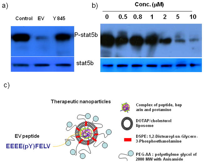
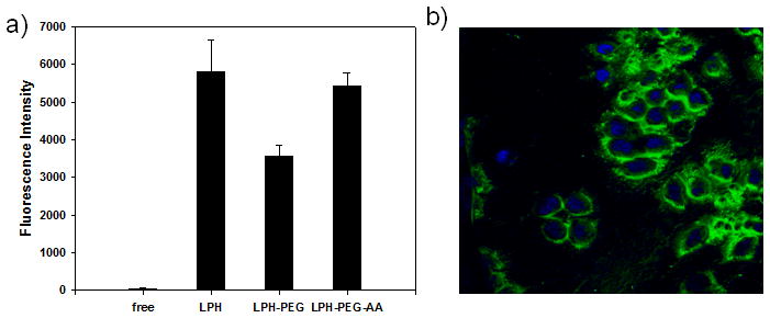
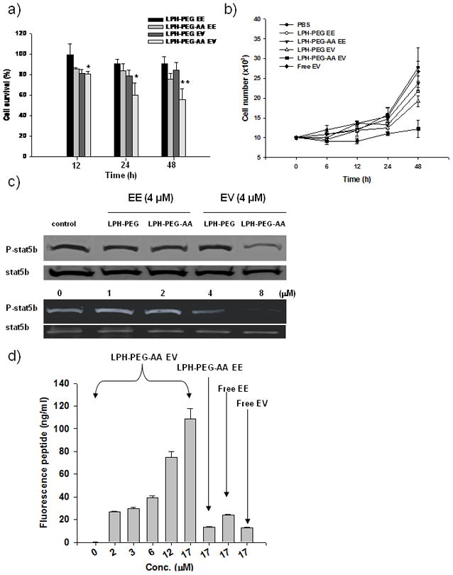
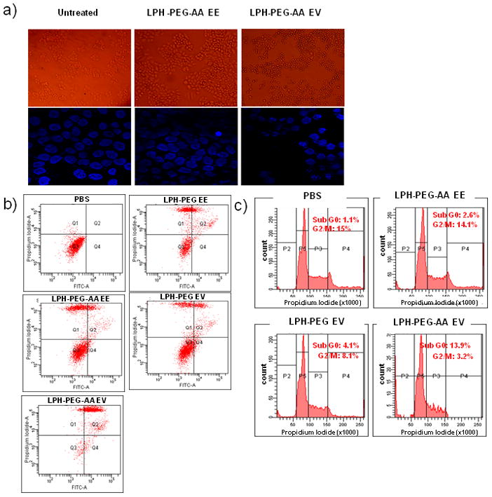
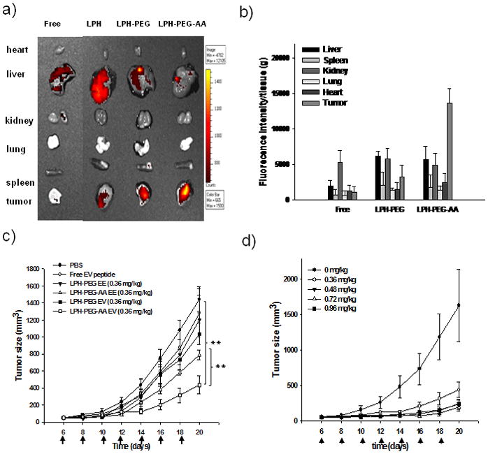
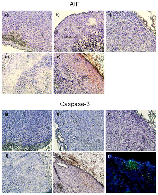
EGFR serves as an important therapeutic target because of its over-expression in many cancers. In this study, we investigated a peptide-based therapy aimed at blocking intracellular protein-protein interactions during EGFR signaling and evaluated a targetable lipid carrier system that can deliver peptides to intracellular targets in human cancer cells. EEEEpYFELV (EV), a nonapeptide mimicking the Y845 site of EGFR which is responsible for STAT5b phosphorylation, was designed to block EGFR downstream signaling. EV was loaded onto LPH nanoparticles that are comprised of a membrane/core structure including a surface-grafted polyethylene glycol (PEG) used to evade the reticuloendothelial system (RES) and anisamide (AA) for targeting the sigma receptor over-expressed in H460 human lung cancer cells. EV formulated with PEGylated and targeted LPH (LPH-PEG-AA) was taken up by the tumor cells and trafficked to the cytoplasm with high efficiency. Using this approach, EV acted as a dominant negative inhibitor of STAT5b phosphorylation, arrested cell proliferation, and induced massive apoptosis. Intravenous administration of EV loaded in LPH-PEG-AA led to efficient EV peptide delivery to the tumor in a xenograft mouse model, and multiple injections inhibited tumor growth in a dose-dependent manner. Our findings offer proof-of-concept for an intracellular peptide-mediated cancer therapy that is delivered by carefully designed nanoparticles.
Copyright © 2011 Elsevier B.V. All rights reserved.
Figures
Similar articles
-
Targeted delivery of EV peptide to tumor cell cytoplasm using lipid coated calcium carbonate nanoparticles.Cancer Lett. 2013 Jul 1;334(2):311-8. doi: 10.1016/j.canlet.2012.07.011. Epub 2012 Jul 14. Cancer Lett. 2013. PMID: 22796364 Free PMC article.
-
Signal transducer and activator of transcription 5b, c-Src, and epidermal growth factor receptor signaling play integral roles in estrogen-stimulated proliferation of estrogen receptor-positive breast cancer cells.Mol Endocrinol. 2008 Aug;22(8):1781-96. doi: 10.1210/me.2007-0419. Epub 2008 Jun 11. Mol Endocrinol. 2008. PMID: 18550772 Free PMC article.
-
Anti-EGFR-iRGD recombinant protein modified biomimetic nanoparticles loaded with gambogic acid to enhance targeting and antitumor ability in colorectal cancer treatment.Int J Nanomedicine. 2018 Aug 31;13:4961-4975. doi: 10.2147/IJN.S170148. eCollection 2018. Int J Nanomedicine. 2018. PMID: 30214200 Free PMC article.
-
In Vivo Delivery of siRNAs Targeting EGFR and BRD4 Expression by Peptide-Modified Redox Responsive PEG-PEI Nanoparticles for the Treatment of Triple-Negative Breast Cancer.Mol Pharm. 2021 Nov 1;18(11):3990-3998. doi: 10.1021/acs.molpharmaceut.1c00282. Epub 2021 Sep 30. Mol Pharm. 2021. PMID: 34591491
-
Nanoparticles targeted with NGR motif deliver c-myc siRNA and doxorubicin for anticancer therapy.Mol Ther. 2010 Apr;18(4):828-34. doi: 10.1038/mt.2009.291. Epub 2010 Jan 12. Mol Ther. 2010. PMID: 20068551 Free PMC article.
Cited by
-
Nanoparticles as Carriers of Proteins, Peptides and Other Therapeutic Molecules.Open Life Sci. 2018 Oct 31;13:285-298. doi: 10.1515/biol-2018-0035. eCollection 2018 Jan. Open Life Sci. 2018. PMID: 33817095 Free PMC article.
-
Multifunctional, stimuli-sensitive nanoparticulate systems for drug delivery.Nat Rev Drug Discov. 2014 Nov;13(11):813-27. doi: 10.1038/nrd4333. Epub 2014 Oct 7. Nat Rev Drug Discov. 2014. PMID: 25287120 Free PMC article. Review.
-
Targeted Liposomal Chemotherapies to Treat Triple-Negative Breast Cancer.Cancers (Basel). 2021 Jul 26;13(15):3749. doi: 10.3390/cancers13153749. Cancers (Basel). 2021. PMID: 34359650 Free PMC article.
-
Current trends in the use of liposomes for tumor targeting.Nanomedicine (Lond). 2013 Sep;8(9):1509-28. doi: 10.2217/nnm.13.118. Nanomedicine (Lond). 2013. PMID: 23914966 Free PMC article. Review.
-
Targeted delivery of EV peptide to tumor cell cytoplasm using lipid coated calcium carbonate nanoparticles.Cancer Lett. 2013 Jul 1;334(2):311-8. doi: 10.1016/j.canlet.2012.07.011. Epub 2012 Jul 14. Cancer Lett. 2013. PMID: 22796364 Free PMC article.
References
-
- Gibbs JB. Mechanism-based target identification and drug discovery in cancer research. Science. 2000;287:1969–1973. - PubMed
-
- Gibbs JB, Oliff A. Pharmaceutical research in molecular oncology. Cell. 1994;79:193–198. - PubMed
-
- Poondra RR, Kumar NN, Bijian K, Prakesch M, Campagna-Slater V, Reayi A, Reddy PT, Choudhry A, Barnes ML, Leek DM, Daroszewska M, Lougheed C, Xu B, Schapira M, Alaoui-Jamali MA, Arya P. Discovery of Indoline-Based, Natural-Product-like Compounds as Probes of Focal Adhesion Kinase Signaling Pathways. J Comb Chem. 1994;11:303–309. - PubMed
-
- Chester KA, Hawkins RE. Clinical issues in antibody design. Trends Biotechnol. 1995;13:294–300. - PubMed
-
- Ladner RC, Sato AK, Gorzelany J, de Souza M. Phage display-derived peptides as therapeutic alternatives to antibodies. Drug Discov Today. 2004;9:525–529. - PubMed
Publication types
MeSH terms
Substances
Grants and funding
LinkOut - more resources
Full Text Sources
Other Literature Sources
Research Materials
Miscellaneous
