. 2011 Nov 21:289:107-15.
doi: 10.1016/j.jtbi.2011.08.017. Epub 2011 Aug 23.

Analysis of the interaction of the Eg5 Loop5 with the nucleotide site

Affiliations

Analysis of the interaction of the Eg5 Loop5 with the nucleotide site

Timothy D Harrington et al. J Theor Biol. .

Abstract

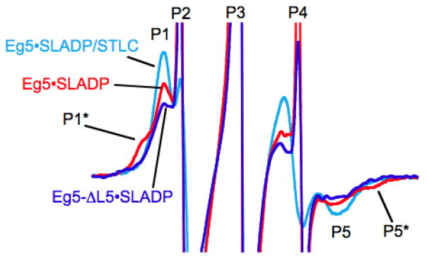
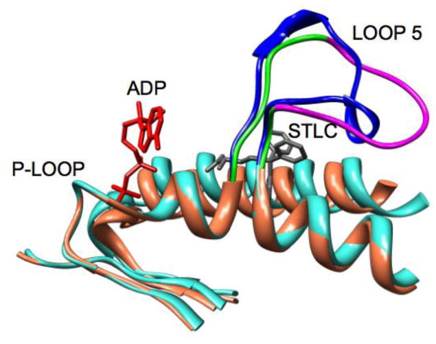
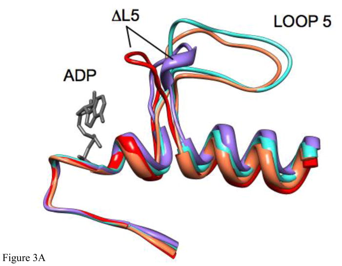
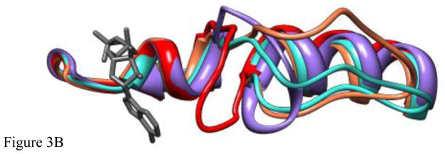
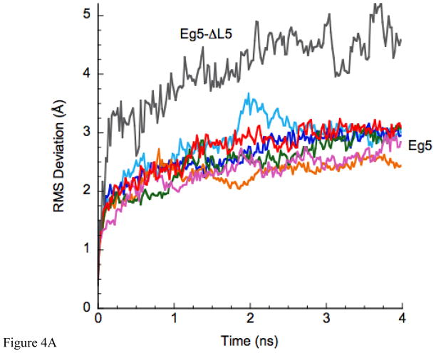
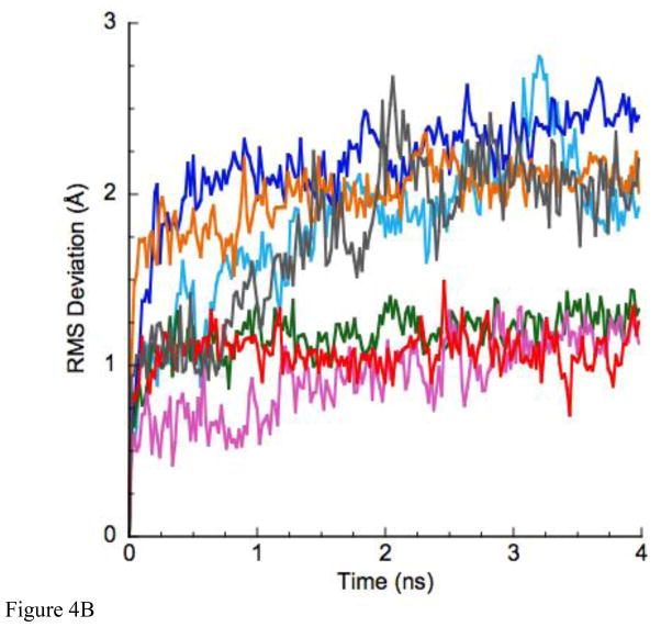
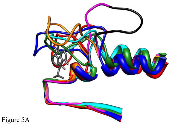
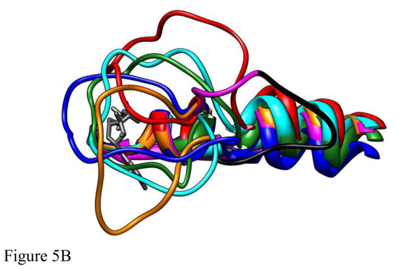
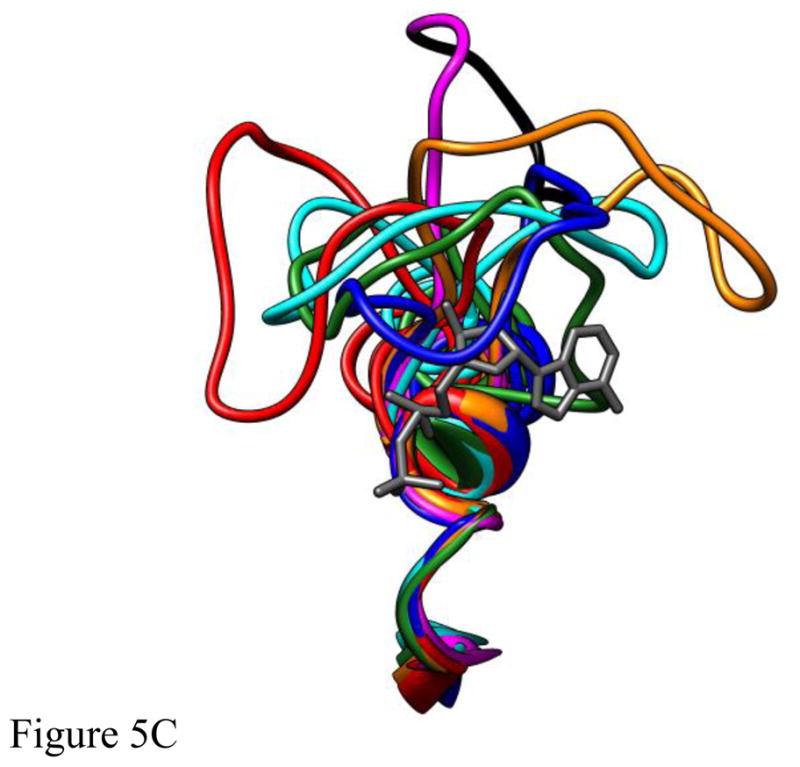
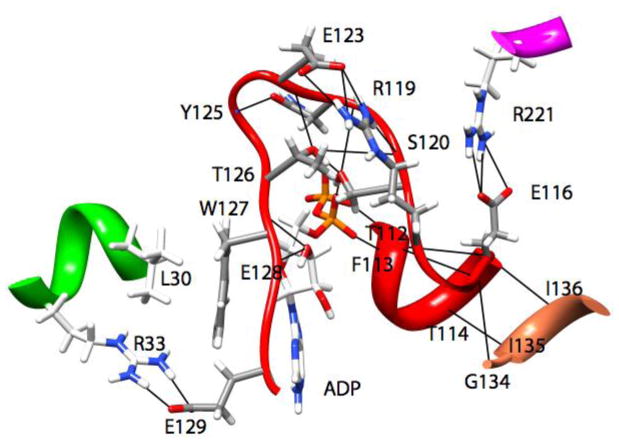
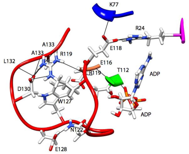
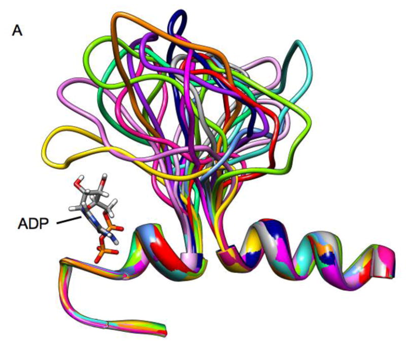
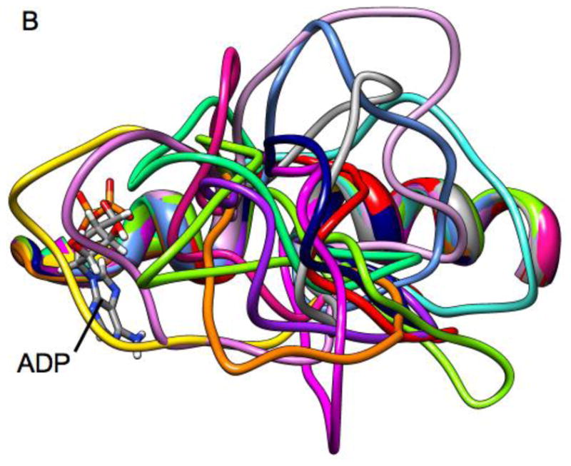
Loop 5 (L5) is a conserved loop that projects from the α2-helix adjacent to the nucleotide site of all kinesin-family motors. L5 is critical to the function of the mitotic kinesin-5 family motors and is the binding site for several kinesin-5 inhibitors that are currently in clinical trials. Its conformational dynamics and its role in motor function are not fully understood. Our previous work using EPR spectroscopy suggested that L5 alters the nucleotide pocket conformation of the kinesin-5 motor Eg5 (Larson et al., 2010). EPR spectra of a spin-labeled nucleotide analog bound at the nucleotide site of Eg5 display a highly immobilized component that is absent if L5 is shortened or if the inhibitor STLC is added (Larson et al., 2010), which X-ray structures suggest stabilizes an L5 conformation pointing away from the nucleotide site. These data, coupled with the proximity of L5 to the nucleotide site suggest L5 could interact with a bound nucleotide, modulating function. Here we use molecular dynamics (MD) simulations of Eg5 to explore the interaction of L5 with the nucleotide site in greater detail. We performed MD simulations in which the L5-domain of the Eg5·ADP X-ray structure was manually deformed via backbone bond rotations. The L5-domain of Eg5 was sufficiently lengthy that portions of L5 could be located in proximity to bound ADP. The MD simulations evolved to thermodynamically stable structures at 300 K showing that L5 can interact directly with bound nucleotide with significant impingement on the ribose hydroxyls, consistent with the EPR spectroscopy results. Taken together, these data provide support for the hypothesis that L5 modulates Eg5 function via interaction with the nucleotide-binding site.


Figures

Figure 1
Figure 1
X-band EPR spectra of 2′,3′-SLADP bound to Eg5 (red), Eg5-ΔL5 (blue) and Eg5 in the presence of the inhibitor, STLC (cyan). The horizontal axis is magnetic field. The vertical axis is the derivative of absorbance. The spectra are 9.0 mT wide with center field at 350 mT. The figure is similar a previously published figure [1] and is present to motivate the computational modeling.
Figure 2
Figure 2
An overlay of ribbon diagrams of the structures of Eg5•ADP [10] and Eg5•ADP with bound STLC [13]. Eg5•ADP is colored coral except for L5, which is colored green and magenta. The magenta segment is the portion of L5 that is removed in the Eg5-ΔL5 construct. Eg5•ADP with bound STLC is colored turquoise, except for L5, which is blue. STLC is gray. ADP is red.
Figure 3
Figure 3
A. The X-ray structure of Eg5•ADP (turquoise), Eg5•ADP following 4ns of MD simulation (coral), the constructed structure of Eg5-ΔL5•ADP distorted to be as close as possible to ADP at the nucleotide site (red), and the structure of Eg5-ΔL5•ADP following 4 ns of MD simulation (purple) are shown as ribbon diagrams. The structures were superimposed using a least-squares distance minimization of the P-loop Cα atoms. ADP from the X-ray structure of Eg5•ADP is in dark gray. Only the regions of the proteins adjacent to the nucleotide site and L5 are shown. Fig. 3A is oriented from the side with the L5 at the top of the panel. B. Same a panel A, but looking down from the top of panel A.
Figure 3
Figure 3
A. The X-ray structure of Eg5•ADP (turquoise), Eg5•ADP following 4ns of MD simulation (coral), the constructed structure of Eg5-ΔL5•ADP distorted to be as close as possible to ADP at the nucleotide site (red), and the structure of Eg5-ΔL5•ADP following 4 ns of MD simulation (purple) are shown as ribbon diagrams. The structures were superimposed using a least-squares distance minimization of the P-loop Cα atoms. ADP from the X-ray structure of Eg5•ADP is in dark gray. Only the regions of the proteins adjacent to the nucleotide site and L5 are shown. Fig. 3A is oriented from the side with the L5 at the top of the panel. B. Same a panel A, but looking down from the top of panel A.
Figure 4
Figure 4
A. The RMS deviation of the MD-simulated structures as a function of time. Color coding: Eg5•ADP (red), Eg5-ΔL5•ADP (gray). The cyan, green, orange, blue and magenta plots are color-coded to the structures in Fig. 5. B. RMS deviation of only the Loop5 domain during the simulation. Color-coding is as in panel A.
Figure 4
Figure 4
A. The RMS deviation of the MD-simulated structures as a function of time. Color coding: Eg5•ADP (red), Eg5-ΔL5•ADP (gray). The cyan, green, orange, blue and magenta plots are color-coded to the structures in Fig. 5. B. RMS deviation of only the Loop5 domain during the simulation. Color-coding is as in panel A.
Figure 5
Figure 5
The structures of five different deformations of L5 in the Eg5•ADP structure (blue, cyan, red, green, orange) interacting with ADP bound at the nucleotide site (dark gray) following 4ns of MD simulation. The structures were superimposed using a least-squares distance minimization of the P-loop Cα atoms. The structure of Eg5•ADP (magenta with the domain deleted for Eg5-ΔL5 in black) is shown for comparison. Fig. 5A is oriented from the side with L5 at the top of the panel. Fig. 5B looks down from the top of panel A. Fig. 5C looks from a direction facing the bound ADP.
Figure 5
Figure 5
The structures of five different deformations of L5 in the Eg5•ADP structure (blue, cyan, red, green, orange) interacting with ADP bound at the nucleotide site (dark gray) following 4ns of MD simulation. The structures were superimposed using a least-squares distance minimization of the P-loop Cα atoms. The structure of Eg5•ADP (magenta with the domain deleted for Eg5-ΔL5 in black) is shown for comparison. Fig. 5A is oriented from the side with L5 at the top of the panel. Fig. 5B looks down from the top of panel A. Fig. 5C looks from a direction facing the bound ADP.
Figure 6
Figure 6
Ribbon representation of the interactions (ionic, hydrogen bonds, and hydrophobic) that result in the forces stabilizing the location of Loop5 (red) in one observed conformation from the MD simulations.
Figure 7
Figure 7
Ribbon representation of the interactions (ionic, hydrogen bonds, and hydrophobic) that result in the forces stabilizing the location of Loop5 (red) in one observed conformation from the MD simulations.
Figure 8
Figure 8
Ribbon diagrams of a superposition of Loop 5 conformations from high temperature molecular dynamics simulation. Individual frames are from a 10 ns simulation with frames separated by 600 ps.
Figure 8
Figure 8
Ribbon diagrams of a superposition of Loop 5 conformations from high temperature molecular dynamics simulation. Individual frames are from a 10 ns simulation with frames separated by 600 ps.

Similar articles

Cited by

References

    1. Larson AG, Naber N, Cooke R, Pate E, Rice SE. The conserved L5 loop establishes the pre-powerstroke conformation of the Kinesin-5 motor, Eg5. Biophys J. 2010;98:2619–2627. - PMC - PubMed
    1. Rayment I, Smith C, Yount RG. The active site of myosin. Annu Rev Physiol. 1996;58:671–702. - PubMed
    1. Kull FJ, Vale RD, Fletterick RJ. The case for a common ancestor: kinesin and myosin motor proteins and G proteins. J Muscle Res Cell Motil. 1998;19:877–886. - PubMed
    1. Vale RD. Switches, latches, and amplifiers: common themes of G proteins and molecular motors. J Cell Biol. 1996;135:291–302. - PMC - PubMed
    1. Walker JE, Saraste M, Runswick MJ, Gay NJ. Distantly related sequences in the alpha- and beta-subunits of ATP synthase, myosin, kinases and other ATP-requiring enzymes and a common nucleotide binding fold. Embo J. 1982;1:945–951. - PMC - PubMed

Publication types

LinkOut - more resources