. 2011 Sep 6;108(36):14873-8.
doi: 10.1073/pnas.1112293108. Epub 2011 Aug 22.

Transcription factor AP4 modulates reversible and epigenetic silencing of the Cd4 gene

Affiliations

Transcription factor AP4 modulates reversible and epigenetic silencing of the Cd4 gene

Takeshi Egawa et al. Proc Natl Acad Sci U S A. .

Abstract

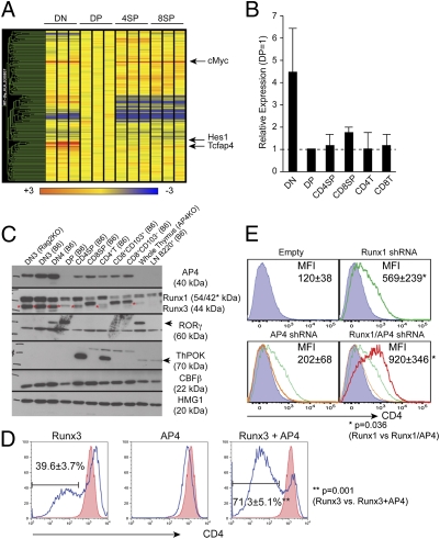
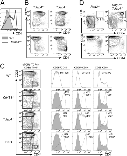
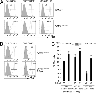
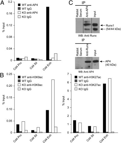
CD4 coreceptor expression is negatively regulated through activity of the Cd4 silencer in CD4(-)CD8(-) double-negative (DN) thymocytes and CD8(+) cytotoxic lineage T cells. Whereas Cd4 silencing is reversed during transition from DN to CD4(+)CD8(+) double-positive stages, it is maintained through heritable epigenetic processes following its establishment in mature CD8(+) T cells. We previously demonstrated that the Runx family of transcription factors is required for Cd4 silencing both in DN thymocytes and CD8(+) T cells. However, additional factors that cooperate with Runx proteins in the process of Cd4 silencing remain unknown. To identify collaborating factors, we used microarray and RNAi-based approaches and found the basic helix-loop-helix ZIP transcription factor AP4 to have an important role in Cd4 regulation. AP4 interacts with Runx1 in cells in which Cd4 is silenced, and is required for Cd4 silencing in immature DN thymocytes through binding to the proximal enhancer. Furthermore, although AP4-deficient CD8(+) T cells appeared to normally down-regulate CD4 expression, AP4 deficiency significantly increased the frequency of CD4-expressing effector/memory CD8(+) T cells in mice harboring point mutations in the Cd4 silencer. Our results suggest that AP4 contributes to Cd4 silencing both in DN and CD8(+) T cells by enforcing checkpoints for appropriate timing of CD4 expression and its epigenetic silencing.


Conflict of interest statement

The authors declare no conflict of interest.

Figures

Fig. 1.
Fig. 1.
Identification of AP4 as a Cd4 silencing factor. (A) Microarray analysis of genes encoding bHLH domain-containing proteins in different thymocyte subsets. Relative signal intensity is normalized against the expression level in DP thymocytes (shown as yellow). (B) Quantitative RT-PCR analysis of AP4 (Tcfap4) expression. Values were normalized against Hprt1 expression levels and are shown relative to the DP expression level. Data represent the average and SD of two independently prepared samples for each cell type. (C) Western blotting analysis of AP4 expression in different subsets of thymocytes and mature T lymphocytes. Anti-Runx, -RORγt, -ThPOK, and -CBFβ immunoblots serve as controls for population purity, and anti-HMG1 serves as a loading control. (D) Effect of AP4 overexpression in a double-positive thymoma cell line. AP4 and/or Runx3 were retrovirally expressed in AKR1 cells, and CD4 expression was assessed in transduced cells at 72 h postinfection (blue lines). Red-filled histograms indicate CD4 expression in empty virus-transduced controls. Proportions of CD4lo/– cells are shown with averages and SDs from five experiments. (E) AP4 and Runx1 synergistically regulate active Cd4 silencing in the 1200M cell line. CD4 expression in cells transduced with shRNAs targeting AP4 (orange), Runx1 (green), or both (red). Mean fluorescence intensities of CD4 staining are shown with averages and SDs from five experiments. Statistical significance was tested by unpaired two-tailed t test with P values shown.
Fig. 2.
Fig. 2.
AP4 is required for Cd4 silencing in immature thymocytes. (A) CD4 expression in sTCRβTCRγδCD8αThy1+ thymocytes from AP4-deficient (open histogram) and WT control (filled histogram) mice. (B) CD4 is robustly up-regulated following β selection in AP4-deficient thymocytes. CD4, CD25, and CD44 expression of sTCRβTCRγδCD8αThy1+ thymocytes from AP4-deficient and WT control mice. (C) AP4 synergizes with Cd4 silencer-binding factors to repress CD4 in immature thymocytes. CD4 expression in preselection DN3 cells (CD25hiCD44), postselection DN3 cells (CD25intCD44) and DN4 cells (CD25CD44) from WT (black lines in the top row and filled histograms), Cd4 silencer-deficient mice (Cd4Sil−/−), AP4-deficient mice (Tcfap4−/−), and mice doubly deficient for the Cd4 silencer and AP4 (DKO) is shown in histograms with mean fluorescence intensity (MFI) indicated. Data shown here are representative of more than three independent experiments. Statistical analysis is shown in Fig. S5C. (D) AP4 deficiency allows Rag2-deficient thymocytes to bypass β selection. CD4, CD8α, CD25, and CD44 expression in Thy1+ thymocytes from Rag2−/− or Rag2−/−Tcfap4−/− mice is shown.
Fig. 3.
Fig. 3.
AP4 modulates variegated CD4 derepression in mice harboring a mutated Cd4 silencer. (A) CD4 and CD8 expression in TCRβhiHSAlo gated mature thymocytes from Tcfap4−/− mice crossed to Cd4 silencer site 3 mutant mice. CD4 expression in site 3 mutant CD8+ mature thymocytes on either AP4-sufficient (Upper) or -deficient (Lower) backgrounds is shown (Right). Filled histograms show CD4 expression in CD8+ mature thymocytes from AP4-sufficient (Upper) or -deficient (Lower) mice with the intact Cd4 silencer. (B and C) CD4 expression in CD8+TCRβ+ lymph node (LN) cells of mice with silencer site 3 mutation, AP4 deficiency, or both. A representative set of data is shown in B and a statistical analysis of frequency of CD4+ T cells within the CD8+ T-cell interval gate in multiple mice is shown in C. Averages and SDs are shown with P values determined by unpaired Student's t test.
Fig. 4.
Fig. 4.
Changes in CD4 expression patterns in different subsets of CD8+ T cells harboring mutated Cd4 silencers. (A) CD4 expression in total CD8+ T cells, CD8+CD103+ and CD8+CD103 subsets from WT (filled histograms), Cd4 silencer-deficient, and Cd4 silencer site 1 mutant (open histograms) mice. Frequencies of CD4+ cells are shown within the interval gates. (B) CD4 expression in CD8+ T-cell subsets from Cd4 silencer site 1 mutant, AP4-sufficient (Upper), or -deficient (Lower) mice. (C) Statistical analysis by unpaired Student's t test of the percentages of CD4+ cells in different subsets of site 1 mutant CD8+ T cells.
Fig. 5.
Fig. 5.
AP4 binding to the Cd4 proximal enhancer and interaction with Runx protein. (A) Chromatin immunoprecipitation analysis showing AP4 binding to the Cd4 proximal enhancer in double-negative thymocytes. Results are representative of four independent experiments. (B) H3K9ac (Left) and H3K27ac (Right) histone modifications at Cd4 regulatory elements in DN thymocytes. Data are representative of two independent experiments. (C) Physical interaction of endogenous AP4 and Runx1 in 1200M cells. Immunoprecipitation (IP) with anti-AP4 (Upper) or anti-Runx (Lower) antibodies was followed by immunoblotting with antibodies specific for the two transcription factors. Results are representative of three independent experiments. WB, Western blot.

Similar articles

Cited by

References

    1. Kioussis D, Ellmeier W. Chromatin and CD4, CD8A and CD8B gene expression during thymic differentiation. Nat Rev Immunol. 2002;2:909–919. - PubMed
    1. Taniuchi I, Sunshine MJ, Festenstein R, Littman DR. Evidence for distinct CD4 silencer functions at different stages of thymocyte differentiation. Mol Cell. 2002;10:1083–1096. - PubMed
    1. Zou YR, et al. Epigenetic silencing of CD4 in T cells committed to the cytotoxic lineage. Nat Genet. 2001;29:332–336. - PubMed
    1. Taniuchi I, et al. Differential requirements for Runx proteins in CD4 repression and epigenetic silencing during T lymphocyte development. Cell. 2002;111:621–633. - PubMed
    1. Setoguchi R, Taniuchi I, Bevan MJ. ThPOK derepression is required for robust CD8 T cell responses to viral infection. J Immunol. 2009;183:4467–4474. - PMC - PubMed

Publication types

MeSH terms