. 2011 Sep 6;108(36):14920-5.
doi: 10.1073/pnas.1111691108. Epub 2011 Aug 22.

Structure of the Newcastle disease virus hemagglutinin-neuraminidase (HN) ectodomain reveals a four-helix bundle stalk

Affiliations

Structure of the Newcastle disease virus hemagglutinin-neuraminidase (HN) ectodomain reveals a four-helix bundle stalk

Ping Yuan et al. Proc Natl Acad Sci U S A. .

Abstract

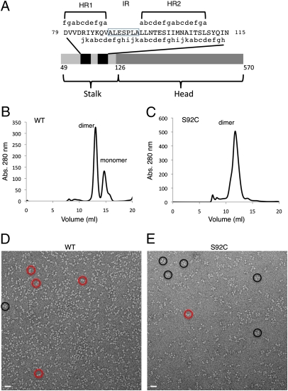
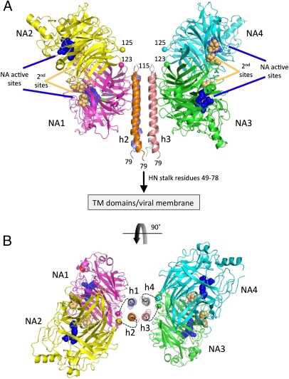
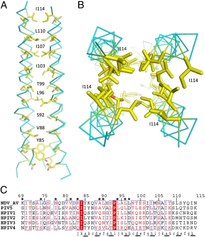
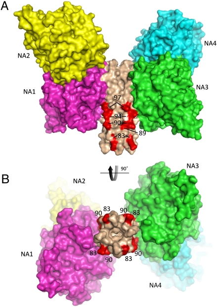
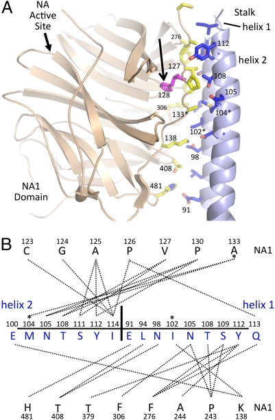
The paramyxovirus hemagglutinin-neuraminidase (HN) protein plays multiple roles in viral entry and egress, including binding to sialic acid receptors, activating the fusion (F) protein to activate membrane fusion and viral entry, and cleaving sialic acid from carbohydrate chains. HN is an oligomeric integral membrane protein consisting of an N-terminal transmembrane domain, a stalk region, and an enzymatically active neuraminidase (NA) domain. Structures of the HN NA domains have been solved previously; however, the structure of the stalk region has remained elusive. The stalk region contains specificity determinants for F interactions and activation, underlying the requirement for homotypic F and HN interactions in viral entry. Mutations of the Newcastle disease virus HN stalk region have been shown to affect both F activation and NA activities, but a structural basis for understanding these dual affects on HN functions has been lacking. Here, we report the structure of the Newcastle disease virus HN ectodomain, revealing dimers of NA domain dimers flanking the N-terminal stalk domain. The stalk forms a parallel tetrameric coiled-coil bundle (4HB) that allows classification of extensive mutational data, providing insight into the functional roles of the stalk region. Mutations that affect both F activation and NA activities map predominantly to the 4HB hydrophobic core, whereas mutations that affect only F-protein activation map primarily to the 4HB surface. Two of four NA domains interact with the 4HB stalk, and residues at this interface in both the stalk and NA domain have been implicated in HN function.


Conflict of interest statement

The authors declare no conflict of interest.

Figures

Fig. 1.
Fig. 1.
Biochemical and EM analysis of WT and S92C mutant NDV HN proteins. (A) Schematic diagram of the NDV HN ectodomain construct and predicted α-helical regions. The black bars indicate the predicted α-helix positions. (B) WT HN migrates as a mixture of dimers and monomers in gel filtration experiments. (C) The S92C mutant migrates primarily as dimers on gel filtration columns. (D) EM pictures of WT and (E) S92C proteins. The red circles indicate potential dimers, and the black circles indicate potential tetramers.
Fig. 2.
Fig. 2.
Structure of the NDV HN (AV) ectodomaim. (A) Two dimers of the NDV HN NA domains flank the 4HB in the stalk. The four NA domains are labeled NA1–NA4. The active sites are marked by three residues shown as blue CPK spheres (E400, R415, and Y525) and labeled accordingly. The secondary sialic acid binding sites located at the NA domain dimer interfaces are marked by residues shown as orange CPK spheres and labeled (second sites). The N termini of the four NA domains, residues 123 and 125, are labeled and indicated by their CA atoms shown in CPK format colored by chain. The connections of the N-terminal region of the stalk to the HN TM domains and viral membrane are indicated. (B) End-on view of the packing of the HN stalk tetramer between two NA domain dimers rotated through 90° as indicated by the curved arrow. Although no electron density was observed to connect the HN stalk helices with the individual NA domains, the dotted lines indicate possible linkages between these domains, with NA1/NA2 and NA3/NA4 forming covalently linked dimers through C123 and C92 in the S92C mutant. The four-stalk helices are indicated as h1–h4.
Fig. 3.
Fig. 3.
The hydrophobic core of the stalk 4HB. (A and B) The 4HB is formed by hydrophobic packing of 11-aa (3, 4-4) repeats. The hydrophobic core is formed by nine residues: Y85, V88, S92, L96, T99, I103, I107, L110, and I114. (C) Sequence alignment of the NDV HN stalk region indicating the 11-residue repeat (a–k), with residues a, d, and h underlined to indicate their location in the hydrophobic core of the 4HB. Residues that specifically affect F activation are indicated by an asterisk above the alignment. I84 and P93 are conserved and highlighted with a red background. Residues colored red and surrounded with a blue box are >70% conserved. Residues 69–78 are not present in the structural model.
Fig. 4.
Fig. 4.
Stalk residues implicated in direct interactions with the NDV F protein. (A and B) Mutations of NDV HN stalk residues R83, A89, L90, L94, and L97 decrease F activation specifically and are implicated in forming direct contacts with the F protein. These residues are colored red and labeled on a surface representation of the HN tetramer model. B is a view rotated by 90°, which is indicated by the arrow. NA domains are colored as in Fig. 2, and the observed packing arrangement against the stalk would sterically restrict access to two sides of the proposed interaction site for F.
Fig. 5.
Fig. 5.
Functional mutations map to the NA to stalk interface. (A) An expanded view of a single NA domain to stalk interface is shown between the NA1 domain (beige) and stalk helices from two subunits (h1 and h2; light blue). Interface residues are shown in stick format, with oxygen atoms colored red and nitrogen atoms colored dark blue. Interface residues in the NA1 domain have carbon atoms colored yellow. Residues in the stalk domain have carbon atoms colored blue. Residue H128, implicated in NDV virulence, is shown in magenta and highlighted with an arrow. (B) Contact map between the NA1 domain and h1/h2 helices generated with the program MONSTER (36). NA1 domain residues are shown above and below a line of h1/h2 stalk residues. Residues in the stalk helices from the two subunits (h1/h2) are separated by a vertical line and indicated. Mutations of residues in this interface (102, 104, and 133) that affect HN NA and fusion activities (Table 1, category IIC) are indicated by asterisks in both A and B. I133 and L244 were truncated to alanine in the model.

References

    1. Lamb RA, Parks GD. Paramyxoviridae: The viruses and their replication. In: Knipe DM, Howley PM, editors. Fields Virology. 5th Ed. Vol 1. Philadelphia: Lippincott Williams & Wilkins; 2007. pp. 1449–1496.
    1. Lamb RA, Jardetzky TS. Structural basis of viral invasion: Lessons from paramyxovirus F. Curr Opin Struct Biol. 2007;17:427–436. - PMC - PubMed
    1. Smith EC, Popa A, Chang A, Masante C, Dutch RE. Viral entry mechanisms: The increasing diversity of paramyxovirus entry. FEBS J. 2009;276:7217–7227. - PMC - PubMed
    1. Crennell S, Takimoto T, Portner A, Taylor G. Crystal structure of the multifunctional paramyxovirus hemagglutinin-neuraminidase. Nat Struct Biol. 2000;7:1068–1074. - PubMed
    1. Takimoto T, Taylor GL, Crennell SJ, Scroggs RA, Portner A. Crystallization of Newcastle disease virus hemagglutinin-neuraminidase glycoprotein. Virology. 2000;270:208–214. - PubMed

Publication types

Associated data

LinkOut - more resources