. 2011 Sep 6;108(36):14855-60.
doi: 10.1073/pnas.1111577108. Epub 2011 Aug 22.

Drosophila Inducer of MEiosis 4 (IME4) is required for Notch signaling during oogenesis

Affiliations

Drosophila Inducer of MEiosis 4 (IME4) is required for Notch signaling during oogenesis

Cintia F Hongay et al. Proc Natl Acad Sci U S A. .

Abstract

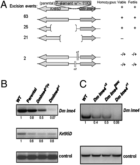
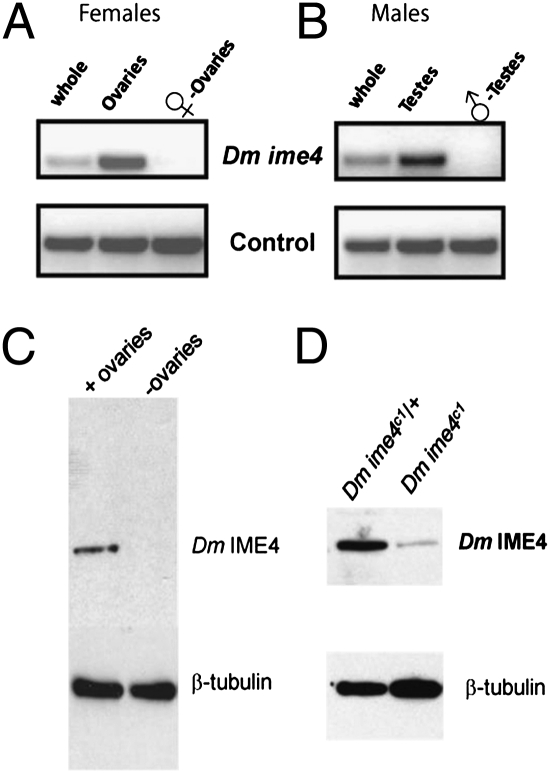
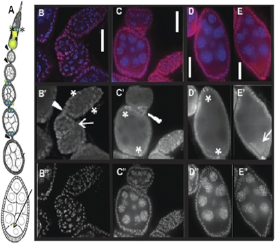
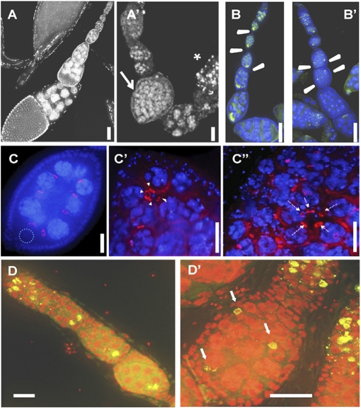
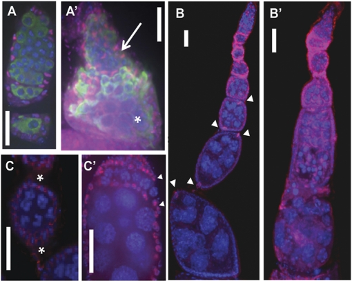
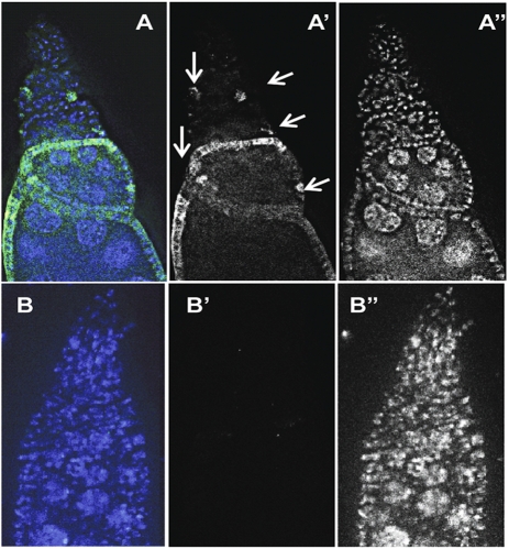
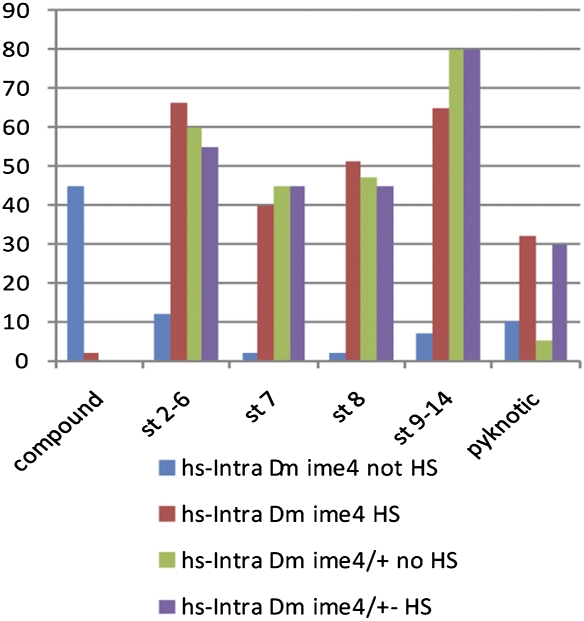
N(6)-methyladenosine is a nonediting RNA modification found in mRNA of all eukaryotes, from yeast to humans. Although the functional significance of N(6)-methyladenosine is unknown, the Inducer of MEiosis 4 (IME4) gene of Saccharomyces cerevisiae, which encodes the enzyme that catalyzes this modification, is required for gametogenesis. Here we find that the Drosophila IME4 homolog, Dm ime4, is expressed in ovaries and testes, indicating an evolutionarily conserved function for this enzyme in gametogenesis. In contrast to yeast, but as in Arabidopsis, Dm ime4 is essential for viability. Lethality is rescued fully by a wild-type transgenic copy of Dm ime4 but not by introducing mutations shown to abrogate the catalytic activity of yeast Ime4, indicating functional conservation of the catalytic domain. The phenotypes of hypomorphic alleles of Dm ime4 that allow recovery of viable adults reveal critical functions for this gene in oogenesis. Ovarioles from Dm ime4 mutants have fused egg chambers with follicle-cell defects similar to those observed when Notch signaling is defective. Indeed, using a reporter for Notch activation, we find markedly reduced levels of Notch signaling in follicle cells of Dm ime4 mutants. This phenotype of Dm ime4 mutants is rescued by inducing expression of a constitutively activated form of Notch. Our study reveals the function of IME4 in a metazoan. In yeast, this enzyme is responsible for a crucial developmental decision, whereas in Drosophila it appears to target the conserved Notch signaling pathway, which regulates many vital aspects of metazoan development.


Conflict of interest statement

The authors declare no conflict of interest.

Figures

Fig. 1.
Fig. 1.
Dm ime4 is essential for viability. (A) P-excision mutagenesis events that deleted the Dm ime4 ORF, partially or in its entirety, did not yield homozygous adult flies. Deletions of the adjacent Krt95D gene are viable. The small deletions 5′ to the Dm ime4 gene greatly reduced female and male fertility. (B) The 5′ deletion in the Dm ime4c1 (shown) specifically affects mRNA levels of Dm ime4 and does not affect mRNA levels of Krt95D. Transcript levels from whole flies were analyzed by RT-PCR in relation to a wild-type Oregon R control (numbers indicate relative abundance after quantification). Parental is the original P-element strain before P-element excision. Dm ime4c1/+ is the heterozygous sibling balanced with TM3. Dm ime4c1 is the homozygous sibling rarely produced in the balanced stock. (C) Knockdown of Dm ime4 via RNAi. pUAST-based RNAi lines (VDRC) were used to knock down Dm ime4 using the ubiquitous act5C-GAL4 driver. mRNA from whole adult flies was analyzed as in B. The strongest phenotypes were observed with the homozygous Dm ime4c1 allele, which resulted in significantly lower levels of Dm ime4 than in the Dm ime4c2 allele or the Dm ime4RNAi knockdown (Fig. S2).
Fig. 2.
Fig. 2.
Dm ime4 is expressed in the gonads of adult flies. mRNA from whole flies, dissected gonads (ovaries or testes, as indicated), or carcasses without gonads was analyzed as in Fig. 1. (A) Adult females. (B) Adult males. (C and D) Western blot using Dm IME4 antisera. (C) Protein extracts from wild-type Oregon R whole flies or their remains after ovary removal showing that Dm IME4 protein can be detected only in whole flies and not in those devoid of ovaries. (D) Protein extracts from ovaries isolated from females of the indicated genotype showing significantly reduced levels of Dm IME4 in homozygous mutant flies.
Fig. 3.
Fig. 3.
Dm IME4 is abundant in follicle cells and also is detectable in early germ-line cysts. (A) Diagram of a Drosophila ovariole in the anterior–posterior orientation, with the germarium at the top. Asterisks at the middle of the germarium show the location of prefollicle cells (marked with asterisks in B′). Arrowheads show stalk cells connecting the egg chambers. Twin green dots marked with green asterisks show the location of anterior and posterior polar cells (marked with asterisks in C′ and D′). Arrow shows the location of the ooplasm (arrow in E′). (BE) Distribution of Dm IME4 protein (red) in ovarioles, showing localization in follicle cells and in germ-line cells. Panels are oriented so that top is anterior and bottom is posterior, as depicted in the cartoon in A. DAPI staining is blue. (B′E′) Dm IME4 single channel. (B′′E′′) DAPI single channel. (B–B′′) Distribution of Dm IME4 protein in germaria showing prefollicle cells (asterisks in B′), stalk (arrowhead in B′), and early egg chambers (stages 2 and 3) where Dm IME4 is abundant in follicle and less abundant but reproducibly observed in germ-line cyst cells (arrow in B′). (CC′′) Germaria and stages 4 and 5, localization of Dm IME4 in stalk (arrowhead in C′) and polar cells (asterisks in C′). (D–D′′) Stage 7, localization of Dm IME4 in all follicle cells and abundantly in polar cells (asterisks in D′) and forming a crescent shape (ooplasm). (E) Stage 8, localization of Dm IME4 in follicle cells, polar cells, and abundantly in ooplasm (arrow in E′). (Scale bars: 10 μm.)
Fig. 4.
Fig. 4.
Oogenesis defects in Dm ime4 mutants. All images are oriented with anterior at the top. (A, B, C, and D) Dm ime4c1/+ control. (A′, B′, C′, C′′, and D’) Dm ime4c1 homozygous. (A and A′) DAPI nuclear staining showing a representative example of the control (A) and a Dm ime4c1 mutant ovariole (A′) with a compound follicle (arrow in A′) and a degenerating previtellogenic egg chamber (asterisk in A′) (Refer to Table S2 for quantification of these phenotypes). (B and B′) Compound follicles in Dm ime4c1 mutants have multiple oocytes [indicated by green Orb staining (arrowheads)] that fail to localize to the posterior end of the follicle. (CC′′) Most compound egg chambers of Dm ime4c1 mutants have multiple 16-cell germ-line cysts (15 nurse cells and one oocyte) from four rounds of mitosis, as indicated by the presence of four ring canals visualized by Kelch antibody (arrowheads in C′) surrounded by a common layer of follicle cells. Dotted circle in C shows position of the posteriorly located oocyte. Five ring canals (arrows in C′′) can be seen around the oocyte at low frequency. (D and D′) Oocytes in Dm ime4c1 compound follicles entered meiosis and assembled the synaptonemal complex (arrows in D′) as visualized by C(3)G antibody (green). DNA was detected with propidium iodide (red). (Scale bars: 10 μm.)
Fig. 5.
Fig. 5.
Defects in cyst encapsulation and follicle-cell specification in Dm ime4 mutants. (A) Control germarium. (A′) Representative germarium of a Dm ime4 homozygote in which cysts fail to follow an orderly pattern as they progress and have incomplete follicle-cell encapsulation (arrow). These trapped germ-line cysts can progress through other developmental stages, as shown by the increased size of the nurse cells at the bottom of this compound germarium (asterisk). Follicle cells were visualized with Traffic Jam (TJ, red). Germ-line cysts were visualized with VASA antibody staining (green). All nuclei were stained with DAPI (blue). (B and B′) Polar cells indicated by arrowheads in control Dm ime4c1 /+ follicles (B) are aberrant or missing in Dm ime4c1 compound follicles (B′). (C and C) Stalks seen in control Dm ime4c1 /+ (asterisks in C) frequently are missing in Dm ime4c1 homozygous mutants (C′), and egg chambers pack atop of each other. (Arrowheads in C′ mark the developmental stage shown between the asterisks in C.) FasIII staining is, red; DAPI staining is blue. (Scale bars: 10 μm.)
Fig. 6.
Fig. 6.
Notch signaling is reduced in Dm ime4 mutants. (A and A′) Notch signaling as detected in Gbe+ Su(H)-lacZ/FM7; Dm ime4C1/TM6b control (arrows indicate sites of Notch activity detected in the control). (B and B′) Gbe+ Su(H)-lacZ/FM7; Dm ime4c1 mutants have significantly lower levels of Notch activity than the sibling Gbe+ Su(H)-lacZ/FM7; Dm ime4c1/TM6b controls as visualized by anti–β-galactosidase staining (green). A and B show merged anti–β-galactosidase (green) and DAPI (blue) channels; A’ and B’ show an anti–β-galactosidase (green) single channel. A′′ and B′′ show a DAPI single channel (white).
Fig. 7.
Fig. 7.
NotchICD rescues egg-chamber defects in Dm ime4 mutants. Bar graph shows quantification of 100 ovarioles for each of the genotypes and conditions assayed as described in the graph color key. Lines carrying the heat shock-inducible NotchICD construct are designated as hs-Intra, heat-shocked treated (HS), or not treated (no HS). Egg chambers in 100 ovarioles were scored and are represented on the x axis as follows: compound egg chambers (compound); previtellogenic stages 2–6 (st 2–6) and stage 7 (st 7); vitellogenic stage 8 (st 8) and stages 9–14 (st 9–14); and degenerating egg chambers (pyknotic).

References

    1. Narayan P, Rottman FM. Methylation of mRNA. Adv Enzymol Relat Areas Mol Biol. 1992;65:255–285. - PubMed
    1. Bokar JA, Shambaugh ME, Polayes D, Matera AG, Rottman FM. Purification and cDNA cloning of the AdoMet-binding subunit of the human mRNA (N6-adenosine)-methyltransferase. RNA. 1997;3:1233–1247. - PMC - PubMed
    1. Shah JC, Clancy MJ. IME4, a gene that mediates MAT and nutritional control of meiosis in Saccharomyces cerevisiae. Mol Cell Biol. 1992;12:1078–1086. - PMC - PubMed
    1. Hongay CF, Grisafi PL, Galitski T, Fink GR. Antisense transcription controls cell fate in Saccharomyces cerevisiae. Cell. 2006;127:735–745. - PubMed
    1. Clancy MJ, Shambaugh ME, Timpte CS, Bokar JA. Induction of sporulation in Saccharomyces cerevisiae leads to the formation of N6-methyladenosine in mRNA: A potential mechanism for the activity of the IME4 gene. Nucleic Acids Res. 2002;30:4509–4518. - PMC - PubMed

Publication types