. 2011 Aug 30;108(35):14416-21.
doi: 10.1073/pnas.1107966108. Epub 2011 Aug 22.

Age-related changes in the plasticity and toughness of human cortical bone at multiple length scales

Affiliations

Age-related changes in the plasticity and toughness of human cortical bone at multiple length scales

Elizabeth A Zimmermann et al. Proc Natl Acad Sci U S A. .

Erratum in

  • Proc Natl Acad Sci U S A. 2012 Jul 17;109(29):11890

Abstract

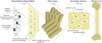
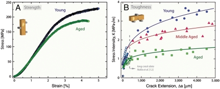
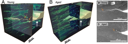
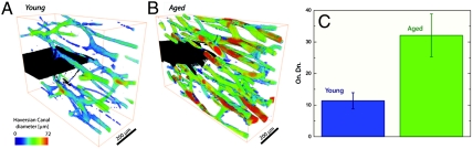
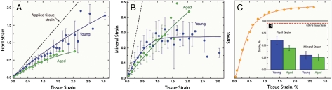
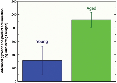
The structure of human cortical bone evolves over multiple length scales from its basic constituents of collagen and hydroxyapatite at the nanoscale to osteonal structures at near-millimeter dimensions, which all provide the basis for its mechanical properties. To resist fracture, bone's toughness is derived intrinsically through plasticity (e.g., fibrillar sliding) at structural scales typically below a micrometer and extrinsically (i.e., during crack growth) through mechanisms (e.g., crack deflection/bridging) generated at larger structural scales. Biological factors such as aging lead to a markedly increased fracture risk, which is often associated with an age-related loss in bone mass (bone quantity). However, we find that age-related structural changes can significantly degrade the fracture resistance (bone quality) over multiple length scales. Using in situ small-angle X-ray scattering and wide-angle X-ray diffraction to characterize submicrometer structural changes and synchrotron X-ray computed tomography and in situ fracture-toughness measurements in the scanning electron microscope to characterize effects at micrometer scales, we show how these age-related structural changes at differing size scales degrade both the intrinsic and extrinsic toughness of bone. Specifically, we attribute the loss in toughness to increased nonenzymatic collagen cross-linking, which suppresses plasticity at nanoscale dimensions, and to an increased osteonal density, which limits the potency of crack-bridging mechanisms at micrometer scales. The link between these processes is that the increased stiffness of the cross-linked collagen requires energy to be absorbed by "plastic" deformation at higher structural levels, which occurs by the process of microcracking.


Conflict of interest statement

The authors declare no conflict of interest.

Figures

Fig. 1.
Fig. 1.
Each level of the hierarchical structure influences the deformation and fracture of human cortical bone; the smaller levels affect the intrinsic toughness, whereas the higher length scales impact the extrinsic toughness. At the nanoscale, the polymeric nature of the collagen molecules allows them to uncoil and slide with respect to one another by breaking sacrificial bonds that absorb energy (2, 6). Sacrificial bonding also exists within higher levels of the hierarchy through shearing/stretching of the interfibrillar matrix and between fibrils (fibrillar sliding) (1, 3, 4). The process of microcracking can act as a plasticity mechanism by dissipating energy at coarser length scales typically exceeding several micrometers (5). Extrinsic mechanisms primarily act at the microstructural level by the interaction of growing cracks with the osteons; the weak boundaries in the secondary osteons absorb energy by microcracking during crack growth to toughen the structure mainly via crack bridging and crack deflection/twist (7, 8, 12).
Fig. 2.
Fig. 2.
In vitro mechanical properties of human cortical bone in 25 °C HBSS as a function of age showing (A) strength and (B) fracture-toughness R-curve properties for the young, middle-aged, and aged groups. The R-curve results encompass (long-crack growth) previous data, measured using crack sizes from approximately 500 µm to several millimeters (12), in addition to the current results on realistically short-crack extensions (< 500 μm) performed in situ in the ESEM. The inset schematics describe the orientation of the osteons with respect to the sample geometry.
Fig. 3.
Fig. 3.
(A and B) X-ray computed microtomography of young and aged bone samples show 3D images of the crack profile after roughly 500 μm of crack growth from a razor-sharpened notch (white arrows). (C and D) SEM images during small-crack R-curve experiments confirm the presence of large-crack bridges (other arrows) in young bone and smaller bridges in aged bone. The increased osteonal density in older bone leads to smaller and less frequent crack bridges and correlates with the marked reduction in the slope of the R curves with age.
Fig. 4.
Fig. 4.
X-ray computed tomography was also used to image the size and density of Haversian canals in (A) young and (B) aged human cortical bone (the color coding indicates the diameter of the Haversian canals). (C) Aged bone has nearly three times the osteonal density—i.e., number of osteons per unit bone area (On.Dn.)—as young bone, implying more cement lines for microcracks to initiate and smaller crack bridges during crack growth.
Fig. 5.
Fig. 5.
The results of the (A) SAXS and (B) WAXD experiments for tensile testing of young and aged cortical bone samples in the longitudinal orientation. For each individual tensile test, the strain values were binned every 0.1% tissue strain; then, for each age group, the average and standard deviation of the binned values were calculated and are shown as the dots and error bars, respectively. C shows a representative stress–strain curve for the tensile tests. (D) At a fixed tissue strain, the individual strain in the fibrils is approximately 25% smaller in aged bone than young bone, although changes in the mineral strain are not significant.
Fig. 6.
Fig. 6.
The accumulation of AGEs was fluorimetrically quantified in the cortex of young and aged bone samples. These results indicate that the aged bone contained nearly three times as many fluorescent cross-links as the young bone.

References

    1. Bailey AJ, Sims TJ, Avery NC, Miles CA. Chemistry of collagen cross-links—glucose-mediated covalent cross-linking of type-IV collagen in lens capsules. Biochem J. 1993;296:489–496. - PMC - PubMed
    1. Buehler MJ. Molecular nanomechanics of nascent bone: Fibrillar toughening by mineralization. Nanotechnology. 2007;18:295102.
    1. Fantner GE, et al. Sacrificial bonds and hidden length dissipate energy as mineralized fibrils separate during bone fracture. Nat Mater. 2005;4:612–616. - PubMed
    1. Gupta HS, et al. Cooperative deformation of mineral and collagen in bone at the nanoscale. Proc Natl Acad Sci USA. 2006;103:17741–17746. - PMC - PubMed
    1. Nalla RK, Kruzic JJ, Ritchie RO. On the origin of the toughness of mineralized tissue: Microcracking or crack bridging? Bone. 2004;34:790–798. - PubMed

Publication types

Substances