. 2011 Sep 6;108(36):14813-8.
doi: 10.1073/pnas.1106420108. Epub 2011 Aug 22.

Topology of human apolipoprotein E3 uniquely regulates its diverse biological functions

Affiliations

Topology of human apolipoprotein E3 uniquely regulates its diverse biological functions

Jianglei Chen et al. Proc Natl Acad Sci U S A. .

Abstract

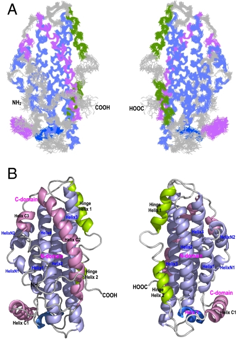
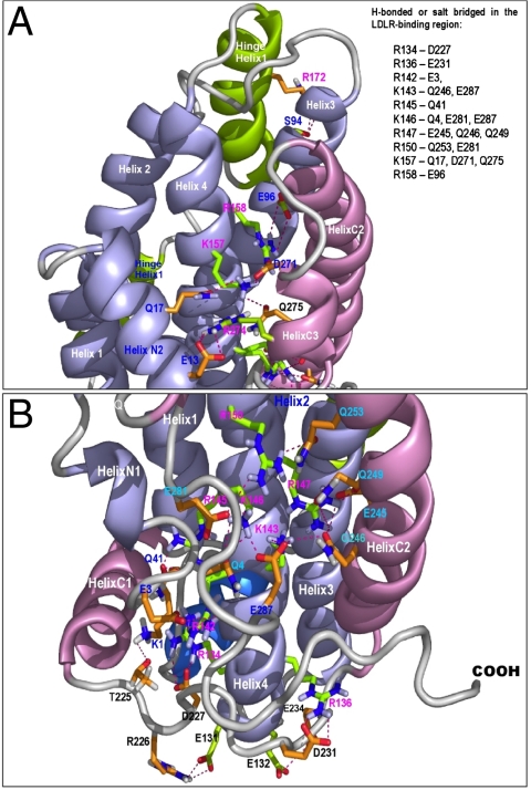
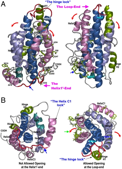
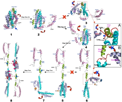
Human apolipoprotein E (apoE) is one of the major determinants in lipid transport, playing a critical role in atherosclerosis and other diseases. Binding to lipid and heparan sulfate proteoglycans (HSPG) induces apoE to adopt active conformations for binding to low-density lipoprotein receptor (LDLR) family. ApoE also interacts with beta amyloid peptide, manifests critical isoform-specific effects on Alzheimer's disease. Despite the importance of apoE in these major human diseases, the fundamental questions of how apoE adjusts its structure upon binding to regulate its diverse functions remain unsolved. We report the NMR structure of apoE3, displaying a unique topology of three structural domains. The C-terminal domain presents a large exposed hydrophobic surface that likely initiates interactions with lipids, HSPG, and beta amyloid peptides. The unique topology precisely regulates apoE tertiary structure to permit only one possible conformational adaptation upon binding and provides a double security in preventing lipid-free and partially-lipidated apoE from premature binding to apoE receptors during receptor biogenesis. This topology further ensures the optimal receptor-binding activity by the fully lipidated apoE during lipoprotein transport in circulation and in the brain. These findings provide a structural framework for understanding the structural basis of the diverse functions of this important protein in human diseases.


Conflict of interest statement

The authors declare no conflict of interest.

Figures

Fig. 1.
Fig. 1.
NMR Structure of apoE3. (A) Superposition of the 20 best-fit NMR structures of apoE3. (B) Ribbon representations of the average structure of apoE3. The N-terminal helices are colored in light blue, the C-terminal helices are in pink, and the hinge domain is colored in green. The Helix1’ is shown in blue. The helices are labeled respectively. The left and right in (A) and (B) are 180° rotation of apoE3 structure, showing both sides of apoE3.
Fig. 2.
Fig. 2.
Buried H-bonds and salt-bridges between apoE3 domains, with a focus on the major LDLR-binding region. The apoE3 is shown in ribbons and the side chains of the interacting residues in stick model. The NT domain is shown in light blue while the CT domain in pink. The side chains of the interacting residues in the major LDLR-binding region are shown in green, whereas the side chains of the other interacting residues are shown in brown. The salt-bridges and H-bonds are indicated with red dashed lines. (A) Detailed interdomain interactions of residues K157 and R158. (B) Detailed interdomain interactions of residues R134, R136, R142, K143, R145, K146, R147, and R150. The interacting residues are summarized in the top box.
Fig. 3.
Fig. 3.
Special structural features that regulate initial conformational adaptation of apoE upon lipid binding. ApoE structures are shown in ribbons with the NT helices colored in blue, the CT helices in pink, and hinge domain in green. The Helix1’ is in dark green. (A) Side views of apoE3 structure. Left: ApoE3 structure with the exposed hydrophobic residues of the apoE-CT shown in green sticks. Right: ApoE3 structure with the buried hydrophobic residues of the apoE-CT shown in green sticks. Two ends of the apoE3 helix bundle are named and labeled as: The Loop-end and the Helix1’-end. A hinge lock is formed by hinge domain that prevents the NT bundle from opening at the Loop-end (Helices 2 and 3 moving away from helices 1 and 4) (B) Left: A bottom view of apoE3 structure (the Helix1’-end view). Helix C1 and the flanking loops form a “Helix C1 lock” (Blue arrow) that prevents the CT domain moving away from the NT domain at the Helix1’-end. Right: A top view of apoE3 structure (the Loop-end view) with the exposed hydrophobic residues of the apoE-CT shown in green sticks. Noticeably, most of hydrophobic residues are exposed in the apoE-CT, forming the only major hydrophobic surface in apoE3 structure (green arrow). The directions of the allowed apoE-CT opening are indicated by red arrows.
Fig. 4.
Fig. 4.
A two-step conformational adaptation of apoE3 upon lipoprotein binding. ApoE3 structures are shown in ribbons and the colors are coded the same as Fig. 2. The helices are labeled respectively. This two-step conformation adaptation of apoE3 upon lipoprotein association follows a specific and precise self-modulation pathway, which is regulated by the special topology of lipid-free apoE3 structure. We assume that there is no major change of the helix locations in apoE3 during lipid-binding.

Similar articles

Cited by

References

    1. Weisgraber KH. Apolipoprotein E: structure-function relationships. Adv Protein Chem. 1994;45:249–302. - PubMed
    1. Mahley RW, Weisgraber KH, Huang Y. Apolipoprotein E: structure determines function, from atherosclerosis to Alzheimer’s disease to AIDS. J Lipid Res. 2009;50(Suppl):S183–S188. - PMC - PubMed
    1. Mahley RW, Ji ZS. Remnant lipoprotein metabolism: key pathways involving cell-surface heparan sulfate proteoglycans and apolipoprotein E. J Lipid Res. 1999;40:1–16. - PubMed
    1. Luc G, et al. Impact of apolipoprotein E polymorphism on lipoproteins and risk of myocardial infarction. The ECTIM Study. Arterioscler Thromb. 1994;14:1412–1419. - PubMed
    1. Mahley RW, Rall SC Jr, editors. The Metabolic and Molecular Base of Inherited Disease. 8 Ed. New York: (McGraw-Hill); 2001. pp. 2835–2862.

Publication types

MeSH terms

Associated data

LinkOut - more resources