. 2011 Aug 30;108(35):14509-14.
doi: 10.1073/pnas.1019386108. Epub 2011 Aug 22.

PI3K/Akt signaling requires spatial compartmentalization in plasma membrane microdomains

Affiliations

PI3K/Akt signaling requires spatial compartmentalization in plasma membrane microdomains

Xinxin Gao et al. Proc Natl Acad Sci U S A. .

Abstract

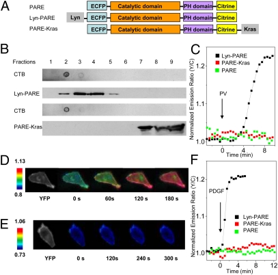
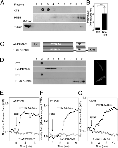
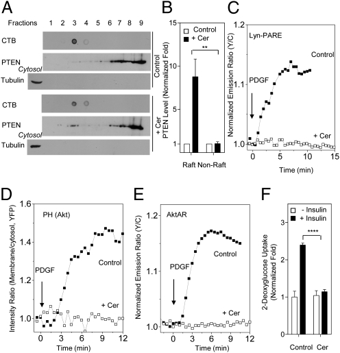
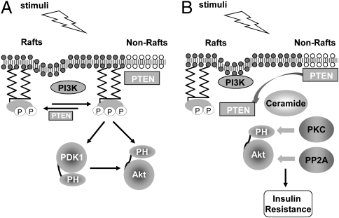
Spatial compartmentalization of signaling pathway components generally defines the specificity and enhances the efficiency of signal transduction. The phosphatidylinositol 3-kinase (PI3K)/Akt pathway is known to be compartmentalized within plasma membrane microdomains; however, the underlying mechanisms and functional impact of this compartmentalization are not well understood. Here, we show that phosphoinositide-dependent kinase 1 is activated in membrane rafts in response to growth factors, whereas the negative regulator of the pathway, phosphatase and tensin homolog deleted on chromosome 10 (PTEN), is primarily localized in nonraft regions. Alteration of this compartmentalization, either by genetic targeting or ceramide-induced recruitment of PTEN to rafts, abolishes the activity of the entire pathway. These findings reveal critical steps in raft-mediated PI3K/Akt activation and demonstrate the essential role of membrane microdomain compartmentalization in enabling PI3K/Akt signaling. They further suggest that dysregulation of this compartmentalization may underlie pathological complications such as insulin resistance.


Conflict of interest statement

The authors declare no conflict of interest.

Figures

Fig. 1.
Fig. 1.
Development and characterization of PDK1 activation reporters. (A) The PDK1 activation reporter PARE was generated with full-length PDK1 sandwiched by a pair of fluorescent proteins, ECFP and citrine. PARE was targeted to membrane rafts by using a targeting motif derived from Lyn kinase and to nonraft regions of the plasma membrane by using a targeting motif derived from Kras. (B) The localization of Lyn-PARE and PARE-Kras was confirmed by sucrose density gradient fractionation. Total cell lysates from HEK 293 cells overexpressing Lyn-PARE or PARE-Kras were subjected to sucrose density gradient fractionation, followed by Western blotting. Cholera toxin subunit B (CTB) was used as a raft marker. The reporters were detected with an anti-GFP antibody. (C) Representative time courses showing the responses of PARE (n = 5), Lyn-PARE (n = 5), and PARE-Kras (n = 4) in serum-starved NIH 3T3 cells after stimulation with 100 μM PV. (D) Pseudocolor images showing the response of Lyn-PARE to 50 ng/mL PDGF in NIH 3T3 cells. The yellow fluorescence image (Left) shows the localization of Lyn-PARE. (E) Pseudocolor images showing the null response of PARE-Kras to 50 ng/mL PDGF in NIH 3T3 cells. The yellow fluorescence image (Left) shows the localization of PARE-Kras. (F) Stimulation of serum-starved NIH 3T3 cells with 50 ng/mL PDGF induced an emission ratio change in Lyn-PARE (n = 7), but not PARE (n = 5) or PARE-Kras (n = 6).
Fig. 2.
Fig. 2.
Genetic targeting of PTEN to membrane rafts abolishes PDK1 activation, Akt membrane recruitment, and Akt activity. (A) PTEN is primarily localized to nonraft regions of the plasma membrane. Crude plasma membranes from HEK 293 cells were solubilized and subjected to sucrose density gradient fractionation, followed by Western blotting with an anti-PTEN antibody. CTB was used as a raft marker. Anti-tubulin was used to ensure the separation of membrane proteins from cytosolic proteins. (B) Statistically significant differences between PTEN levels in rafts and nonraft regions (***P < 0.001; n = 3). Densitometric analysis indicated that the majority of the membrane PTEN resides in nonraft regions. (C) PTEN A4, fused with a C-terminal fluorescent protein (FP), was targeted to membrane rafts and nonraft regions with a Lyn or Kras motif. (D) The membrane localization of Lyn-PTEN A4 or PTEN A4-Kras was verified by sucrose density gradient fractionation of total cell lysates of HEK 293 cells expressing Lyn-PTEN A4-YFP or PTEN A4-YFP-Kras. CTB was used as a raft marker. Targeted PTEN A4 was detected with an anti-GFP antibody. Yellow fluorescence images show the membrane localization of PTEN. (E) Representative time courses indicating that the response of Lyn-PARE was abolished by Lyn-PTEN A4 (n = 5; PTEN fused with mCherry), but not PTEN A4-Kras (n = 7). (F) Representative time courses demonstrating that the membrane translocation of the Akt PH domain was abolished by Lyn-PTEN A4 (n = 4; PTEN fused with mCherry), but not PTEN A4-Kras (n = 4). (G) Representative time courses indicating that the response of AktAR was abolished by Lyn-PTEN A4 (n = 7; PTEN fused with mCherry), but not PTEN A4-Kras (n = 6).
Fig. 3.
Fig. 3.
Ceramide recruits PTEN to membrane rafts and suppresses PDK1 activation, Akt membrane recruitment, and Akt activity. (A) Ceramide recruits PTEN to membrane rafts. Crude plasma membranes from 3T3 L1 adipocytes (in the presence or absence of 50 μM C2-ceramide) were solubilized and subjected to sucrose density gradient fractionation, followed by Western blotting with an anti-PTEN antibody. CTB was used as a raft marker. Anti-tubulin was used to ensure the separation of membrane proteins from cytosolic proteins. (B) Densitometric analysis indicated a statistically significant difference between the raft PTEN levels in the presence of C2-ceramide and those in its absence (**P < 0.01; n = 3). (C) Representative time courses showing that the response of Lyn-PARE (n = 7) in 3T3 L1 preadipocytes was abolished by preincubation with 50 μM C2-ceramide (n = 6). (D) Representative time courses showing that membrane translocation of Akt PH domain (n = 3) in 3T3 L1 preadipocytes was abolished by preincubation with 50 μM C2-ceramide (n = 3). (E) Representative time courses showing that the response of AktAR (n = 6) in 3T3 L1 preadipocytes was abolished by preincubation of 50 μM C2-ceramide (n = 5). (F) Ceramide-mediated suppression of insulin-induced glucose uptake in 3T3 L1 adipocytes. Preincubation of 50 μM C2-ceramide with 3T3 L1 adipocytes for 60 min inhibited insulin-induced glucose uptake in these cells (****P < 0.0001; n = 3).
Fig. 4.
Fig. 4.
Compartmentalized PDK1 and PTEN activity in membrane microdomains is essential for proper PI3K/Akt signaling. (A) Preferential activation of Akt in membrane rafts is mediated by activated PDK1 in rafts and lack of PTEN-mediated down-regulation in these microdomains. (B) Dysregulation of the raft-localized PI3K/Akt signaling as the underlying mechanism for insulin resistance. Ceramide may inhibit Akt signaling through promoting PKCζ phosphorylation of Akt or activating the Akt phosphatase PP2A. The ceramide-induced mislocalization of PTEN to membrane rafts critically contributes to the inhibitory effect of ceramide on PI3K/Akt signaling.

Similar articles

Cited by

References

    1. Franke TF. PI3K/Akt: Getting it right matters. Oncogene. 2008;27:6473–6488. - PubMed
    1. Wong KK, Engelman JA, Cantley LC. Targeting the PI3K signaling pathway in cancer. Curr Opin Genet Dev. 2010;20:87–90. - PMC - PubMed
    1. Farese RV, Sajan MP, Standaert ML. Insulin-sensitive protein kinases (atypical protein kinase C and protein kinase B/Akt): Actions and defects in obesity and type II diabetes. Exp Biol Med (Maywood) 2005;230:593–605. - PubMed
    1. Bozulic L, Hemmings BA. PIKKing on PKB: Regulation of PKB activity by phosphorylation. Curr Opin Cell Biol. 2009;21:256–261. - PubMed
    1. Mora A, Komander D, van Aalten DM, Alessi DR. PDK1, the master regulator of AGC kinase signal transduction. Semin Cell Dev Biol. 2004;15:161–170. - PubMed

Publication types

MeSH terms

Substances