. 2011 Dec;157(Pt 12):3292-3305.
doi: 10.1099/mic.0.051987-0. Epub 2011 Aug 26.

Structure-function analysis of HsiF, a gp25-like component of the type VI secretion system, in Pseudomonas aeruginosa

Affiliations

Structure-function analysis of HsiF, a gp25-like component of the type VI secretion system, in Pseudomonas aeruginosa

Nadine S Lossi et al. Microbiology (Reading). 2011 Dec.

Abstract

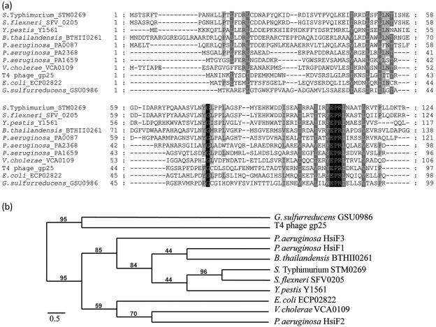
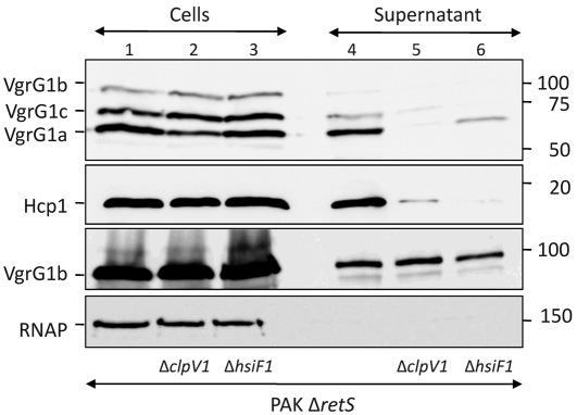
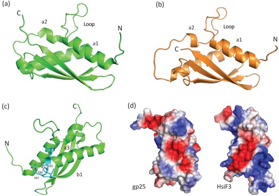
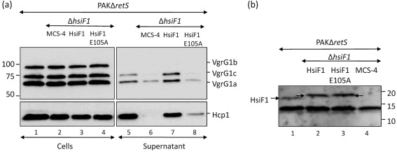
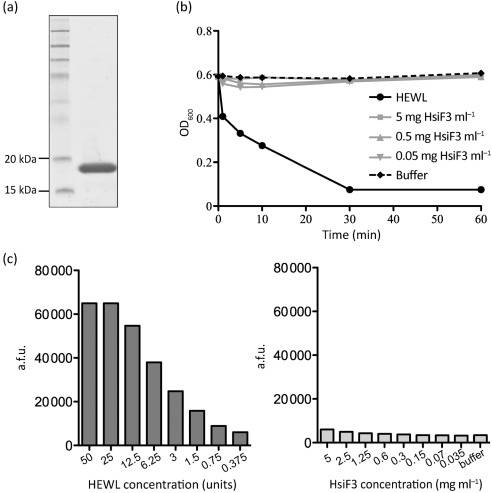
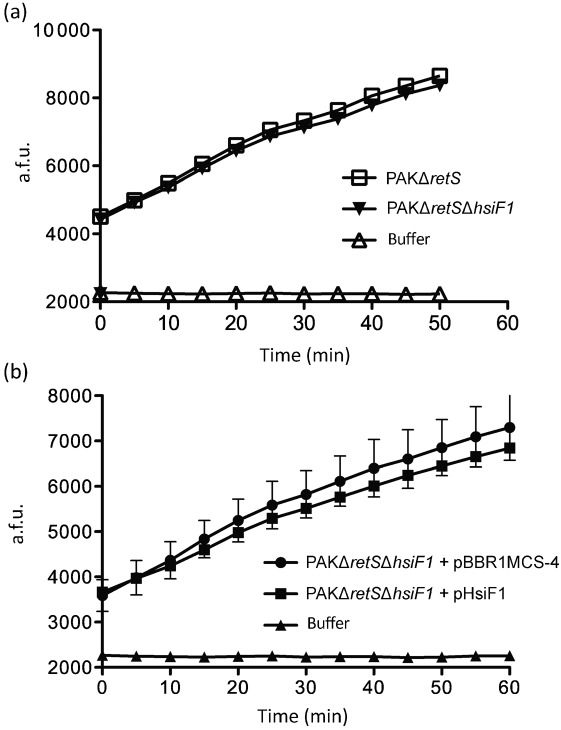
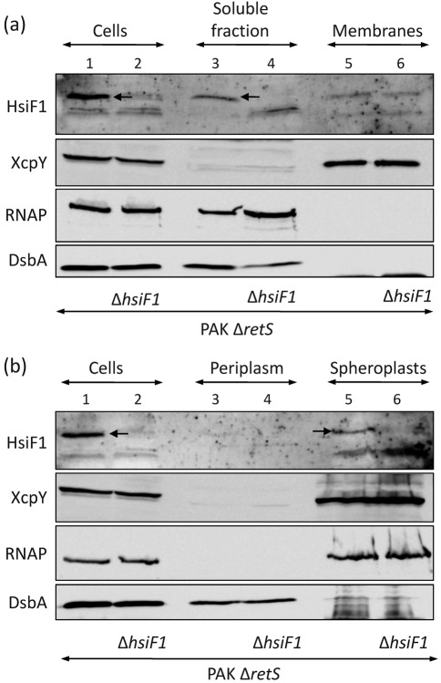
Bacterial pathogens use a range of protein secretion systems to colonize their host. One recent addition to this arsenal is the type VI secretion system (T6SS), which is found in many Gram-negative bacteria. The T6SS involves 12-15 components, including a ClpV-like AAA(+) ATPase. Moreover, the VgrG and Hcp components have been proposed to form a puncturing device, based on structural similarity to the tail spike components gp5/gp27 and the tail tube component gp19 of the T4 bacteriophage, respectively. Another T6SS component shows similarity to a T4 phage protein, namely gp25. The gp25 protein has been proposed to have lysozyme activity. Other T6SS components do not exhibit obvious similarity to characterized T4 phage components. The genome of Pseudomonas aeruginosa contains three T6SS gene clusters. In each cluster a gene encoding a putative member of the gp25-like protein family was identified, which we called HsiF. We confirmed this similarity by analysing the structure of the P. aeruginosa HsiF proteins using secondary and tertiary structure prediction tools. We demonstrated that HsiF1 is crucial for the T6SS-dependent secretion of Hcp and VgrG. Importantly, lysozyme activity of HsiF proteins was not detectable, and we related this observation to the demonstration that HsiF1 localizes to the cytoplasm of P. aeruginosa. Finally, our data showed that a conserved glutamate, predicted to be required for proper HsiF folding, is essential for its function. In conclusion, our data confirm the central role of HsiF in the T6SS mechanism, provide information on the predicted HsiF structure, and call for reconsideration of the function of gp25-like proteins.


Figures

Fig. 1.
Fig. 1.
Amino acid sequence comparison and phylogenetic relationship among members of the gp25 family. (a) Identification of conserved residues in members of the gp25-like protein family. Amino acid sequence alignments of HsiF proteins of P. aeruginosa (HsiF1/PA0087, HsiF2/PA1659 and HsiF3/PA2368), T4 phage gp25, G. sulfurreducens GSU0986 and HsiF homologues found in other Gram-negative bacteria associated with a T6SS gene cluster (clustal w2, GeneDoc). Different levels of shading indicate the level of conservation (100 %, black; >80 %, dark grey; >60 %, light grey; <60 %, white). The amino acid position is indicated at the beginning and the end of each line. (b) Phylogenetic analysis of gp25-like proteins. Neighbour-joining tree based on HsiF proteins, gp25 of the T4 bacteriophage, GSU0986 of G. sulfurreducens and several HsiF orthologues found in Gram-negative bacteria that are associated with T6SS gene clusters. Indicated bootstrap values correspond to 100 replicates. Phylogenetic analysis was carried out using http://www.phylogeny.fr.
Fig. 2.
Fig. 2.
HsiF1 is essential for H1-T6SS function. PAKΔretS, PAKΔretSΔclpV1 and PAKΔretSΔhsiF1 were grown in TSB for 5 h. Whole-cell lysates (lanes 1–3) and 10× concentrated Pseudomonas culture supernatant (lanes 4–6) were analysed by SDS-PAGE and Western blotting using polyclonal antibodies directed against VgrG1a (first row), Hcp1 (second row) or VgrG1b (third row). Monoclonal anti-RNAP antibody was used to control for cell lysis in the supernatant fraction (fourth row). Molecular mass markers are indicated on the right (kDa). The positions of VgrG1a, VgrG1b, VgrG1c, Hcp1 and RNAP are indicated on the left.
Fig. 3.
Fig. 3.
Homology modelling of HsiF homologues based on the crystal structure of GSU0986 of G. sulfurreducens. (a) Ribbon representation of the crystal structure of Geobacter GSU0986 (PDB2ia7), showing the unusual loop conformation between α1 and α2. N and C termini are labelled. (b) Ribbon representation of the HsiF3 homology model based on GSU0986 as an exemplar of the HsiF homologues. (c) Ribbon representation of GSU0986, highlighting identical residues between GSU0986 and HsiF homologues. The E85-P86-R87 forms a tight turn between α3 and β1, with E85 and R87 forming a salt bridge (dotted black line). This turn is essential for stabilizing the positions of α1, α3 and β1 and in establishing the overall gp25 fold. Other conserved residues not labelled include W84 and I88, both of which form hydrophobic interactions that also stabilize the GSU0986 fold. (d) Representations of the electrostatic surface potential for GSU0986/gp25 and HsiF3, with positive potential shown in blue and negative in red. Note the striking striated pattern of positive and negative surface potential for HsiF3 versus GSU0986/gp25. All figures were made using PyMOL (http://www.pymol.org).
Fig. 4.
Fig. 4.
The conserved glutamate residue E105 is required for HsiF1 function. (a) PAKΔretS (lanes 1 and 5), PAKΔretSΔhsiF1 strains containing pBBR1MCS-4 (labelled MCS-4, lanes 2 and 6), pHsiF1 (lanes 3 and 7) and pHsiF1E105A (lanes 4 and 8), as indicated, were grown to late exponential phase. Whole-cell lysates (lanes 1–4) and 10× concentrated Pseudomonas culture supernatant (lanes 5–8) were analysed by SDS-PAGE and Western blotting using polyclonal anti-VgrG1a and anti-Hcp1 antibodies. Molecular mass markers are shown on the left (kDa). (b) HsiF1 and HsiF1E105A are produced at similar levels in P. aeruginosa. PAKΔretS (lane 1) and PAKΔretSΔhsiF1 containing pBBR1MCS-4 (lane 2), pHsiF1 (lane 3) or pHsiF1E105A (lane 4) were grown to late exponential phase, and production of HsiF1 and HsiF1E105A was analysed by Western blotting using polyclonal anti-HsiF1 antibody. Chromosomal levels of HsiF1 as well as HsiF1 and HsiF1E105A produced from pHsiF1 and pHsiF1E105A, respectively, are indicated by arrows. HsiF1 and HsiF1E105A contain a C-terminal 6×His tag, and the corresponding band therefore runs slightly more slowly than the chromosomally expressed HsiF. Molecular mass markers are indicated on the right (kDa). The band below the 15 kDa marker is a non-specific cross-reacting protein.
Fig. 5.
Fig. 5.
Purified HsiF3 protein does not exhibit detectable lysozyme activity. (a) Recombinant HsiF3 used for in vitro analysis. HsiF3 was expressed and purified as described in Methods. Purified HsiF3 was analysed by SDS-PAGE and stained by Coomassie blue. (b) Recombinant HsiF3 (5, 0.5 or 0.05 mg ml−1), HEWL (0.5 mg ml−1) or assay buffer was incubated with a suspension of M. lysodeikticus in 96-well plates. The decrease in turbidity was monitored at 595 nm over 60 min at 37 °C. (c) HEWL (bar chart to the left; dilution series from left to right 50 to 0.375 U per assay point) or recombinant HsiF3 (bar chart to the right; dilution series 5 to 0.035 mg ml−1 final enzyme concentration per assay point) was incubated with fluorescently labelled M. lysodeikticus for 60 min at 37 °C before fluorescence was measured as described in Methods. Emission of fluorescence is represented by arbitrary fluorescence units (a.f.u.). One unit of HEWL is defined by the manufacturer as the amount of enzyme required to produce a change in the A450 of 0.001 U min−1 at pH 6.24 and 25 °C, using a suspension of M. lysodeikticus as the substrate.
Fig. 6.
Fig. 6.
HsiF1 does not exhibit detectable lysozyme activity when expressed in P. aeruginosa. (a) Cell lysates of P. aeruginosa PAKΔretS and PAKΔretSΔhsiF1, grown to late exponential phase as described in Methods, were incubated with fluorescently labelled M. lysodeikticus for 50 min at 37 °C, during which fluorescence was measured every 5 min. Assay buffer was incubated with M. lysodeikticus as a negative control. Emission of fluorescence is represented by arbitrary fluorescence units (a.f.u.). (b) Cell lysates of P. aeruginosa PAKΔretSΔhsiF1 containing pBBR1MCS-4 or pHsiF1, grown to late exponential phase, were incubated with fluorescently labelled M. lysodeikticus for 60 min at 37 °C, during which fluorescence was measured every 5 min. Assay buffer was incubated with M. lysodeikticus as a negative control. Error bars, sd derived from triplicates of each assay point. Emission of fluorescence is represented by a.f.u.
Fig. 7.
Fig. 7.
HsiF1 does not localize to the periplasm of P. aeruginosa. Subcellular localization of HsiF1 was analysed in PAKΔretS and PAKΔretSΔhsiF1 grown to late exponential phase. Cell fractions were analysed by SDS-PAGE and immunoblotting. Successful fractionation was verified using antibodies directed against periplasmic DsbA (bottom row), the inner-membrane protein XcpY (second row) and cytosolic RNAP (third row). HsiF1 was detected using polyclonal anti-HsiF1 antibody (first row). (a) HsiF1 localizes to the soluble fraction in P. aeruginosa. The soluble (lanes 3 and 4) and insoluble fractions (indicated as membranes, lanes 5 and 6) of PAKΔretS and PAKΔretSΔhsiF1 were obtained by sonication followed by ultracentrifugation as described in Methods, and were analysed by immunoblotting using the controls described above. (b) HsiF1 does not localize to the periplasm in P. aeruginosa. Western blot analysis of whole-cell lysate (lanes 1 and 2), periplasmic fraction (lanes 3 and 4) and spheroplasts (representing a combination of cytosol and bacterial membranes; lanes 5 and 6) was performed using the antibodies described above.

Similar articles

Cited by

References

    1. Aschtgen M. S., Bernard C. S., De Bentzmann S., Lloubès R., Cascales E. (2008). SciN is an outer membrane lipoprotein required for type VI secretion in enteroaggregative Escherichia coli. J Bacteriol 190, 7523–7531. 10.1128/JB.00945-08 - DOI - PMC - PubMed
    1. Aschtgen M. S., Gavioli M., Dessen A., Lloubès R., Cascales E. (2010a). The SciZ protein anchors the enteroaggregative Escherichia coli type VI secretion system to the cell wall. Mol Microbiol 75, 886–899. 10.1111/j.1365-2958.2009.07028.x - DOI - PubMed
    1. Aschtgen M. S., Thomas M. S., Cascales E. (2010b). Anchoring the type VI secretion system to the peptidoglycan: TssL, TagL, TagP... what else? Virulence 1, 535–540. 10.4161/viru.1.6.13732 - DOI - PubMed
    1. Ballister E. R., Lai A. H., Zuckermann R. N., Cheng Y., Mougous J. D. (2008). In vitro self-assembly of tailorable nanotubes from a simple protein building block. Proc Natl Acad Sci U S A 105, 3733–3738. 10.1073/pnas.0712247105 - DOI - PMC - PubMed
    1. Bingle L. E., Bailey C. M., Pallen M. J. (2008). Type VI secretion: a beginner’s guide. Curr Opin Microbiol 11, 3–8. 10.1016/j.mib.2008.01.006 - DOI - PubMed

Publication types

MeSH terms

Substances

LinkOut - more resources