. 2011 Oct 21;286(42):36331-9.
doi: 10.1074/jbc.M111.272146. Epub 2011 Aug 26.

Inhibition of c-Jun-N-terminal kinase increases cardiac peroxisome proliferator-activated receptor alpha expression and fatty acid oxidation and prevents lipopolysaccharide-induced heart dysfunction

Affiliations

Inhibition of c-Jun-N-terminal kinase increases cardiac peroxisome proliferator-activated receptor alpha expression and fatty acid oxidation and prevents lipopolysaccharide-induced heart dysfunction

Konstantinos Drosatos et al. J Biol Chem. .

Abstract

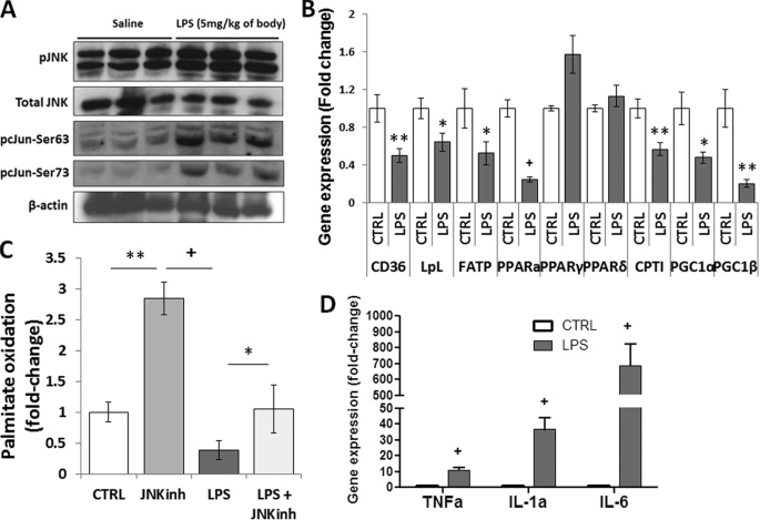
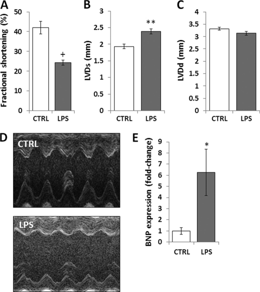
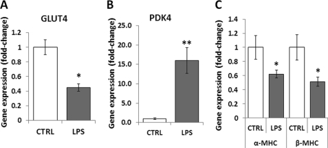
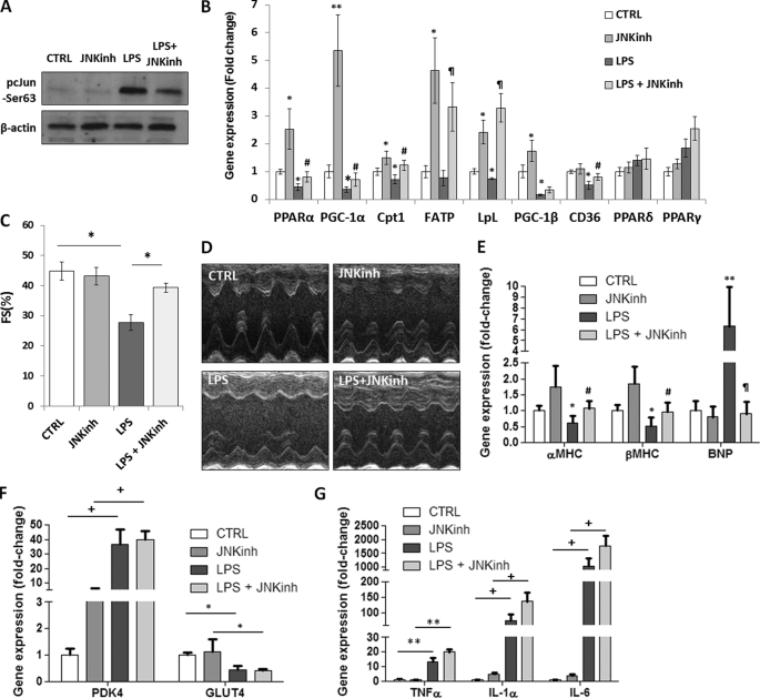
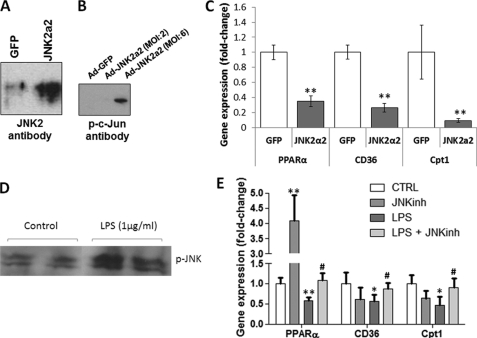
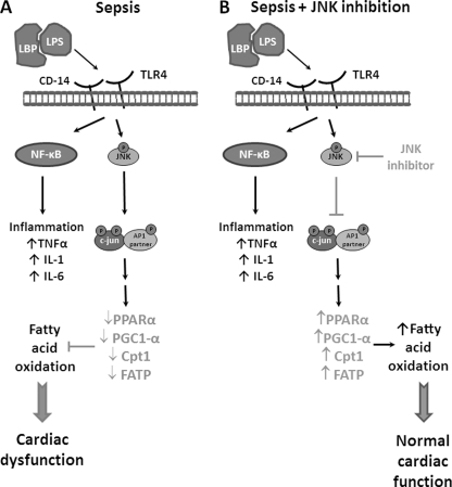
Septic shock results from bacterial infection and is associated with multi-organ failure, high mortality, and cardiac dysfunction. Sepsis causes both myocardial inflammation and energy depletion. We hypothesized that reduced cardiac energy production is a primary cause of ventricular dysfunction in sepsis. The JNK pathway is activated in sepsis and has also been implicated in impaired fatty acid oxidation in several tissues. Therefore, we tested whether JNK activation inhibits cardiac fatty acid oxidation and whether blocking JNK would restore fatty acid oxidation during LPS treatment. LPS treatment of C57BL/6 mice and adenovirus-mediated activation of the JNK pathway in cardiomyocytes inhibited peroxisome proliferator-activated receptor α expression and fatty acid oxidation. Surprisingly, none of the adaptive responses that have been described in other types of heart failure, such as increased glucose utilization, reduced αMHC:βMHC ratio or induction of certain microRNAs, occurred in LPS-treated mice. Treatment of C57BL/6 mice with a general JNK inhibitor (SP600125) increased fatty acid oxidation in mice and a cardiomyocyte-derived cell line. JNK inhibition also prevented LPS-mediated reduction in fatty acid oxidation and cardiac dysfunction. Inflammation was not alleviated in LPS-treated mice that received the JNK inhibitor. We conclude that activation of JNK signaling reduces fatty acid oxidation and prevents the peroxisome proliferator-activated receptor α down-regulation that occurs with LPS.


Figures

FIGURE 1.
FIGURE 1.
LPS activates JNK, inhibits fatty acid oxidation, and induces inflammation. A, Western blot analysis of pJNK, total JNK, pc-Jun-Ser-63, pc-Jun-Ser-73, and β-actin protein levels obtained from hearts of 10- to 12-week-old C57BL/6 mice that were treated with 5 mg/kg LPS. (B) CD36, LpL, FATP, PPARα, PPARγ, PPARδ, CPT-1β, PGC-1α, and PGC-1β mRNA levels in hearts of 10- to 12-week-old C57BL/6 mice that were treated with 5 mg/kg LPS. n = 5; *, p < 0.05; **, p < 0.01; +, p < 0.001. C, [3H]palmitic acid oxidation in cardiac muscle of C57BL/6 mice treated with 5 mg/kg JNK inhibitor (JNKinh) (SP600125), 5 mg/kg LPS, or a combination of LPS and JNK inhibitor. n = 4; *, p < 0.05. D, TNFα, IL-1α, and IL-6 mRNA levels in hearts of 10- to 12-week-old C57BL/6 mice that were treated with 5 mg/kg LPS. n = 5; +, p < 0.001.
FIGURE 2.
FIGURE 2.
Treatment of C57BL/6 mice with LPS impairs cardiac function. A–D, fractional shortening (A), left ventricular systolic diameter (LVDs) (B), and left ventricular diastolic diameter (LVDd) (C) as measured by two-dimensional echocardiography of 10- to 12-week-old C57BL/6 mice that were treated with 5 mg/kg LPS. D, photographs of echocardiograms from LPS-treated C57BL/6 mice. E, cardiac BNP mRNA levels of 10- to 12-week-old C57BL/6 mice that were treated with 5 mg/kg LPS. Control mice were treated with saline. n = 6–8; *, p < 0.05; **, p < 0.01; +, p < 0.001.
FIGURE 3.
FIGURE 3.
Mechanisms of cardiac dysfunction in sepsis differ from chronic heart failure. A–C, GLUT4 (A), PDK4 (B), and αMHC and βMHC (C) mRNA levels in hearts of 10- to 12-week-old C57BL/6 mice that were treated with 5 mg/kg LPS. Control mice were treated with saline. n = 6–8; *, p < 0.05; **, p < 0.01.
FIGURE 4.
FIGURE 4.
JNK inhibition prevents LPS-mediated reduction in cardiac fatty acid oxidation and improves heart function despite elevated inflammation. A, Western blot analysis of pc-Jun-Ser-63 and β-actin protein levels obtained from hearts of 10- to 12-week-old C57BL/6 mice that were treated with 5 mg/kg LPS or a combination of LPS and 5 mg/kg JNK inhibitor (JNKinh) (SP600125). Control cells were treated with saline. B, PPARα, PGC-1α,CPT-1β, FATP, LpL, PGC-1β, CD36, PPARδ, and PPARγ mRNA levels in hearts of 10- to 12-week-old C57BL/6 mice that were treated with 5 mg/kg LPS or a combination of LPS and 5 mg/kg JNK inhibitor (SP600125). n = 5; *, p < 0. 05 versus control; **, p < 0.01 versus control; #, p < 0.05 versus LPS; ¶, p < 0.01 versus LPS. C, fractional shortening as measured by two-dimensional echocardiography of 10- to 12-week-old C57BL/6 mice that were treated with 5 mg/kg LPS or a combination of 5 mg/kg LPS and 5 mg/kg JNK inhibitor (SP600125). n = 5; *, p < 0.05. D, photographs of echocardiograms of 10- to 12-week-old C57BL/6 mice that were treated with 5 mg/kg LPS or a combination of 5 mg/kg LPS and 5 mg/kg JNK inhibitor (SP600125). E, cardiac αMHC, βMHC, and BNP mRNA levels of 10- to 12-week-old C57BL/6 mice that were treated with 5 mg/kg LPS or a combination of LPS and 5 mg/kg JNK inhibitor (SP600125). n = 4; *, p < 0.05 versus control; **, p < 0.01 versus control; #, p < 0.05 versus LPS; ¶, p < 0.01 versus LPS. F, cardiac PDK4 and GLUT4 mRNA levels of 10- to 12-week-old C57BL/6 mice that were treated with 5 mg/kg LPS or a combination of LPS and 5 mg/kg JNK inhibitor (SP600125). n = 4; *, p < 0.05; +, p < 0.001. G, TNFα, IL-1α, and IL-6 mRNA levels in hearts of 10- to 12-week-old C57BL/6 mice that were treated with 5 mg/kg LPS or a combination of 5 mg/kg LPS and 5 mg/kg JNK inhibitor (SP600125). n = 5; **, p < 0.01; +, p < 0.001.
FIGURE 5.
FIGURE 5.
Adenovirus- or LPS-mediated activation of JNK in cardiomyocytes alters the expression of genes that are associated with fatty acid metabolism. A, Western blot analysis of JNK2 in AC-16 cells that were treated with adenovirus expressing constitutively active JNK2α2. Control cells were treated with adenovirus expressing GFP. B, Western blot analysis of pc-Jun in AC-16 cells that were treated with adenovirus expressing constitutively active JNK2α2 at multiplicity of infection 2 or 6. Control cells were treated with adenovirus expressing GFP. C, PPARα, CD36 and CPT-1β mRNA levels determined by quantitative RT-PCR analysis of AC16 cells treated with adenovirus expressing JNK2α2. Control cells were treated with adenovirus (multiplicity of infection 6) expressing GFP. n = 4. *, p < 0.05; **, p < 0.01 as compared with cells that were treated with Ad-GFP. D, Western blot analysis of pJNK in AC16 cells that were treated with LPS. E, PPARα, CD36, and CPT-1β mRNA levels in AC16 cells that were treated with 100 nm JNK inhibitor (SP600125), LPS, or a combination of LPS and the JNK inhibitor (SP600125). The mRNA levels were determined by quantitative RT-PCR analysis. n = 6. *, p < 0.05; **, p < 0.01 as compared with cells that were not treated with LPS; #, p < 0.05 as compared with cells that were treated with LPS only.
FIGURE 6.
FIGURE 6.
Proposed model. A, schematic model that explains the role of the JNK signaling pathway in the inhibition of fatty acid oxidation and relevant gene expression that leads to cardiac dysfunction during sepsis. Binding of the complex that consists of LPS and lipopolysaccharide binding protein (LBP) on the TLR4 and CD14 receptors activates the JNK signaling pathway. In addition, inflammatory pathways are activated via stimulation of the NF-κB signaling pathway. The JNK signaling pathway leads to down-regulation of the expression of fatty acid oxidation-associated genes and eventually causes cardiac dysfunction. B, inhibition of JNK prevents sepsis-mediated down-regulation of cardiac fatty acid oxidation, thus it protects cardiac function in sepsis despite increased levels of inflammatory cytokines.

References

    1. Annane D., Bellissant E., Cavaillon J. M. (2005) Lancet 365, 63–78 - PubMed
    1. Levy M. M., Fink M. P., Marshall J. C., Abraham E., Angus D., Cook D., Cohen J., Opal S. M., Vincent J. L., Ramsay G. (2003) Crit. Care Med. 31, 1250–1256 - PubMed
    1. Ballard F. B., Danforth W. H., Naegle S., Bing R. J. (1960) J. Clin. Invest. 39, 717–723 - PMC - PubMed
    1. Neubauer S. (2007) N. Engl. J. Med. 356, 1140–1151 - PubMed
    1. Tessier J. P., Thurner B., Jüngling E., Lückhoff A., Fischer Y. (2003) Cardiovasc. Res. 60, 119–130 - PubMed

Publication types

MeSH terms

Associated data