. 2011 Nov 4;286(44):37919-37931.
doi: 10.1074/jbc.M111.251777. Epub 2011 Aug 26.

Ca2+ influx through reverse mode Na+/Ca2+ exchange is critical for vascular endothelial growth factor-mediated extracellular signal-regulated kinase (ERK) 1/2 activation and angiogenic functions of human endothelial cells

Affiliations

Ca2+ influx through reverse mode Na+/Ca2+ exchange is critical for vascular endothelial growth factor-mediated extracellular signal-regulated kinase (ERK) 1/2 activation and angiogenic functions of human endothelial cells

Petros Andrikopoulos et al. J Biol Chem. .

Abstract

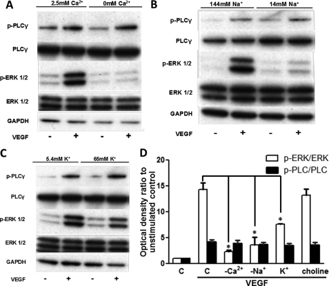
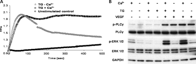
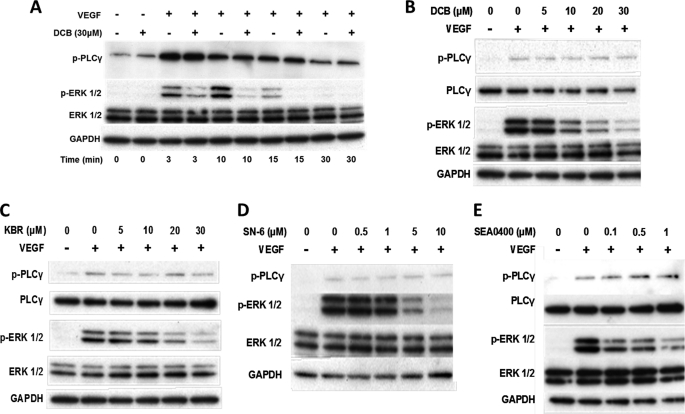
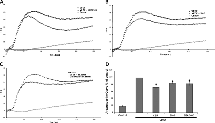
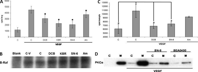
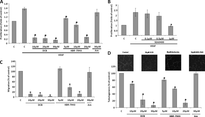
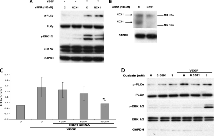
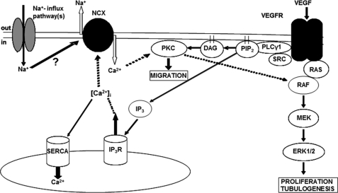
VEGF is a key angiogenic cytokine and a major target in anti-angiogenic therapeutic strategies. In endothelial cells (ECs), VEGF binds VEGF receptors and activates ERK1/2 through the phospholipase γ (PLCγ)-PKCα-B-Raf pathway. Our previous work suggested that influx of extracellular Ca(2+) is required for VEGF-induced ERK1/2 activation, and we hypothesized that this could occur through reverse mode (Ca(2+) in and Na(+) out) Na(+)-Ca(2+) exchange (NCX). However, the role of NCX activity in VEGF signaling and angiogenic functions of ECs had not previously been described. Here, using human umbilical vein ECs (HUVECs), we report that extracellular Ca(2+) is required for VEGF-induced ERK1/2 activation and that release of Ca(2+) from intracellular stores alone, in the absence of extracellular Ca(2+), is not sufficient to activate ERK1/2. Furthermore, inhibitors of reverse mode NCX suppressed the VEGF-induced activation of ERK1/2 in a time- and dose-dependent manner and attenuated VEGF-induced Ca(2+) transients. Knockdown of NCX1 (the main NCX isoform in HUVECs) by siRNA confirmed the pharmacological data. A panel of NCX inhibitors also significantly reduced VEGF-induced B-Raf activity and inhibited PKCα translocation to the plasma membrane and total PKC activity in situ. Finally, NCX inhibitors reduced VEGF-induced HUVEC proliferation, migration, and tubular differentiation in surrogate angiogenesis functional assays in vitro. We propose that Ca(2+) influx through reverse mode NCX is required for the activation and the targeting of PKCα to the plasma membrane, an essential step for VEGF-induced ERK1/2 phosphorylation and downstream EC functions in angiogenesis.


Figures

FIGURE 1.
FIGURE 1.
Effect of ionic substitutions on phospho-ERK1/2. A–C, HUVECs were serum-starved in complete physiological medium for 45 min, the medium was aspirated, and the cells were incubated for 15 min in media with the following ionic substitutions: Ca2+ omitted from the buffer (A), Na+ ions isotonically substituted with choline chloride to 14 mm (B), and buffer contains 65 mm K+ (C). HUVECs were stimulated with 50 ng/ml VEGF for 10 min. ERK1/2 and PLCγ activation was determined by Western blot. Total ERK1/2 and GAPDH protein levels were used to demonstrate protein loading. Representative immunoblots are shown (n = 3). D, optical densities of the p-ERK1/2 and p-PLCγ bands in A–C and supplemental Fig. S1 were determined and normalized against the corresponding total protein loading controls. The normalized optical density of the unstimulated control (bar C) sample for each experiment is arbitrarily set to 1. The mean value of the ratio for each condition is expressed as fold × unstimulated control value. The bars represent the means ± S.E. from three independent experiments. *, p < 0.05 versus the VEGF stimulated control.
FIGURE 2.
FIGURE 2.
Role of extracellular Ca2+ as opposed to Ca2+ release from internal stores in ERK1/2 phosphorylation. A, representative time courses of Ca2+- sensitive Fluo-4NW fluorescence recorded during TG stimulation of HUVECs. Serum-starved HUVECs were stimulated with TG (4 μm) at time 0 in the presence (blue squares) or absence (red triangles) of extracellular Ca2+. The unstimulated control (black diamonds) is also shown. Fluorescence intensity was measured every 2 s at 37 °C on a FLIPR plate reader (Molecular Devices) with excitation of 485 nm and emission wavelength of 525 nm. n = 3 in triplicate. B, HUVECs were serum-starved in a complete physiological medium for 45 min, the medium was aspirated, and the cells were incubated for a further 15 min in a medium where Ca2+ was omitted as indicated. HUVECs were then stimulated with 4 μm TG for 2 min. Subsequently, where indicated, 50 ng/ml VEGF was applied for 10 min. ERK1/2 and PLCγ activation was probed as described in the legend of Fig. 1. Representative immunoblots are shown (n = 3).
FIGURE 3.
FIGURE 3.
Effect of NCX inhibitors on VEGF-induced ERK1/2 phosphorylation. A, serum-starved HUVECs were preincubated (for 30 min) with 30 μm DCB or vehicle (0.5% v/v Me2SO) prior to VEGF stimulation (50 ng/ml) for the times indicated. B, ERK1/2 and PLCγ activation was analyzed by Western blot as described in Fig. 1. The effect of DCB was also dose-dependent. C–E, serum-starved HUVECs were preincubated for 30 min with various concentrations of the reverse mode Na+-Ca2+ exchanger inhibitors KBR-7943 (C), SN-6 (D), or SEA0400 (E) prior to stimulation with VEGF (50 ng/ml) for 10 min. ERK1/2 and PLCγ phosphorylation was analyzed by Western blot. Representative immunoblots are shown (n = 3).
FIGURE 4.
FIGURE 4.
Effect of reverse mode NCX inhibitors on Ca2+ transients. Representative time courses of Ca2+-sensitive Fluo-4NW fluorescence recorded during VEGF stimulation of HUVECs are shown. Serum-starved HUVECs were stimulated with VEGF (50 ng/ml) at time 0. A–C, the following traces are shown: unstimulated control (red circles), VEGF-stimulated control (black squares), 20 min of preincubation with 10 μm KBR7943 (A), 20 min of preincubation with 10 μm SN-6 (B), 20 min of preincubation with 1 μm SEA0400 prior to VEGF stimulation (blue triangles) (C). Fluorescence intensity was measured every 2 s at 37 °C on a FLIPR plate reader (Molecular Devices) with excitation of 485 nm and emission wavelength of 525 nm (n = 3 in triplicate). D, the area under the curve of the Ca2+ trace was calculated for each condition. The area under the curve of the VEGF-stimulated control was set to 100%. The bars represent the means ± S.E. (n = 3 in triplicate). *, p < 0.05 versus VEGF-stimulated control.
FIGURE 5.
FIGURE 5.
Effect of NCX inhibitors on B-Raf and PKC activity. A, serum-starved HUVECs were preincubated for 30 min with 30 μm DCB, 20 μm KBR, 10 μm SN-6, or 30 μm amiloride (Am) as a negative control or vehicle (0.5% v/v Me2SO) prior to VEGF (50 ng/ml) stimulation for 10 min. B-Raf was immunoprecipitated overnight from total cell extracts. Subsequently, B-Raf activity was assayed using an immunoprecipitation kinase cascade kit (Upstate) by determining radioactivity incorporated into the substrate (myelin basic protein) by scintillation counting. Specific B-Raf activity was determined by subtracting the background radioactivity in a control sample (Blank) containing no anti-B-Raf Ab. The bars represent mean counts per min per milligram of input protein per sample ± S.E. from four independent experiments. *, p < 0.05 versus the VEGF-stimulated control. B, B-Raf immunoprecipitation equivalence in different samples was assessed by Western blot. Sample abbreviations are as in A; the control sample with no anti-B-Raf antibody (Blank) is also included. A representative immunoblot is shown (n = 2). C, serum-starved HUVECs were pretreated with vehicle (bar C) (0.5% v/v Me2SO), 30 μm DCB, 10 μm SN-6, or amiloride (Am) (30 μm) for 30 min and stimulated with VEGF (50 ng/ml) for 10 min. PKC activity, in situ, was determined by measuring the radioactivity incorporated into a PKC peptide substrate with scintillation counting as described under “Experimental Procedures.” The bars represent mean counts per sample ± S.E. from three independent experiments. *, p < 0.05 versus the VEGF-stimulated control. D, serum-starved HUVECs were incubated for 30 min with 10 μm SN-6 or 1 μm SEA0400 prior to stimulation with VEGF (50 ng/ml) for 10 min. Subsequently, the cells were lysed in a detergent-free buffer and ultracentrifuged for 1 h (105 × gav). The supernatant was collected as the cytosolic fraction (lanes C), and the pellet was resuspended in lysis buffer (membrane fraction, lanes M). PKCα distribution to each fraction was determined by Western blot and subsequent probing with an anti-PKCα monoclonal antibody. A representative immunoblot from three independent repeats is shown.
FIGURE 6.
FIGURE 6.
Effect of NCX inhibitors on angiogenesis functional assays. A, HUVECs were exposed to VEGF (50 ng/ml) for 48 h in the presence of the indicated concentrations of DCB and KBR-7943, in MCDB-131 containing 0.1%w/v BSA. Bar C, control; bar Am, amiloride. The number of viable cells was determined using an alkaline phosphatase assay. The absorbance of the control sample (no VEGF stimulation) is arbitrarily set as 1. The bars represent the means ± S.E. from four independent experiments in triplicate. *, p < 0.05 versus the control. B, the effect of SEA0400 on HUVEC proliferation was determined as in A (n = 3). C, HUVEC migration toward VEGF was assessed in a FluoroblokTM assay in the presence of the indicated concentrations of DCB, KBR-7943, or amiloride, as described under “Experimental Procedures.” 50 ng/ml VEGF was used as a chemoattractant in the lower chamber. Bar C, control (0.1% v/v Me2SO); bar Am, amiloride. After 16 h, the migrated cells were fixed with 4% w/v paraformaldehyde and quantified. The mean number of migrating cells in the control wells is arbitrarily set at 100%. The bars represent the means ± S.E. from three independent experiments in duplicate. *, p < 0.05 versus the control. D, HUVECs (prelabeled with CellTracker green) were seeded in 24-well plates precoated with MatrigelTM in the presence of the indicated concentrations of DCB, KBR-7943, or amiloride in complete medium. Bar C, control (0.1% v/v Me2SO); bar Am, amiloride. The images were obtained after 3 h with an inverted fluorescent microscope and tubule lengths quantified with ImagePro software. The mean tubule length in control (C) wells is arbitrarily set at 100%. The bars represent the means ± S.E. from three independent experiments in duplicate. *, p < 0.05 versus the control. Representative images are shown in the inset, as indicated.
FIGURE 7.
FIGURE 7.
Effect of NCX siRNA on phospho-ERK1/2 and HUVEC proliferation. A, HUVECs transfected with control nontargeting siRNA or NCX1 targeting siRNA (100 nm for 48 h) were serum-starved and subsequently challenged with VEGF (50 ng/ml) for 10 min. ERK1/2 and PLCγ phosphorylation were determined as described for Fig. 1 (n = 3). B, in a parallel experiment NCX1 was immunoprecipitated from 500 μg of control or NCX1 siRNA-treated HUVECs (48 h). NCX1 knockdown was monitored by Western blot, and protein loading was demonstrated by GAPDH expression of the input cell lysates prior to immunoprecipitation (n = 3). C, HUVECs, transfected with NCX1-siRNA or control siRNA were exposed to VEGF (50 ng/ml) for 48 h, and proliferation was determined as in Fig. 6A. Bar C, control. The absorbance of the control sample (no VEGF stimulation) is arbitrarily set as 1. The bars represent the means ± S.E. from three independent experiments in triplicate. *, p < 0.05 versus the control. D, serum-starved HUVECs were pretreated with 100 nm or 1 mm of the Na+-K+ ATPase inhibitor ouabain for 1 min prior to application of VEGF (50 ng/ml) for 2 min. ERK1/2 and PLCγ phosphorylation was determined by Western blot. A representative immunoblot of three repeats is shown.
FIGURE 8.
FIGURE 8.
Proposed mechanism of reverse mode NCX involvement in VEGF-induced ERK1/2 phosphorylation. Bold lines show direct interactions. Dashed lines show indirect interactions. Activation of PLCγ downstream of VEGFR leads to the generation of diacylglycerol (DAG) and inositol 1,4,5-triphosphate (IP3). Inositol 1,4,5-triphosphate leads to Ca2+ release from the internal stores activating the NCX. Ca2+ influx through the exchanger targets PKCα to the plasma membrane and ultimately results in ERK1/2 activation. IP3R, inositol triphosphate receptor; SERCA, sarcoendoplasmic reticulum Ca2+-ATPase.

References

    1. Chung A. S., Lee J., Ferrara N. (2010) Nat. Rev. Cancer 10, 505–514 - PubMed
    1. Adams R. H., Alitalo K. (2007) Nat. Rev. Mol. Cell Biol. 8, 464–478 - PubMed
    1. Olsson A. K., Dimberg A., Kreuger J., Claesson-Welsh L. (2006) Nat. Rev. Mol. Cell Biol. 7, 359–371 - PubMed
    1. Takahashi T., Yamaguchi S., Chida K., Shibuya M. (2001) EMBO J. 20, 2768–2778 - PMC - PubMed
    1. Gerber H. P., McMurtrey A., Kowalski J., Yan M., Keyt B. A., Dixit V., Ferrara N. (1998) J. Biol. Chem. 273, 30336–30343 - PubMed

Publication types

MeSH terms