. 2011 Dec;301(6):L836-46.
doi: 10.1152/ajplung.00385.2010. Epub 2011 Aug 26.

Mechanisms of lung endothelial barrier disruption induced by cigarette smoke: role of oxidative stress and ceramides

Affiliations

Mechanisms of lung endothelial barrier disruption induced by cigarette smoke: role of oxidative stress and ceramides

Kelly S Schweitzer et al. Am J Physiol Lung Cell Mol Physiol. 2011 Dec.

Abstract

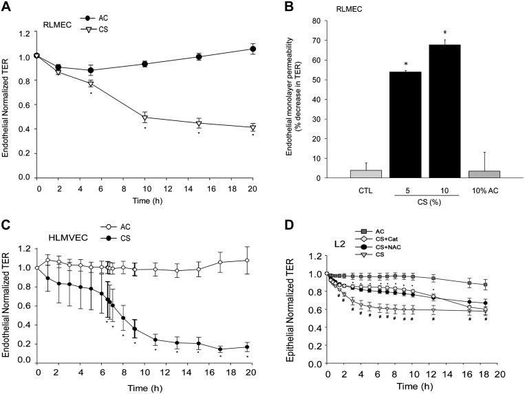
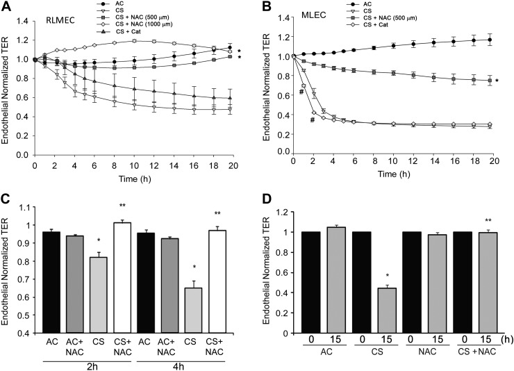
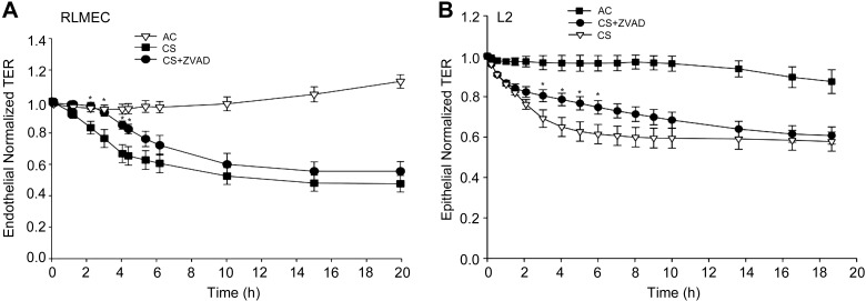
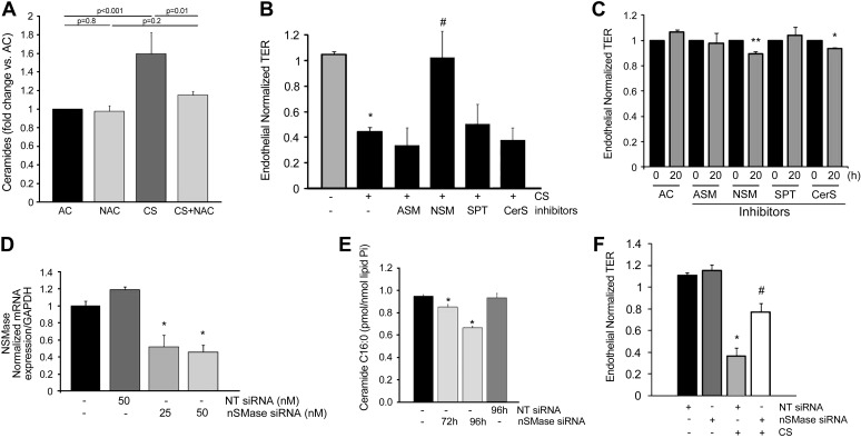
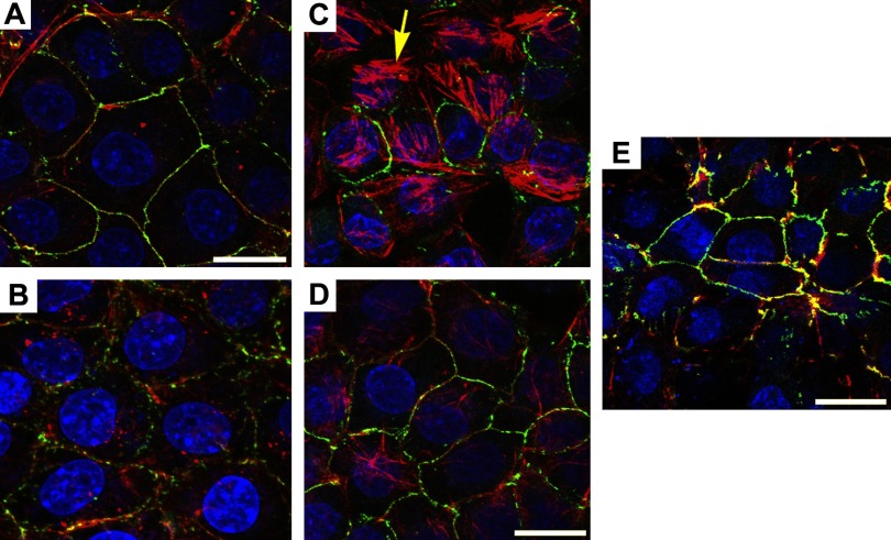
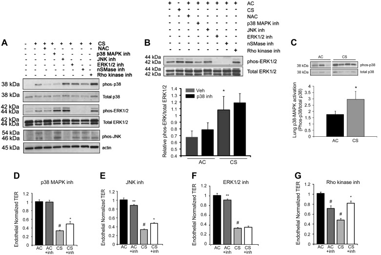
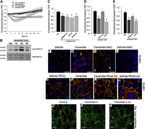
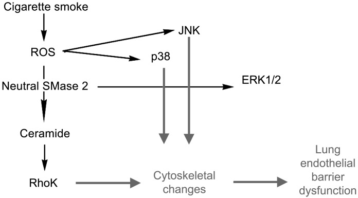
The epithelial and endothelial cells lining the alveolus form a barrier essential for the preservation of the lung respiratory function, which is, however, vulnerable to excessive oxidative, inflammatory, and apoptotic insults. Whereas profound breaches in this barrier function cause pulmonary edema, more subtle changes may contribute to inflammation. The mechanisms by which cigarette smoke (CS) exposure induce lung inflammation are not fully understood, but an early alteration in the epithelial barrier function has been documented. We sought to investigate the occurrence and mechanisms by which soluble components of mainstream CS disrupt the lung endothelial cell barrier function. Using cultured primary rat microvascular cell monolayers, we report that CS induces endothelial cell barrier disruption in a dose- and time-dependent manner of similar magnitude to that of the epithelial cell barrier. CS exposure triggered a mechanism of neutral sphingomyelinase-mediated ceramide upregulation and p38 MAPK and JNK activation that were oxidative stress dependent and that, along with Rho kinase activation, mediated the endothelial barrier dysfunction. The morphological changes in endothelial cell monolayers induced by CS included actin cytoskeletal rearrangement, junctional protein zonula occludens-1 loss, and intercellular gap formation, which were abolished by the glutathione modulator N-acetylcysteine and ameliorated by neutral sphingomyelinase inhibition. The direct application of ceramide recapitulated the effects of CS, by disrupting both endothelial and epithelial cells barrier, by a mechanism that was redox and apoptosis independent and required Rho kinase activation. Furthermore, ceramide induced dose-dependent alterations of alveolar microcirculatory barrier in vivo, measured by two-photon excitation microscopy in the intact rat. In conclusion, soluble components of CS have direct endothelial barrier-disruptive effects that could be ameliorated by glutathione modulators or by inhibitors of neutral sphingomyelinase, p38 MAPK, JNK, and Rho kinase. Amelioration of endothelial permeability may alleviate lung and systemic vascular dysfunction associated with smoking-related chronic obstructive lung diseases.


Figures

Fig. 1.
Fig. 1.
Cigarette smoke extract (CS) disrupts endothelial monolayer integrity. A: transendothelial resistance (TER) measured in primary rat lung microvascular endothelial cells (RLMEC) following treatment with CS (10%) compared with ambient air control (AC; 10%); means ± SE; n = 11–13, *P < 0.05; Student's t-test. B: dose-dependent increase in RLMEC permeability in response to CS (5% or 10%; 20 h) compared with AC (10%); means ± SE; n = 3–7; *P < 0.05 vs. AC; ANOVA. C: kinetics of normalized TER across primary human lung microvascular endothelial cells monolayers in response to CS (3%) compared with AC (3%); means ± SE; n = 4–6, *P < 0.05; Student's t-test. D: normalized TER of primary rat lung epithelial cell (L2) monolayers treated with AC (4%) or CS (4%), CS + N-acetylcysteine (NAC) (500 μM), or CS + catalase (Cat) (1 mM); means ± SE; n = 4–15, *P < 0.05 vs. CS; #P < 0.05 vs. AC; ANOVA.
Fig. 2.
Fig. 2.
Effects of antioxidants NAC and catalase on CS-induced decrease in TER. A: TER of primary RLMEC monolayers treated with AC (10%), CS (10%), CS + NAC (500 μM, or 1 mM), or CS + Cat (1.5 mM); means ± SE; n = 3–11, *P < 0.05 vs. CS alone all time points; ANOVA. B: TER measured in mouse lung endothelial cell (MLEC) monolayers treated with AC (4%) or CS (4%), CS + NAC (500 μM), or CS + Cat (1 mM); means ± SE; n = 3–8, *P < 0.05 vs. CS at all time points; #P < 0.05 vs. CS at the indicated time points; ANOVA. C and D: bar graphs with mean TER at the indicated early (C) or late (D) time points (0 h indicating the start of TER measurements) in RLMEC monolayers treated with AC (10%; n = 14), CS (10%; n = 19), AC + NAC (500 μM; n = 14), or CS + NAC (n = 13); means ± SE; *P < 0.001 vs. AC, **P < 0.001 vs. CS; ANOVA.
Fig. 3.
Fig. 3.
Effect of caspase inhibition on CS-induced endothelial and epithelial barrier disruption. A: effect of the general caspase inhibitor Z-VAD-FMK (ZVAD) on RLMEC barrier treated with CS (10%), CS + ZVAD (250 nM), compared with AC (10%); means ± SE; n = 3–11 *P < 0.05 vs. CS; ANOVA. B: normalized TER across primary rat lung epithelial cell (L2) monolayers exposed to AC (4%), CS (4%), or to CS + ZVAD (250 nM); means ± SE; n = 4–6; *P < 0.05 vs. CS; ANOVA.
Fig. 4.
Fig. 4.
Ceramide synthesis involvement in CS-induced permeability. A: total ceramide levels (normalized by lipid phosphorus, Pi) in RLMEC treated with AC (10%), AC + NAC (500 μM), CS (10%; 4 h), or CS + NAC (500 μM); means + SE; n = 3–6. B: normalized TER (20 h) RLMEC monolayers following treatment with AC (10%; n = 14), CS (10%; n = 19), or CS and each of the following inhibitors (n = 7–8): acid sphingomyelinase (ASM) inhibitor (Imipramine, 50 μM), neutral sphingomyelinase (NSM) inhibitor (GW4869, 15 μM), serine palmitoyl transferase (SPT) inhibitor (myriocin; 50 nM), or ceramide synthase (CerS) inhibitor (fumonisin B1, FB1, 1 μM); means ± SE; *P < 0.05 vs. AC; #P < 0.05 vs. CS; ANOVA. C: mean normalized TER (0 or 20 h) of RLMEC monolayers following treatment with AC alone (n = 14), or AC and each of the following inhibitors (n = 3–15): ASM inhibitor (Imipramine, 50 μM), NSM inhibitor (GW4869, 15 μM), SPT inhibitor (myriocin; 50 nM), or CerS inhibitor (FB1, 1 μM); means ± SE; *P < 0.05 vs. AC; **P < 0.01 vs. AC; ANOVA. D: neutral SMase (nSMase) mRNA expression in RLMEC treated with specific siRNA or nontarget (NT) siRNA for 72 h and measured by real-time PCR; means + SE; n = 3, *P < 0.01 vs. NT siRNA. E: ceramide C16:0 levels (normalized by lipid, Pi) in RLMEC treated with AC (10%) alone or with specific nSMase siRNA or NT siRNA (100 nM) for the indicated time; means + SE; n = 3, *P < 0.05 vs. AC; ANOVA. F: TER in RLMEC following exposure to AC (10%; 20 h) or CS (10%; 20 h) in the presence of NT siRNA or nSMase siRNA (50 nM; 96 h); means ± SE; n = 4–9, *P < 0.05 vs. AC; #P < 0.05 vs. CS 10%; ANOVA.
Fig. 5.
Fig. 5.
Cytoskeletal changes induced by CS in lung endothelial cells. Representative 3D-reconstructed images comprised of 4–17 confocal slices captured with the fluorescence microscope of RLMEC treated with AC (A; 10%; 4 h), AC + NAC (B; 500 μM), CS (C; 10%; 4 h), CS + NAC (D), or CS + GW4869 (E; 15 μM), and stained for actin (red), zonula occludens (ZO)-1 (green; arrowhead), and nuclei (blue). Note the actin reorganization in stress fibers (arrow), intercellular gap formation, and disruption of ZO-1 expression in CS-treated cells and their amelioration with antioxidant or nSMase inhibitor treatment. Scale bar = 17 μm.
Fig. 6.
Fig. 6.
Role of MAPK activation in CS-induced barrier dysfunction in endothelial cells. A: Western blot of p38, ERK1/2, and JNK phosphorylation in RLMEC exposed to AC (10%; 1 h), CS (10%; 1 h), or CS + NAC (500 μM), p38 inhibitor SB203580 (5 μM), JNK inhibitor SP600125 (50 μM), ERK1/2 inhibitor PD98059 (50 μM), nSMase inhibitor GW4869 (15 μM), or Rho kinase inhibitor Y27632 (3 μM). Representative of n = 5 independent experiments. B: top: Western blot of ERK1/2 phosphorylation in RLMEC exposed to AC (10%; 1 h), CS (10%; 1 h), or AC + NAC (500 μM), p38 inhibitor SB203580 (5 μM), JNK inhibitor SP600125 (50 μM), ERK1/2 inhibitor PD98059 (50 μM), nSMase inhibitor GW4869 (15 μM), or Rho kinase inhibitor Y27632 (3 μM). Representative of n = 3 independent experiments. Bottom: densitometry ERK1/2 phosphorylation in response to AC (10%, 1 h), AC + p38 MAPK inhibitor SB203580 (5 μM), CS (10%, 1 h), and CS + p38 MAPK inhibitor; means + SE, n = 12, *P < 0.05 vs. AC. C: Western blot of p38 phosphorylation in total lung homogenates isolated from DBA/2J mice exposed to CS for 4 mo compared with AC control with densitometric analysis of Western blot data; means + SE; n = 6, *P < 0.05, Student's t-test. DG: normalized TER measurements of RLMEC exposed to AC (10%) or CS (10%; 15 h), without or with a p38 MAPK inhibitor SB203580 (D; 5 μM; n = 3–8; #P < 0.001 vs. AC; *P < 0.01 vs. CS), JNK1/3 inhibitor SP600125 (E; 50 μM; n = 3–8; #P < 0.001 vs. AC; *P < 0.01 vs. CS; **P < 0.05 vs. AC), ERK1/2 inhibitor PD98059 (F; 50 μM; n = 3–8; #P < 0.001 vs. AC; **P < 0.05 vs. AC), or Rho kinase inhibitor Y27632 (G; 3 μM; n = 6–17; #P < 0.001 vs. AC; *P < 0.05 vs. CS).
Fig. 7.
Fig. 7.
Effect of ceramides on endothelial barrier function in vitro and in vivo. A: normalized TER in RLMEC treated with ceramide C6:0 (20 μM), ceramide C16:0 (20 μM), or their respective vehicles methanol (%) or polyethylene glycol (PEG)2000 (10 μl); means ± SE; *P < 0.05 vs. vehicle, n = 3–15. B: Western blot of phosphorylated and total ERK1/2 in RLMEC treated with ceramide 6:0 (20 μM) for the indicated time. C: normalized TER measurements of RLMEC exposed to vehicle (methanol) or ceramide 6:0 (20 μM; 19 h) alone or with the p38 MAPK inhibitor SB203580 (5 μM), JNK1/3 inhibitor SP600125 (50 μM), or ERK1/2 inhibitor U0126 (20 μM); means + SE; #P < 0.05 vs. vehicle. D: mean normalized TER in RLMEC treated with vehicle (methanol), ceramide 6:0 (20 μM; 19 h), ceramide plus Rho Kinase inhibitor Y27632 (4 μM), or ceramide plus NAC (500 μM); n = 7–15; #P < 0.001 vs. vehicle; *P < 0.005 vs. ceramide. E: normalized TER in RLMEC treated with vehicle (PEG2000), ceramide-16:0 (20 μM; 5 h), ceramide plus Rho Kinase inhibitor Y27632 (4 μM), or ceramide plus NAC (500 μM); means + SE; n = 7–12; #P < 0.001 vs. vehicle; *P < 0.05 vs. ceramide. FI: representative confocal fluorescence micrographs of RLMEC treated with vehicle (PEG2000; 10 μM; 2 h), ceramide C16:0-PEG (10 μM; 2 h), ceramide + NAC (500 μM; 2 h), or vehicle + NAC (500 μM; 2 h) and stained for actin (red), ZO-1 (green; arrowhead), and nuclei with 4,6-diamidino-2-phenyl indole dihydrochloride (DAPI) (blue). Note patchy actin reorganization in stress fibers (arrow) and the marked disruption of ZO-1 expression in ceramide-treated cells and lack of amelioration with NAC treatment. Scale bar = 17 μm. JM: representative immunofluorescence micrographs of RLMEC treated with vehicle (PEG2000; 10 μM; 2 h), ceramide C16:0-PEG (10 μM; 2 h), ceramide + RhoK inhibitor (0.5 μM; 2 h), or vehicle + RhoK inhibitor and stained for actin (red), ZO-1 (green; arrowhead), and nuclei with DAPI (blue). Note that the marked disruption of ZO-1 expression in ceramide-treated cells was markedly inhibited in cells treated with RhoK inhibitor. Scale bar = 10 μm. NP: effect of ceramide on the lung microcirculation captured in real time in the pulmonary microvasculature of a living rat (Supplemental Movies). Three-dimensional reconstruction of FITC-labeled vessels (green) surrounding alveoli (dark regions) and Rho-G6-labeled neutrophils (red) imaged via intravital 2-photon microscopy before (N) and after (O and P) intravenous administration of ceramide C16:0 (10 mg/kg). Nuclei were stained with intravenous Hoechst (blue). Note increasing neutrophil trafficking and plasma extravasation into airspaces captured 6 min postceramide administration (O), and then 2 min following a second dose of ceramide (P). Scale bars = 25 μm.
Fig. 8.
Fig. 8.
Schematic with the proposed mechanisms by which soluble components of CS induce barrier dysfunction in lung endothelial cells. ROS, reactive oxygen species.

References

    1. Bartalesi B, Cavarra E, Fineschi S, Lucattelli M, Lunghi B, Martorana PA, Lungarella G. Different lung responses to cigarette smoke in two strains of mice sensitive to oxidants. Eur Respir J 25: 15–22, 2005. - PubMed
    1. Batinic-Haberle I, Cuzzocrea S, Reboucas JS, Ferrer-Sueta G, Mazzon E, Di Paola R, Radi R, Spasojevic I, Benov L, Salvemini D. Pure MnTBAP selectively scavenges peroxynitrite over superoxide: comparison of pure and commercial MnTBAP samples to MnTE-2-PyP in two models of oxidative stress injury, an SOD-specific Escherichia coli model and carrageenan-induced pleurisy. Free Radic Biol Med 46: 192–201, 2009. - PMC - PubMed
    1. Bligh EG, Dyer WJ. A rapid method of total lipid extraction and purification. Can J Med Sci 37: 911–917, 1959. - PubMed
    1. Borbiev T, Birukova A, Liu F, Nurmukhambetova S, Gerthoffer WT, Garcia JG, Verin AD. p38 MAP kinase-dependent regulation of endothelial cell permeability. Am J Physiol Lung Cell Mol Physiol 287: L911–L918, 2004. - PubMed
    1. Boucher RC, Johnson J, Inoue S, Hulbert W, Hogg JC. The effect of cigarette smoke on the permeability of guinea pig airways. Lab Invest 43: 94–100, 1980. - PubMed

Publication types

MeSH terms