. 2012 May;61(5):753-64.
doi: 10.1136/gutjnl-2011-300007. Epub 2011 Aug 26.

Inhibition of histidine decarboxylase ablates the autocrine tumorigenic effects of histamine in human cholangiocarcinoma

Affiliations

Inhibition of histidine decarboxylase ablates the autocrine tumorigenic effects of histamine in human cholangiocarcinoma

Heather Francis et al. Gut. 2012 May.

Abstract

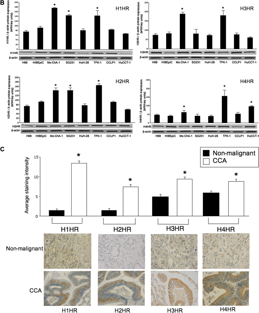
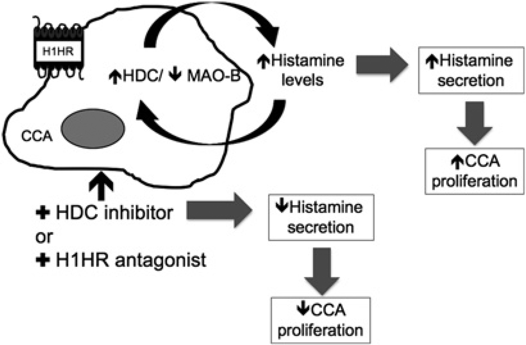
Background: In several tumours the endogenous activity of histidine decarboxylase (HDC), the enzyme stimulating histamine synthesis, sustains the autocrine trophic effect of histamine on cancer progression. Cholangiocarcinoma is a biliary cancer with limited treatment options. Histamine interacts with four G-protein coupled receptors, H1-H4 histamine receptors (HRs).

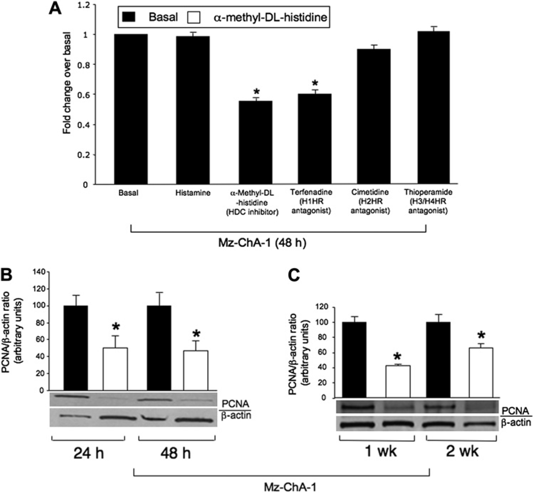
Objective: To determine the effects of histamine stimulation and inhibition of histamine synthesis (by modulation of HDC) on cholangiocarcinoma growth.

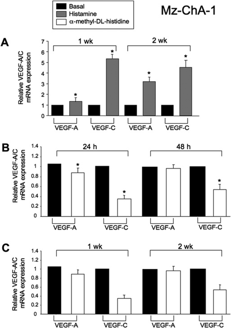
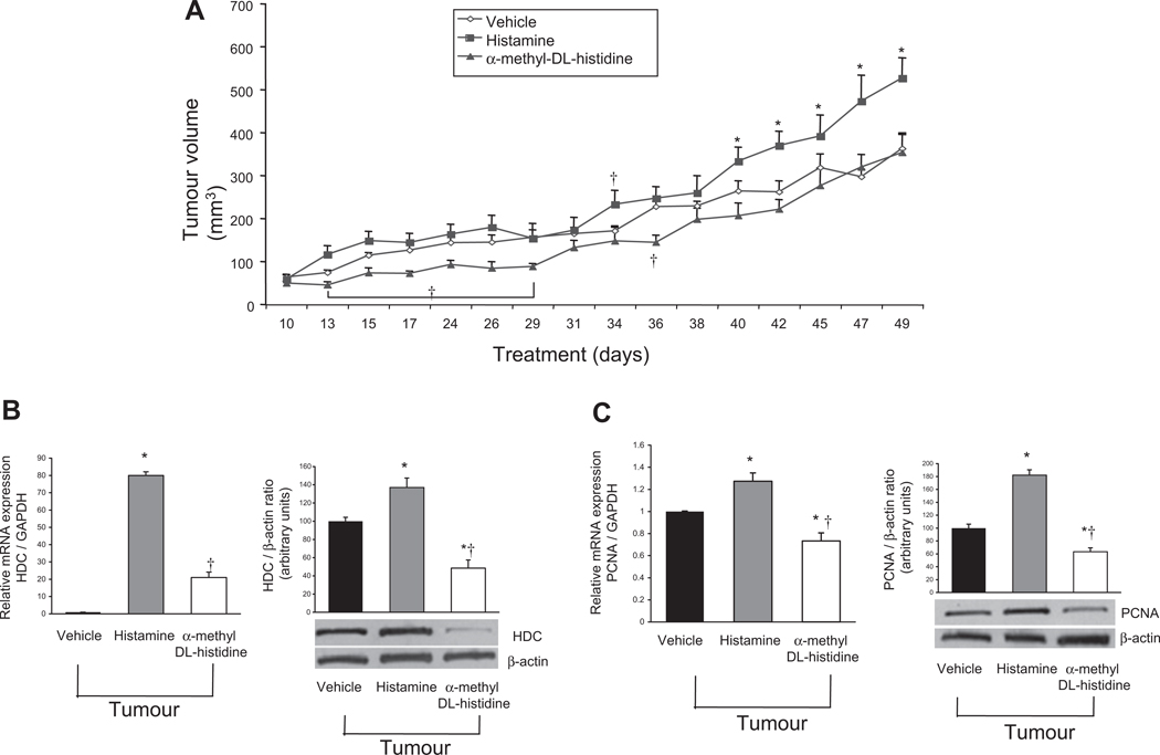
Methods: In vitro studies were performed using multiple human cholangiocarcinoma lines. The expression levels of the histamine synthetic machinery and HRs were evaluated along with the effects of histamine stimulation and inhibition on cholangiocarcinoma proliferation. A xenograft tumour model was used to measure tumour volume after treatment with histamine or inhibition of histamine synthesis by manipulation of HDC. Vascular endothelial growth factor (VEGF) expression was measured in cholangiocarcinoma cells concomitant with the evaluation of the expression of CD31 in endothelial cells in the tumour microenvironment.

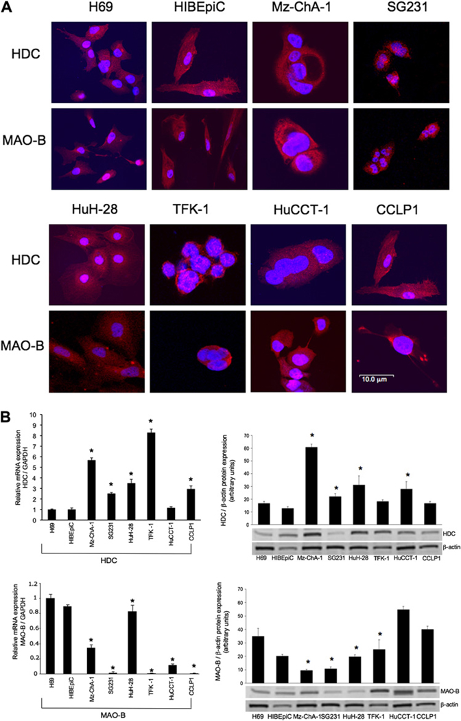
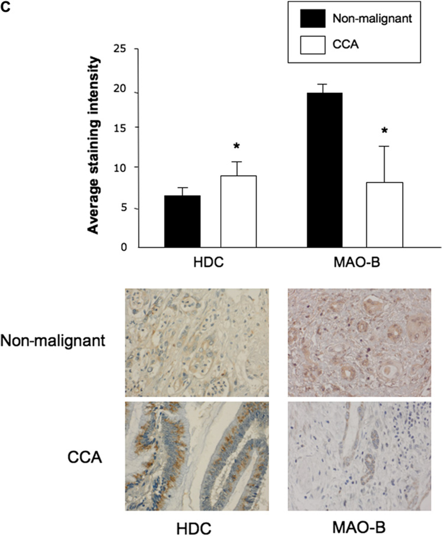
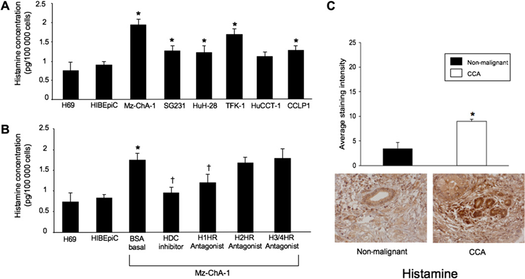
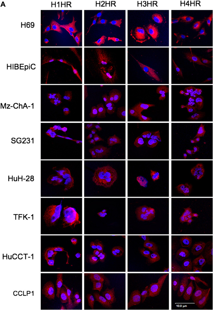
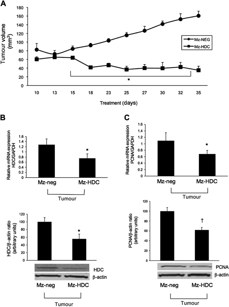
Results: Cholangiocarcinoma cells display (1) enhanced HDC and decreased monoamine oxidase B expression resulting in increased histamine secretion; and (2) increased expression of H1-H4 HRs. Inhibition of HDC and antagonising H1HR decreased histamine secretion in Mz-ChA-1 cells. Long-term treatment with histamine increased proliferation and VEGF expression in cholangiocarcinoma that was blocked by HDC inhibitor and the H1HR antagonist. In nude mice, histamine increased tumour growth (up to 25%) and VEGF expression whereas inhibition of histamine synthesis (by reduction of HDC) ablated the autocrine stimulation of histamine on tumour growth (~80%) and VEGF expression. No changes in angiogenesis (evaluated by changes in CD31 immunoreactivity) were detected in the in vivo treatment groups.

Conclusion: The novel concept that an autocrine loop (consisting of enhanced histamine synthesis by HDC) sustains cholangiocarcinoma growth is proposed. Drug targeting of HDC may be important for treatment of patients with cholangiocarcinoma.


Figures

Figure 1
Figure 1
(A) By immunofluorescence, non-malignant cholangiocytes and cholangiocarcinoma cells express histidine decarboxylase (HDC) and monoamine oxidase B (MAO-B). Merged staining for nuclei (blue) and enzyme (red) is shown for each cell line. Bar=10 µm. (B) By both real-time PCR and immunoblots, HDC was expressed at higher levels in some of the cholangiocarcinoma lines (whereas MAO-B levels decreased) compared with non-malignant cholangiocytes. Data are mean±SEM of three experiments for real-time PCR and eight experiments for immunoblots (*p<0.05 vs the corresponding value of non-malignant cholangiocytes). (C) HDC immunoreactivity was increased in cholangiocarcinoma (CCA) samples whereas MAO-B immunoreactivity was significantly decreased compared with non-malignant samples (*p<0.05 vs non-malignant). Data are mean±SEM of 10 blinded evaluations of 10 randomly selected fields of three slides. Original magnification ×40.
Figure 1
Figure 1
(A) By immunofluorescence, non-malignant cholangiocytes and cholangiocarcinoma cells express histidine decarboxylase (HDC) and monoamine oxidase B (MAO-B). Merged staining for nuclei (blue) and enzyme (red) is shown for each cell line. Bar=10 µm. (B) By both real-time PCR and immunoblots, HDC was expressed at higher levels in some of the cholangiocarcinoma lines (whereas MAO-B levels decreased) compared with non-malignant cholangiocytes. Data are mean±SEM of three experiments for real-time PCR and eight experiments for immunoblots (*p<0.05 vs the corresponding value of non-malignant cholangiocytes). (C) HDC immunoreactivity was increased in cholangiocarcinoma (CCA) samples whereas MAO-B immunoreactivity was significantly decreased compared with non-malignant samples (*p<0.05 vs non-malignant). Data are mean±SEM of 10 blinded evaluations of 10 randomly selected fields of three slides. Original magnification ×40.
Figure 2
Figure 2
Histamine levels were evaluated by ELISA in the medium of non-malignant and cholangiocarcinoma (CCA) cell lines. (A) Histamine levels increased in all cholangiocarcinoma cell lines except HuCCT-1 compared with non-malignant cholangiocytes. (B) Mz-ChA-1 cells secreted almost twice as much histamine as non-malignant cells. In Mz-ChA-1 cells, histamine secretion was inhibited by the histidine decarboxylase (HDC) inhibitor and the H1 histamine receptor (H1HR) antagonist terfenadine but not the H2 or H3/4 HR antagonists (*p<0.05 vs H69; †p<0.05 vs bovine serum albumin (BSA)-treated Mz-ChA-1 cell). Data are mean±SEM of 12 experiments. (C) Immunohistochemistry in tissue array samples for histamine in human liver biopsies from healthy controls and patients with cholangiocarcinoma. Histamine immunoreactivity significantly increased in cholangiocarcinoma biopsy samples compared with non-malignant samples (*p<0.05 vs non-malignant cells). Data are mean±SEM of 10 blinded evaluations of 10 randomly selected fields of three slides.
Figure 3
Figure 3
(A) By immunofluorescence, non-malignant and cholangiocarcinoma (CCA) cells were positive for four histamine receptors (HRs), H1–H4 HRs. Merged staining for nuclei (blue) and receptor (red) is shown for each cell line. Bar=10 µm. (B) By immunoblots, the protein expression of H1–H4 HRs was increased in multiple cholangiocarcinoma lines compared with normal cholangiocytes. Data are mean±SEM of six experiments (*p<0.05 vs corresponding values of non-malignant cells). (C) By tissue array analysis, the immunoreactivity for H1–H4 HRs increased in tumour liver biopsies compared with non-malignant controls (*p<0.05 vs non-malignant cells). Data are mean±SEM of 10 blinded evaluations of one randomly selected field of three slides.
Figure 3
Figure 3
(A) By immunofluorescence, non-malignant and cholangiocarcinoma (CCA) cells were positive for four histamine receptors (HRs), H1–H4 HRs. Merged staining for nuclei (blue) and receptor (red) is shown for each cell line. Bar=10 µm. (B) By immunoblots, the protein expression of H1–H4 HRs was increased in multiple cholangiocarcinoma lines compared with normal cholangiocytes. Data are mean±SEM of six experiments (*p<0.05 vs corresponding values of non-malignant cells). (C) By tissue array analysis, the immunoreactivity for H1–H4 HRs increased in tumour liver biopsies compared with non-malignant controls (*p<0.05 vs non-malignant cells). Data are mean±SEM of 10 blinded evaluations of one randomly selected field of three slides.
Figure 4
Figure 4
(A) Short-term effect of histamine, the histidine decarboxylase (HDC) inhibitor and H1–H4 histamine receptor (HR) antagonists on the proliferation of cholangiocarcinoma cell lines. While histamine alone had no effect on cholangiocarcinoma growth, the HDC inhibitor α-methyl-D,L-histidine dihydrochloride and the H1HR antagonist terfenadine decreased cholangiocarcinoma growth. Neither the H2HR antagonist cimetidine nor the H3/H4 antagonist thioperamide affected cholangiocarcinoma growth. (B,C) Mz-ChA-1 cells were treated with the HDC inhibitor (3 mM) every day for up to 2 weeks before measuring cellular proliferation by proliferating cellular nuclear antigen (PCNA) immunoblots. Treatment with the HDC inhibitor reduced Mz-ChA-1 proliferation at all time points studied up to 2 weeks (*p<0.05 vs corresponding basal values). Data are mean±SEM of eight experiments.
Figure 5
Figure 5
(A) After stimulation with histamine for 1 and 2 weeks there was a significant increase in the expression of vascular endothelial growth factor (VEGF)-A and VEGF-C. (B) In Mz-ChA-1 cells stimulated with the histidine decarboxylase (HDC) inhibitor there was a significant decrease in VEGF-A expression at 24 h but not at 48 h. VEGF-C expression was significantly decreased at both 24 and 48 h. (C) There was a significant decrease in VEGF-A mRNA expression at 1 week and VEGF-C mRNA expression at 1 and 2 weeks after treatment of Mz-ChA-1 cells with the HDC inhibitor. Data are mean±SEM of three experiments. *p<0.05 vs VEGF mRNA expression of basal-treated cells.
Figure 6
Figure 6
In vivo evaluation of xenograft tumour volume/growth over time after chronic histamine stimulation or inhibition. (A) Using two-way ANOVA analysis, histamine significantly increased tumour volume at days 13, 34 and 40–49 compared with vehicle (0.9% NaCl) and the histidine decarboxylase (HDC) inhibitor significantly decreased tumour volume at days 15–29, 36, 40, 42 and 45 compared with vehicle. Inhibition of HDC by α-methyl-DL-histidine significantly decreased tumour volume at all time points except day 10 compared with histamine-induced tumour volume. (B,C) Histamine increased HDC and proliferating cellular nuclear antigen (PCNA) expression compared with vehicle treatment. Treatment with the HDC inhibitor decreased protein but not mRNA expression of HDC and also decreased mRNA and protein expression of PCNA compared with both vehicle and histamine treatment. Data are mean±SEM of three experiments (for real-time PCR) and six experiments (for immunoblots). *p<0.05 HDC and PCNA expression vs vehicle treatment. †p<0.05 vs mRNA expression of histamine treatment.
Figure 7
Figure 7
(A) Xenograft tumour growth over time after implantation of genetically modified Mz-ChA-1 cells. After 18 days the Mz-HDC tumours decreased in volume and remained similar throughout the measurement time compared with the Mz-neg tumours which continued steadily to increase in volume. By two-way ANOVA, tumour growth in Mz-HDC was significantly lower (p<0.001) than in Mz-neg at all time points except day 13. (B,C) By real-time PCR and immunoblots in RNA and protein samples from tumours extracted from both Mz-neg and Mz-HDC, a significant decrease was found in histidine decarboxylase (HDC) expression in Mz-HDC tumour cells compared with Mz-neg tumour cells and proliferating cellular nuclear antigen (PCNA) expression (*p<0.05 vs Mz-neg cells; †p<0.01 vs Mz-neg cells). Data are mean±SEM of three experiments (real-time PCR) and eight experiments (immunoblotting).
Figure 8
Figure 8
Schematic diagram of working model. Histamine secretion increases after histidine decarboxylase (HDC) expression is enhanced during cholangiocarcinogenesis. Increased HDC and histamine levels induce the growth of cholangiocarcinoma (CCA). Use of the HDC inhibitor or the H1 histamine receptor (H1HR) antagonist decreases histamine secretion levels and tumour growth.

References

    1. Alpini G, Prall RT, LaRusso NF. The pathobiology of biliary epithelia. In: Arias IM, Boyer JL, Chisari FV, et al., editors. The Liver; Biology & Pathobiology. 4th edn. Philadelphia, PA: Lippincott Williams & Wilkins; 2001. pp. 421–435.
    1. Gores GJ. Cholangiocarcinoma: current concepts and insights. Hepatology. 2003;37:961–969. - PubMed
    1. Sirica AE. Cholangiocarcinoma: molecular targeting strategies for chemoprevention and therapy. Hepatology. 2005;41:5–15. - PubMed
    1. Aljiffry M, Walsh MJ, Molinari M. Advances in diagnosis, treatment and palliation of cholangiocarcinoma: 1990–2009. World J Gastroenterol. 2009;15:4240–4262. - PMC - PubMed
    1. Rosenthaler J, Guirard BM, Chang GW, et al. Purification and properties of histidine decarboxylase from lactobacillus 30a. Proc Natl Acad Sci U S A. 1965;54:152–158. - PMC - PubMed

Publication types

MeSH terms

Substances