. 2011 Oct 1;187(7):3493-8.
doi: 10.4049/jimmunol.1100714. Epub 2011 Aug 26.

Cutting edge: accelerated autoimmune diabetes in the absence of LAG-3

Affiliations

Cutting edge: accelerated autoimmune diabetes in the absence of LAG-3

Maria Bettini et al. J Immunol. .

Abstract

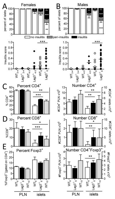
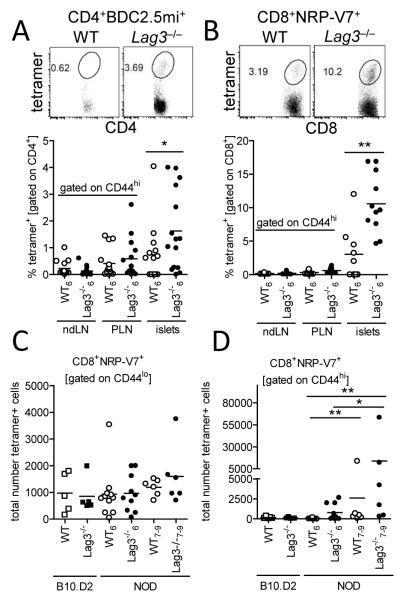
Lymphocyte activation gene-3 (LAG-3; CD223) is a CD4 homolog that is required for maximal regulatory T cell function and for the control of CD4(+) and CD8(+) T cell homeostasis. Lag3(-)(/)(-) NOD mice developed substantially accelerated diabetes with 100% incidence. Adoptive transfer experiments revealed that LAG-3 was primarily responsible for limiting the pathogenic potential of CD4(+) T cells and, to a lesser extent, CD8(+) T cells. Lag3(-)(/)(-) mice exhibited accelerated, invasive insulitis, corresponding to increased CD4(+) and CD8(+) T cell islet infiltration and intraislet proliferation. The frequencies of islet Ag-reactive chromogranin A-specific CD4(+) T cells and islet specific glucose-6-phosphatase-specific CD8(+) T cells were significantly increased in the islets of Lag3(-)(/)(-) mice, suggesting an early expansion of pathogenic clones that is normally restrained by LAG-3. We conclude that LAG-3 is necessary for regulating CD4(+) and CD8(+) T cell function during autoimmune diabetes, and thus may contribute to limiting autoimmunity in disease-prone environments.


Figures

Figure 1
Figure 1
Accelerated diabetes in the absence of LAG-3. Diabetes incidence was monitored in Lag3−/− NOD females (A) and males (B) together with littermate heterozygous Lag3+/– and Lag3+/+(WT) mice (♀ +/+ n=53, +/− n=46, −/− n=29; ♂ +/+ n=40, +/− n=39, −/− n=31). C, NOD WT female mice (7 weeks old) were treated with blocking antibodies to LAG-3, PD-1, control IgG1, or PBS on days 0, 5, and 7 and monitored for diabetes (n=9). D-E, Splenocytes from 5-6 week old mice (D - WT; E - Lag3−/−) were MACS depleted of CD4+ or CD8+ T cells, reconstituted with either CD4+ or CD8+ T cells from age-matched Lag3−/− (D) or WT (E) female mice, and transferred into female NOD.scid recipients (n=11-14).
Figure 2
Figure 2
Increased insulitis and numbers of infiltrating CD4+ and CD8+ T cells in the islets of Lag3−/− mice. Histological assessment of insulitis performed in female (A) and male (B) Lag3−/− and WT mice at three different ages (3-4 weeks, ♀ n=2-6 ♂ n=2-4; 4-5 weeks, ♀ n=3-11 ♂ n=4-9; and 6 weeks old, ♀ n=16-18 ♂ n=20-22). In the top panel: percent of islets exhibiting no insulitis (white), peri-insulitis (grey) and invasive insulitis (black) (***p≤0.0009, **p=0.0014). Percent and number of CD4+ (C; n=14-23), CD8+ (D; n=5-12), and CD4+Foxp3+ (E; n=9-18) T cells based on flow cytometric analysis of pancreatic LN (PLN) and islets from 6 week old wild type (white), 6 week old Lag3−/− (black) or 10 week old Lag3−/− (grey) female mice (***p≤0.0004, **p≤0.088, *p=0.0399).
Figure 3
Figure 3
CD4+ and CD8+ T cells exhibit increased proliferation in the islets of Lag3−/− mice. A, Proliferation was assessed based on intracellular Ki67 staining of CD8+, CD4+Foxp3 and CD4+Foxp3+ T cells in 6 week old Lag3−/− female mice (n=7) and 6 (n=10) or 10 week old (n=5) WT female mice (*p≤0.0229). B, CXCR3 cell surface expression on CD4+ T cells was measured by flow cytometric analysis of 6-7 week old female Lag3−/− mice (n=7; *p=0.0262). C, Intracellular IFNγ expression was assessed in the organs of 6 week old female and 7 week old male Lag3−/− and WT mice after 5 hour stimulation with PMA and ionomycin (n=7-10).
Figure 4
Figure 4
Accumulation of islet antigen specific clones in the islets of Lag3−/− NOD mice. Frequency of chromogranin A mimotope (BDC2.5mi) tetramer positive CD4+ (A; n=14-15; *p=0.0495) and IGRP mimotope (NRP-V7) tetramer positive CD8+ (B; n=9-11; **p=0.0024) T cells was assessed in the organs of Lag3−/− mice. Representative islet FACS plots are shown. Numbers of CD44lo precursor (C) and CD44hi activated/expanded (D) IGRP reactive CD8+ T cells were calculated after enrichment for NRP-V7 tetramer+ cells via anti-PE MACS beads and flow cytometric analysis (n=5-9; **p≤0.0022, *p=0.04).

References

    1. Lehuen A, Diana J, Zaccone P, Cooke A. Immune cell crosstalk in type 1 diabetes. Nat Rev Immunol. 10:501–513. - PubMed
    1. Bettini M, Vignali DA. Regulatory T cells and inhibitory cytokines in autoimmunity. Curr Opin Immunol. 2009;21:612–618. - PMC - PubMed
    1. Feuerer M, Shen Y, Littman DR, Benoist C, Mathis D. How punctual ablation of regulatory T cells unleashes an autoimmune lesion within the pancreatic islets. Immunity. 2009;31:654–664. - PMC - PubMed
    1. Ansari MJ, Salama AD, Chitnis T, Smith RN, Yagita H, Akiba H, Yamazaki T, Azuma M, Iwai H, Khoury SJ, Auchincloss H, Jr., Sayegh MH. The programmed death-1 (PD-1) pathway regulates autoimmune diabetes in nonobese diabetic (NOD) mice. J Exp Med. 2003;198:63–69. - PMC - PubMed
    1. Workman CJ, Cauley LS, Kim IJ, Blackman MA, Woodland DL, Vignali DA. Lymphocyte activation gene-3 (CD223) regulates the size of the expanding T cell population following antigen activation in vivo. J Immunol. 2004;172:5450–5455. - PubMed

Publication types