. 2011 Oct 1;187(7):3888-94.
doi: 10.4049/jimmunol.1101629. Epub 2011 Aug 26.

An acquired defect in IgG-dependent phagocytosis explains the impairment in antibody-mediated cellular depletion in Lupus

Affiliations

An acquired defect in IgG-dependent phagocytosis explains the impairment in antibody-mediated cellular depletion in Lupus

Anupama Ahuja et al. J Immunol. .

Abstract

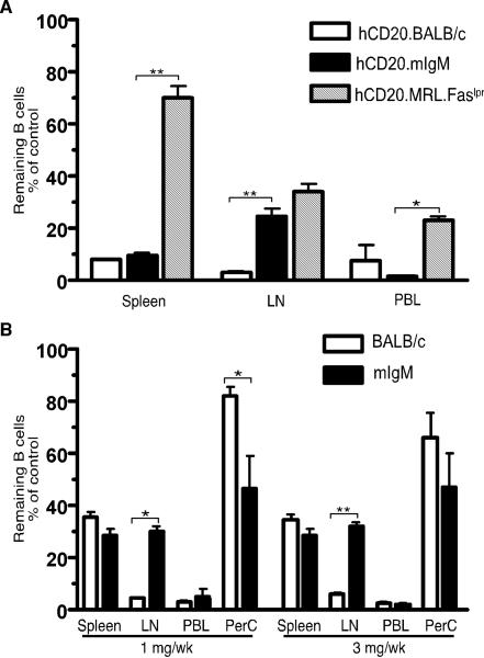
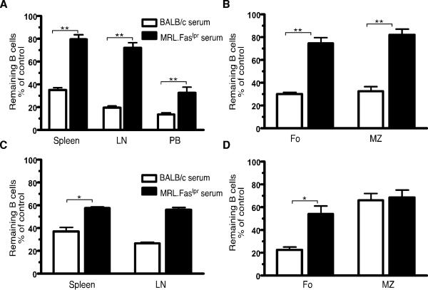
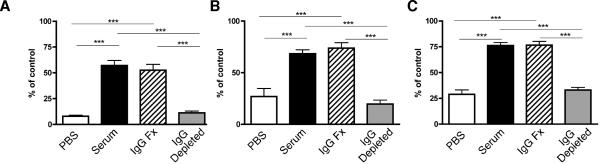
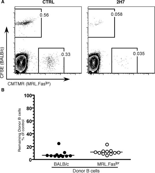
B cells play important roles in autoimmune diseases ranging from multiple sclerosis to rheumatoid arthritis. B cells have also long been considered central players in systemic lupus erythematosus. However, anti-CD20-mediated B cell depletion was not effective in two clinical lupus studies, whereas anti-B lymphocyte stimulator, which inhibits B cell survival, was effective. Others and we previously found that anti-CD20-based depletion was surprisingly ineffective in tissues of lupus-prone mice, but that persistent high doses eventually led to depletion and ameliorated lupus. Lupus patients might also have incomplete depletion, as suggested in several studies, and which could have led to therapeutic failure. In this study, we investigated the mechanism of resistance to Ab-mediated cellular depletion in murine lupus. B cells from lupus-prone mice were easily depleted when transferred into normal environments or in lupus-prone mice that lacked serum Ig. Serum from lupus-prone mice transferred depletion resistance, with the active component being IgG. Because depletion is FcγR-dependent, we assayed macrophages and neutrophils exposed to lupus mouse serum, showing that they are impaired in IgG-mediated phagocytosis. We conclude that depletion resistance is an acquired, reversible phagocytic defect depending on exposure to lupus serum IgG. These results have implications for optimizing and monitoring cellular depletion therapy.


Figures

Figure 1
Figure 1
Efficient depletion of B cells in MRL.Faslpr mice lacking circulating Ab. (A) Six-to-eight wk old hCD20.BALB/c (n=2), hCD20.MRL.Faslpr (n=5) and hCD20.mIgM.MRL.Faslpr (n=6 for treated, n=4 for control, referred to as hCD20.mIgM on the figure label) mice were treated with 4mg/wk of 2H7 Ab i.p., given in divided doses twice/wk for 2 wks. Residual CD22+ B cells in spleen, LN and PB, as percentage of the average B cells in IgG treated, age-matched control mice are shown. The hCD20.BALB/c and hCD20.MRL.Faslpr data are derived from ref. (10) Fig. 2c and Fig. 3b, respectively, and are shown for comparison. (B) Six-to-eight wk old mIgM (n>6) and BALB/c (n>3) mice were treated with either 1 mg/wk or 3 mg/wk of 18B12 mAb i.p., twice/wk for 2 wks. Residual CD19+ B cells are plotted as in panel A for all sites including peritoneal cavity (PerC). The BALB/c data are derived from ref. (10) Fig. 3c, and are shown for comparison. Data are from 1 experiment for hCD20.mIgM and 2 for mIgM mice. Error bars represent SEM. *, p < 0.05; **, p < 0.01 by Mann-Whitney Test.
Figure 2
Figure 2
Serum from lupus-prone mice blocks B-cell depletion in BALB/c mice and MRL.Faslpr mice lacking serum Ab. (A, B) Serum from BALB/c or aged MRL.Faslpr mice was transferred into eight-to-ten wk old hCD20.BALB/c (n=5) prior to treatment with a single dose of 0.5 mg 2H7 i.v. on d7. Residual CD22+ B cells in spleen, LN and PB (A), and splenic CD19+ B cell subpopulations (Fo: Follicular cells and MZ: Marginal zone cells, defined as CD23hiCD21lo and CD23loCD21hi respectively) (B) were analyzed on d11. (C, D) Serum from BALB/c or aged MRL.Faslpr mice was transferred into nine-to-eleven wk old mIgM MRL.Faslpr Tg mice (n>3) prior to treatment with a single dose of 0.5 mg 18B12 i.v. on d7. Residual CD22+ B cells in spleen and LN (C), and splenic B cell subpopulations (D) were analyzed on d11. Residual B cells in all cases are calculated as percentage of the average B cells in control-IgG treated, age-matched mice (infused with same serum). Error bars represent SEM. *, p < 0.05; **, p < 0.01 by Mann-Whitney Test.
Figure 3
Figure 3
The active component in serum contains IgG. Serum from older MRL.Faslpr mice was collected and separated into IgG and non-IgG containing fractions. Unfractionated serum (black bars), IgG fraction (hatched bars), non-IgG fraction (gray bars) or PBS (open bars) was then infused into hCD20.BALB/c mice as described in Methods. Mice were then treated with 2H7 and the fraction of B cells in the indicated tissues was measured by FACS as previously reported (10) and is plotted as a percent of the average of the value of untreated control mice analyzed simultaneously. (A) PB, (B) LN, and (C) Spleen. Bars show the mean and error bars the SEM. Data combined from two independent experiments with n=7-8 mice per group. ***, p <0.0001 by Student's unpaired t test.
Figure 4
Figure 4
Resistance to depletion is not a B cell intrinsic trait. B cells from six-to-eight wk old hCD20.BALB/c and hCD20.MRL.Faslpr mice were purified; labeled with fluorescent dyes, CFSE and CMTMR respectively; and co-transferred into seven-to-nine wk old H2d x k heterozygous BALB/c mice. On d1, the recipient mice were treated with 100 μg of a single dose of either 2H7 Ab (n=11) or a control IgG Ab (n=8) and the mice were analyzed on d3. (A) Representative FACS plots of CD19+ gated B cells showing residual donor B cells after the CTRL Ab (Left panel) or 2H7 (Right Panel) treatment. (B) Comparison of the residual donor B cells in spleen of recipient mice, as percentage of the average frequency of the donor B cells in IgG treated mice. Each symbol is an individual recipient mouse. hCD20.BALB/c donor, closed circles; hCD20.MRL.Faslpr donor, open circles. Lines represent Median. Data are combined from two independent experiments.
Figure 5
Figure 5
Fc receptor-mediated phagocytosis is impaired in macrophages and granulocytes from MRL.Faslpr mice relative to MRL.Faslpr .Jh-/- mice. Splenic CD11b+ cells were incubated with opsonized or non-opsonized, PKH26-labeled SRBCs for 60 min. Engulfment of SRBCs by (A) macrophages (F4/80hi, GR-1int) and (B) granulocytes (GR-1hi, F4/80lo) was measured by flow-cytometry (line represents median) by scoring for positive staining in the red PKH26 channel, as shown in Supplemental Fig. 2. Comparisons are between Jh-/- mice (closed circles, each symbol an individual mouse) and MRL.Faslpr mice (open circles). Data are combined from two independent experiments. **, p<0.01 by Student's t test.

Similar articles

Cited by

References

    1. Martin F, Chan AC. B cell immunobiology in disease: evolving concepts from the clinic. Ann. Rev. Immunol. 2006;24:467–496. - PubMed
    1. Browning JL. B cells move to centre stage: novel opportunities for autoimmune disease treatment. Nat Rev Drug Discov. 2006;5:564–576. - PubMed
    1. Chan OT, Madaio MP, Shlomchik MJ. B cells are required for lupus nephritis in the polygenic, Fas-intact MRL model of systemic autoimmunity. J Immunol. 1999;163:3592–3596. - PubMed
    1. Shlomchik MJ, Madaio MP, Ni D, Trounstein M, Huszar D. The role of B cells in lpr/lpr-induced autoimmunity. J Exp Med. 1994;180:1295–1306. - PMC - PubMed
    1. Looney RJ. B cells as a therapeutic target in autoimmune diseases other than rheumatoid arthritis. Rheumatology (Oxford) 2005;44(Suppl 2):ii13–ii17. - PubMed

Publication types

MeSH terms