. 2011 Oct;60(10):2571-7.
doi: 10.2337/db10-1711. Epub 2011 Aug 26.

Donor islet endothelial cells in pancreatic islet revascularization

Affiliations

Donor islet endothelial cells in pancreatic islet revascularization

Daniel Nyqvist et al. Diabetes. 2011 Oct.

Abstract

Objective: Freshly isolated pancreatic islets contain, in contrast to cultured islets, intraislet endothelial cells (ECs), which can contribute to the formation of functional blood vessels after transplantation. We have characterized how donor islet endothelial cells (DIECs) may contribute to the revascularization rate, vascular density, and endocrine graft function after transplantation of freshly isolated and cultured islets.

Research design and methods: Freshly isolated and cultured islets were transplanted under the kidney capsule and into the anterior chamber of the eye. Intravital laser scanning microscopy was used to monitor the revascularization process and DIECs in intact grafts. The grafts' metabolic function was examined by reversal of diabetes, and the ultrastructural morphology by transmission electron microscopy.

Results: DIECs significantly contributed to the vasculature of fresh islet grafts, assessed up to 5 months after transplantation, but were hardly detected in cultured islet grafts. Early participation of DIECs in the revascularization process correlated with a higher revascularization rate of freshly isolated islets compared with cultured islets. However, after complete revascularization, the vascular density was similar in the two groups, and host ECs gained morphological features resembling the endogenous islet vasculature. Surprisingly, grafts originating from cultured islets reversed diabetes more rapidly than those originating from fresh islets.

Conclusions: In summary, DIECs contributed to the revascularization of fresh, but not cultured, islets by participating in early processes of vessel formation and persisting in the vasculature over long periods of time. However, the DIECs did not increase the vascular density or improve the endocrine function of the grafts.


Figures

FIG. 1.
FIG. 1.
DIECs contribute to the vasculature of fresh but not cultured islet grafts. AD: Ex vivo imaging of fresh islet grafts located under the kidney capsule, 1 (A, n = 5) and 5 months (BD, n = 4) after transplantation. A: Extensive contribution of GFP-fluorescent (green) DIECs is found among the engrafted endocrine cells (blue); note also the contribution to long vessel segments. The image is a projection of an image z-stack corresponding to 30 µm. B: GFP-fluorescent DIECs contribute to islet graft vessels. C: Perfusion of the graft-bearing kidney with Texas Red (red) shows that vessels with contributing DIECs are connected to the circulation. D: DIECs and the perfused vessels are shown together with the reflection of endocrine cells. BD: The images are projections of image z-stacks corresponding to 11 µm. Scale bars correspond to 100 μm. E: Quantification of the percentage contribution of DIECs (GFP area) to the graft vasculature (CD31 area). F: Quantification of the total vascular area of the graft (CD31 area). Two hundred islets were transplanted per mouse. All values are presented as mean ± SEM; **P < 0.01. (A high-quality digital representation of this figure is available in the online issue.)
FIG. 2.
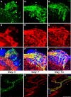
FIG. 2.
Characterization of DIEC participation in islet revascularization. AI: Projections of image z-stacks captured at 3, 7, and 14 days during revascularization of a fresh islet graft in the AC. AC: At day 3, elongated GFP-fluorescent (green) DIECs contribute to the wall of vessels established outside of the islets (A, arrows). Round GFP-fluorescent cells are found in the vicinity of areas of diffused Texas Red dye, as well as distant from perfused regions (A, arrowheads). DF: At day 7, mostly elongated DIECs contributing to established vessels are found (D). GI: At day 14, structural refinements of the vessels are observed with only small changes for the DIECs. The images are representatives of five to seven islets imaged per time point. JL: Projections of an image z-stack captured 14 days after transplantation of freshly isolated islets. GFP (J), Texas Red (K), and an overlay image of GFP, Texas Red, and reflection (L). One islet graft is localized in the upper right image corner (arrow) and another one is localized in the lower left corner (arrow). Large vessels in the iris connecting to the islets show extensive contribution of DIECs. Thirty islets were transplanted per mouse. Scale bars correspond to 100 μm. (A high-quality digital representation of this figure is available in the online issue.)
FIG. 3.
FIG. 3.
Vessel density in fresh and cultured islets during engraftment in the AC. A: Image projections of one freshly isolated islet (top) and two cultured islets (bottom) during the process of revascularization are shown. The left column displays the reflection image captured at day 14. The vasculature was visualized by intravenously administered Texas Red, 70 kDa, at the indicated time points. Scale bar corresponds to 150 μm. B: Quantification of vessel density in fresh and cultured islet grafts at indicated time points after transplantation. The number of analyzed islet grafts is indicated within parentheses at the respective time points. The number of recipients was six with fresh islets and five with cultured islets. Thirty islets were transplanted per mouse. All values are presented as mean ± SEM; *P = 0.05.
FIG. 4.
FIG. 4.
Ultrastructural and metabolic evaluation of islets engrafted in the AC. AD: TEM images of a native islet in the pancreas (A and C) and an islet engrafted in the AC (B and D). A and B: The same ultrastructure of the ECs of the endogenous and engrafted islets and the close location of endocrine cells and capillaries. C and D: Thin ECs of capillaries, which are only separated by a basement membrane (arrows) from the islet cells. In addition, the characteristic abundant diaphragm-covered fenestrations (arrowheads) of the ECs are found in both endogenous and engrafted islets. Thirty islets were transplanted per mouse. E and F: Metabolic evaluation of AC-engrafted syngeneic islets. E: Nonfasting glycemic values in recipients of a syngeneic marginal islet mass transplantation (150 islet equivalents/mouse). At the end of the study, hyperglycemia promptly resumed after enucleation of the graft-bearing eye. F: Days needed for return to normoglycemia of diabetic recipients receiving fresh (○) and cultured (●) syngeneic islets.

References

    1. Shapiro AM, Ricordi C, Hering BJ, et al. International trial of the Edmonton protocol for islet transplantation. N Engl J Med 2006;355:1318–1330 - PubMed
    1. Vajkoczy P, Olofsson AM, Lehr HA, et al. Histogenesis and ultrastructure of pancreatic islet graft microvasculature. Evidence for graft revascularization by endothelial cells of host origin. Am J Pathol 1995;146:1397–1405 - PMC - PubMed
    1. Mattsson G, Jansson L, Carlsson PO. Decreased vascular density in mouse pancreatic islets after transplantation. Diabetes 2002;51:1362–1366 - PubMed
    1. Davalli AM, Scaglia L, Zangen DH, Hollister J, Bonner-Weir S, Weir GC. Vulnerability of islets in the immediate posttransplantation period. Dynamic changes in structure and function. Diabetes 1996;45:1161–1167 - PubMed
    1. Lai Y, Schneider D, Kidszun A, et al. Vascular endothelial growth factor increases functional beta-cell mass by improvement of angiogenesis of isolated human and murine pancreatic islets. Transplantation 2005;79:1530–1536 - PubMed

Publication types

MeSH terms