. 2011 Oct 10;52(11):8006-15.
doi: 10.1167/iovs.11-7693.

Quantification of peripapillary sparing and macular involvement in Stargardt disease (STGD1)

Affiliations

Quantification of peripapillary sparing and macular involvement in Stargardt disease (STGD1)

Tomas R Burke et al. Invest Ophthalmol Vis Sci. .

Abstract

Purpose: To quantify and compare structure and function across the macula and peripapillary area in Stargardt disease (STGD1).

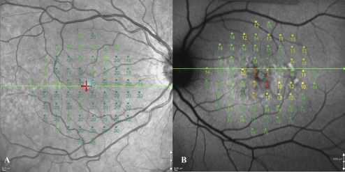
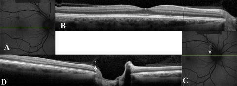
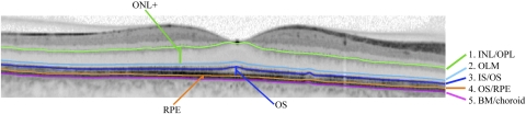
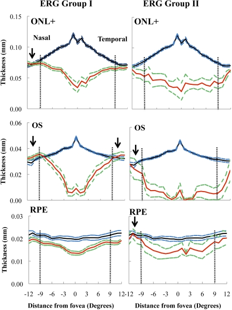
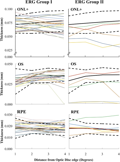
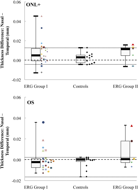
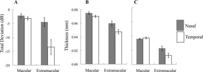
Methods: Twenty-seven patients (27 eyes) and 12 age-similar controls (12 eyes) were studied. Patients were classified on the basis of full-field electroretinogram (ERG) results: Fundus autofluorescence (FAF) and spectral domain-optical coherence tomography (SD-OCT) horizontal line scans were obtained through the fovea and peripapillary area. The thicknesses of the outer nuclear layer plus outer plexiform layer (ONL+), outer segment (OS), and retinal pigment epithelium (RPE) were measured through the fovea, and peripapillary areas from 1° to 4° temporal to the optic disc edge using a computer-aided, manual segmentation technique. Visual sensitivities in the central 10° were assessed using microperimetry and related to retinal layer thicknesses.

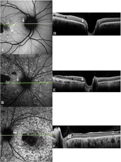
Results: Compared to the central macula, the differences between controls and patients in ONL+, OS, and RPE layer thicknesses were less in the nasal and temporal macula. Relative sparing of the ONL+ and/or OS layers was detected in the nasal (i.e., peripapillary) macula in 8 of 13 patients with extramacular disease on FAF; relative functional sparing was also detected in this subgroup. All 14 patients with disease confined to the central macula, as detected on FAF, showed ONL+ and OS layer thinning in regions of normal RPE thickness.

Conclusions: Relative peripapillary sparing was detected in STGD1 patients with extramacular disease on FAF. Photoreceptor thinning may precede RPE degeneration in STGD1.


Figures

Figure 1.
Figure 1.
(A) Microperimetry results from a control subject registered to the IR image. There was stable fixation. Note the position of the line scan (green horizontal line through the fovea). (B) Microperimetry results from a patient with stable fixation, registered to an FAF image.
Figure 2.
Figure 2.
FAF (A, C) and corresponding SD-OCT scans (B, D) of a control. Horizontal green line: the position of the SD-OCT scan. In this case, the horizontal macular line scan (B) did not bisect the optic disc. Therefore, a second horizontal SD-OCT line scan which bisected the optic disc was acquired (D). White-dashed arrow: position of the optic disc edge.
Figure 3.
Figure 3.
An example of a segmented SD-OCT scan through the fovea is presented. The ONL+ (from OLM to INL/OPL), OS (from OS/RPE to IS/OS), and RPE (from BM/choroid to OS/RPE) layers are demonstrated.
Figure 4.
Figure 4.
(AC) The various patterns of peripapillary autofluorescence in STGD1; (DF) the corresponding SD-OCT line scans (position indicated on FAF image by the horizontal green line). Peripapillary sparing (white arrows) is seen in (A) and (D) and (B) and (E). Peripapillary atrophy (double white arrows) is seen in (C) and (F). Black double-headed arrows: regions where there was loss of IS/OS in the macula.
Figure 5.
Figure 5.
Thicknesses of the ONL+, OS, and RPE layers across the macula in patients with ERG group I or II disease. Solid black and dashed blue lines: mean thickness ±1 SE for the 12 controls. Solid red lines: mean values for the patients with ERG groups I and II disease; green dashed lines: ±1 SE for both groups. Vertical dashed horizontal lines: an eccentricity of 9° in the nasal (−) and temporal (+) macula. Arrows: regions where the mean thickness is equal to or greater than that of controls in the nasal or temporal macula.
Figure 6.
Figure 6.
Peripapillary ONL+, OS, and RPE layer thicknesses in 20 patients with ERG group I and 5 patients with ERG group II disease (colored lines). Black lines: the mean ± 95% CI of the controls (solid, mean; dashed, CI).
Figure 7.
Figure 7.
Box-and-whisker plots of the ONL+ and OS layer mean thicknesses differences between the nasal and temporal macula, at eccentricities from 9° to 12°, in the controls and in both ERG groups. The median values of each subgroup are presented together, with the first and third quartiles and the minimum and maximum values. Colored symbols: individual patient mean thickness differences values at these eccentricities. Central black dashed line: 0 difference; top and bottom dashed lines: the highest and lowest differences detected between the nasal and temporal macula, respectively, in the controls.
Figure 8.
Figure 8.
The mean TD (A), from 7° and 9° from the fovea, in the nasal and temporal macula in patients with macular disease only and in those with extramacular disease. Error bars, mean ± 1 SE. Data are also shown for the mean ONL+ (B) and OS layer (C) thicknesses in these subgroups, at the same eccentricities.

References

    1. Allikmets R, Singh N, Sun H, et al. A photoreceptor cell-specific ATP-binding transporter gene (ABCR) is mutated in recessive Stargardt's macular dystrophy. Nat Genet. 1997;15:236–246 - PubMed
    1. Cideciyan AV, Sider M, Aleman TS, et al. ABCA4-associated retinal degenerations spare structure and function of the human parapapillary retina. Invest Ophthalmol Vis Sci. 2005;46:4739–4746 - PMC - PubMed
    1. Schwoerer J, Secrétan M, Zografos L, Piguet B. Indocyanine green angiography in fundus flavimaculatus. Ophthalmologica. 2000;214:240–245 - PubMed
    1. Klein R, Lewis RA, Meyers SM, Myers FL. Subretinal neovascularization associated with fundus flavimaculatus. Arch Ophthalmol. 1978;96:2054–2057 - PubMed
    1. Lois N, Halfyard AS, Bird AC, Holder GE, Fitzke FW. Fundus autofluorescence in Stargardt macular dystrophy-fundus flavimaculatus. Am J Ophthalmol. 2004;138:55–63 - PubMed

Publication types

MeSH terms

Substances