. 2011 Aug 26;30(19):4071-83.
doi: 10.1038/emboj.2011.293.

A hippocampal insulin-growth factor 2 pathway regulates the extinction of fear memories

Affiliations

A hippocampal insulin-growth factor 2 pathway regulates the extinction of fear memories

Roberto Carlos Agis-Balboa et al. EMBO J. .

Abstract

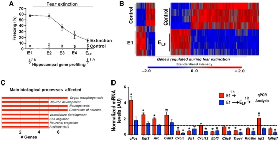
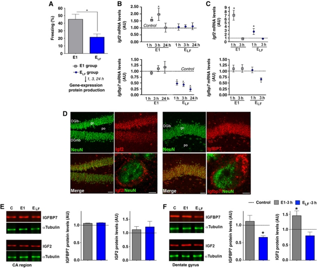
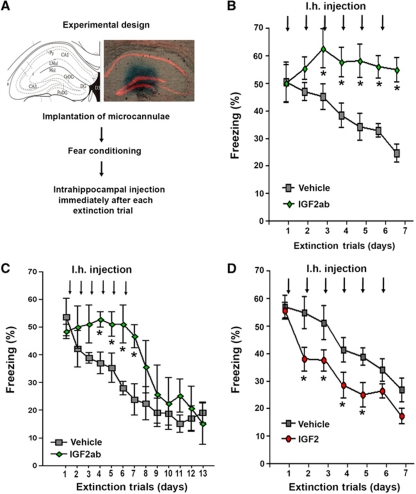
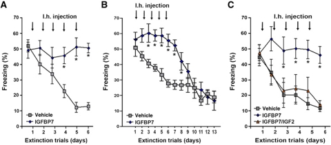
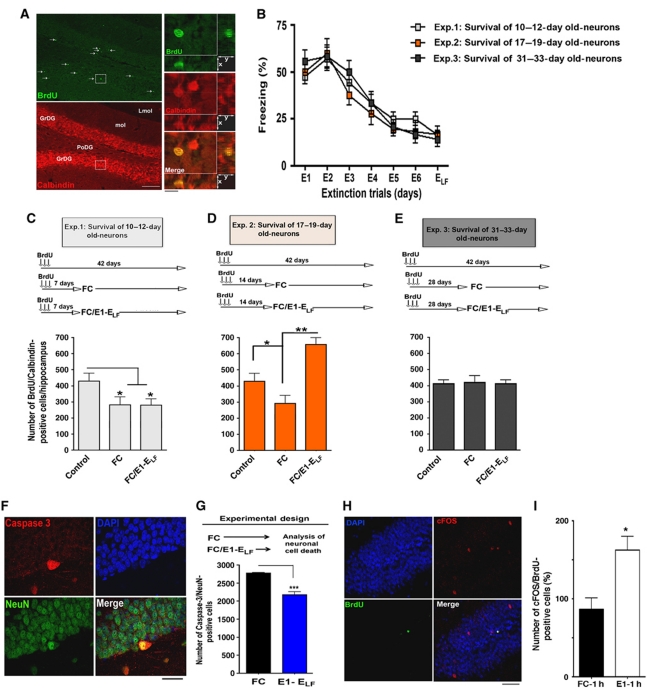
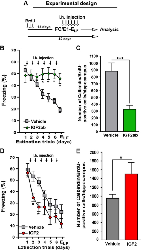
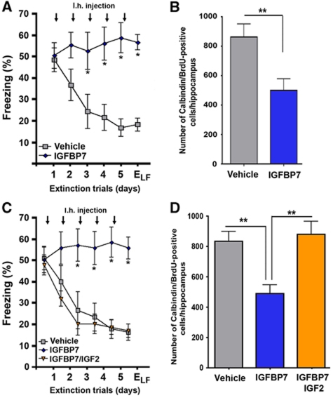
Extinction learning refers to the phenomenon that a previously learned response to an environmental stimulus, for example, the expression of an aversive behaviour upon exposure to a specific context, is reduced when the stimulus is repeatedly presented in the absence of a previously paired aversive event. Extinction of fear memories has been implicated with the treatment of anxiety disease but the molecular processes that underlie fear extinction are only beginning to emerge. Here, we show that fear extinction initiates upregulation of hippocampal insulin-growth factor 2 (Igf2) and downregulation of insulin-growth factor binding protein 7 (Igfbp7). In line with this observation, we demonstrate that IGF2 facilitates fear extinction, while IGFBP7 impairs fear extinction in an IGF2-dependent manner. Furthermore, we identify one cellular substrate of altered IGF2 signalling during fear extinction. To this end, we show that fear extinction-induced IGF2/IGFBP7 signalling promotes the survival of 17-19-day-old newborn hippocampal neurons. In conclusion, our data suggest that therapeutic strategies that enhance IGF2 signalling and adult neurogenesis might be suitable to treat disease linked to excessive fear memory.


Conflict of interest statement

The authors declare that they have no conflict of interest.

Figures

Figure 1
Figure 1
Fear extinction regulates hippocampal gene expression. (A) Experimental design. Mice (n=5/group) were subjected to contextual fear conditioning (FC) followed by extinction training on consecutive days. Hippocampal tissue was prepared for gene array analysis 1 h after E1 and ELF. Mice that were subjected to the conditioning context but did not undergo fear extinction served as control groups. (B) Heat map showing differential gene expression during fear extinction. (C) GO-term analysis of genes regulated during fear extinction. (D) The expression of selected genes upon fear extinction was analysed via qPCR. *P<0.05 versus control; E, extinction trial; ELF, extinction trial low freezing; (n=5/group); error bars indicate s.e.m.
Figure 2
Figure 2
Fear extinction regulates hippocampal Igf2/Igfbp7 expression. (A) Mice were subjected to fear extinction. Behavioural analysis confirmed that freezing behaviour was significantly reduced upon ELF exposure, when compared to E1. Mice that were subjected to the conditioning context but not subjected to fear extinction served as control (*P<0.05 versus E1; n=15/group). (B) qPCR analysis of Igf2 and Igfbp7 expression in the total hippocampus at different time points after fear extinction training (*P<0.05 versus control; n=5/group). (C) qPCR analysis of Igf2 and Igfbp7 expression in the dentate gyrus (*P<0.05 versus control; n=5/group). (D) Representative images showing immunostaining for IGF2 and IGFBP7 protein levels in the dentate gyrus. Scale bars low magnification images: 50 μm; high magnification: 2 μm. (E) Left panel: Representative immunoblot analysis of IGF2 and IGFBP7 protein levels in the CA region 3 h after exposure to E1 or ELF. Right panel. Quantification (n=5/group). (F) Left panel: Representative immunoblot analysis of IGF2 and IGFBP7 protein levels in the dentate gyrus region 3 h after exposure to E1 or ELF. Right panel. Quantification (*P<0.05 versus control, n=5/group). DGlb, dentate gyrus lateral blade; DGmb, dentate gyrus medial blade; E, extinction trial; ELF, extinction trial low freezing; gc, granular cell layer of the dentate gyrus; Po, polymorph layer of the dentate gyrus; error bars indicate s.e.m.
Figure 3
Figure 3
Hippocampal IGF2 signalling regulates fear extinction. (A) Experimental design. The image shows the position of the injection cannulae as visualized by methylene blue injection. (B) Fear extinction was significantly impaired in mice that were injected immediately after each extinction trial with IGF2ab when compared to the vehicle group (*P<0.05 versus vehicle, n=10/group). (C) In an independent experiment, we found that IGF2ab-treated mice were able to successfully extinct fear memories once IGF2ab treatment was stopped. (D) Fear extinction was significantly enhanced in mice that were injected immediately after each extinction trial with IGF2 when compared to the vehicle group (*P<0.05 versus vehicle, n=10/group). Py, pyramidal cell layer; rad, stratum radiatum; Lmol, lacunosum moleculare; CA1, subfield CA1; CA3, subfield CA3; DG, dentate gyrus. Error bars indicate s.e.m.
Figure 4
Figure 4
Hippocampal IGFBP7 signalling regulates fear extinction. (A) Fear extinction was significantly impaired in mice that were injected immediately after each extinction trial with IGFBP7 when compared to the vehicle group (*P<0.05 versus vehicle) (n=10/group). (B) In an independent experiment, we observed that once IGFBP7 treatment was stopped, those mice were able to successfully extinct fear memories. (C) Mice were subjected to fear extinction and subsequently injected with IGFBP7, IGFBP7 and IGF2 or vehicle. IGF2 injection rescued the effect of IGFBP7 (*P<0.05 versus vehicle) (n=10/group). Error bars indicate s.e.m.
Figure 5
Figure 5
Fear extinction promotes the survival of immature newly generated neurons. (A) Representative images showing BrdU/Calbindin-positive cells that indicate maturation of newborn hippocampal neurons. Arrows indicate BrdU-positive cells. Scale bar low magnification images: 100 μm. Scale bar high magnification images: 20 μm. (B) Fear extinction, as indicated by the gradual decline of aversive freezing behaviour throughout extinction trials, was similar among the experimental groups that were used to analyse the survival of newly generated hippocampal neurons during fear extinction (n=8/group). (CE) Upper panels show the experimental design to analyse the effect of fear extinction on 10–12-day-old (C), 17–19-day-old (D) or 31–33-day-old (E) hippocampal neurons. Lower panels display the quantification of BrdU/Calbindin-positive cells under each experimental condition. Note that successful fear extinction significantly enhanced the survival of 17–19-day-old newly generated neurons when compared to the control (*P<0.05) or fear conditioning group (**P<0.01). (F) Representative images showing active caspase-3-positive cells, indicative of apoptotic cell death, in the hippocampal dentate gyrus region. Scale bar: 20 μm. (G) Mice were subjected to fear conditioning or fear extinction training. The number of active caspase-3-positive cells in the dentate gyrus was significantly reduced in mice that underwent extinction training when compared to mice that were only exposed to fear conditioning (***P<0.0001). (H) Representative image showing a cFOS-positive 17–19-day-old neuron in the hippocampus of a mouse 1 h after exposure to E1. Scale bar: 50 μm. (I) Mice were subjected to fear conditioning or fear extinction training. The number of cFOS/BrdU-positive cells was analysed 1 h after exposure to E1 or 1 h after FC. The data are normalized to the FC group (P=0.0154). GrDG, granular cell layer of the dentate gyrus; Lmol, lacunosum moleculare. Error bars indicate s.e.m.
Figure 6
Figure 6
IGF2 promotes the survival of immature neurons during fear extinction. (A) Experimental design. Mice were injected with BrdU on three consecutive days and subjected to fear extinction training 14 days later. Immediately after each extinction trial, mice were injected with vehicle IGF2 or IGF2ab. Neuronal survival was analysed 42 days after the first BrdU injection by counting BrdU/Calbindin-positive cells. (B) Fear extinction was significantly impaired in mice injected intrahippocampally with an IGF2ab (*P<0.05). (C) Survival of 17–19-day-old neurons was impaired in IGF2ab-treated mice (***P<0.0001 versus vehicle). (D) Fear extinction was significantly facilitated in the IGF2-treated mice (*P<0.05 versus vehicle). (E) Survival of 17–19-day-old neurons was enhanced in IGF2-treated mice (*P<0.05; **P<0.01). n=8/group; Error bars indicate s.e.m.
Figure 7
Figure 7
IGFBP7 regulates the survival of immature neurons during fear extinction in an IGF2-dependent manner. (A) Using the same experimental approach as shown in Figure 6A, we found that fear extinction was significantly impaired in mice injected intrahippocampally with IGFBP7 (*P<0.05 versus vehicle). (B) Survival of 17–19 days was impaired in IGFBP7-treated mice (**P<0.001 versus vehicle). (C) Mice were injected with either vehicle, IGFBP7 or co-injected with IGFBP7 and IGF2. While fear extinction was impaired in IGFBP7-injected mice, co-injection with IGF2 was able to rescue this effect. (D) IGF2 rescued IGFBP7-mediated impairment of neuronal survival during fear extinction (**P<0.001 versus Igfbp7/vehicle). n=8/group; Error bars indicate s.e.m.

References

    1. Aberg MA, Aberg ND, Palmer TD, Alborn AM, Carlsson-Skwirut C, Bang P, Rosengren LE, Olsson T, Gage FH, Eriksson PS (2003) IGF-I has a direct proliferative effect in adult hippocampal progenitor cells. Mol Cell Neurosci 24: 23–40 - PubMed
    1. Aimone JB, Wiles J, Gage FH (2006) Potential role for adult neurogenesis in the encoding of time in new memories. Nat Neurosci 9: 723–727 - PubMed
    1. Aimone JB, Wiles J, Gage FH (2009) Computational influence of adult neurogenesis on memory encoding. Neuron 61: 187–202 - PMC - PubMed
    1. Alexa A, Rahnenfuhrer J, Lengauer T (2006) Improved scoring of functional groups from gene expression data by decorrelating GO graph structure. Bioinformatics 22: 1600–1607 - PubMed
    1. Bateman JM, McNeill H (2006) Insulin/IGF signalling in neurogenesis. Cell Mol Life Sci 63: 1701–1705 - PMC - PubMed

Publication types

Substances