. 2011 Aug 28;18(9):1060-7.
doi: 10.1038/nsmb.2109.

Mechanism of actin filament nucleation by Vibrio VopL and implications for tandem W domain nucleation

Affiliations

Mechanism of actin filament nucleation by Vibrio VopL and implications for tandem W domain nucleation

Suk Namgoong et al. Nat Struct Mol Biol. .

Abstract

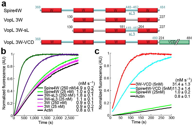
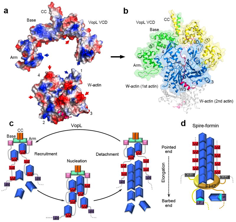
Pathogen proteins targeting the actin cytoskeleton often serve as model systems to understand their more complex eukaryotic analogs. We show that the strong actin filament nucleation activity of Vibrio parahaemolyticus VopL depends on its three W domains and on its dimerization through a unique VopL C-terminal domain (VCD). The VCD shows a previously unknown all-helical fold and interacts with the pointed end of the actin nucleus, contributing to the nucleation activity directly and through duplication of the W domain repeat. VopL promotes rapid cycles of filament nucleation and detachment but generally has no effect on elongation. Profilin inhibits VopL-induced nucleation by competing for actin binding to the W domains. Combined, the results suggest that VopL stabilizes a hexameric double-stranded pointed end nucleus. Analysis of hybrid constructs of VopL and the eukaryotic nucleator Spire suggest that Spire may also function as a dimer in cells.


Figures

Figure 1
Figure 1
Nucleation activity of VopL constructs. (a) Domain organization of VopL, constructs used in this study and alignment of W domains illustrating the sites of point mutations. (b) Time course of polymerization of 2 μM Mg-ATP-actin (6% pyrene-labeled) alone or in the presence of 25 nM VopL constructs (color-coded). Polymerization rates are reported as mean and s.e.m. values (number of measurements ≥ 3). (c) Effect of VopL construct concentration on the polymerization rate of pyrene-actin (see also Supplementary Fig. 2). (d) Time course of actin polymerization by VopL constructs containing point mutations in the W domains (shown in part a). (e) Polymerization of 1.5 μM Mg-ATP-actin (33% Oregon Green-labeled) alone or in the presence of 0.1 nM VopL constructs visualized by TIRF microscopy on NEM-myosin II-coated coverslips, including time-lapse micrographs (see Supplementary Videos 1–4) of representative 19.5 μm2 fields (left) and plots of the growth of 20 filament barbed-ends over time (right). Circles and arrowheads indicate the pointed and barbed ends of representative filaments. The average number of filaments in a 133 μm2 field of view at 300 s from two independent experiments is indicated as f μm-2. Errors are ±s.d.
Figure 2
Figure 2
VCD mediates dimerization and binds to the pointed end of the polymerization nucleus. (a) SEC-MALS mass measurements of VopL constructs (theoretical masses in parenthesis). Supplementary Table 1 lists the masses of other constructs. (b) Time course of polymerization of 2 μM Mg-ATP-actin alone or induced by 25 nM VopL constructs. (c,d) Gel electrophoresis of supernatant (S) and pellet (P) fractions after co-sedimentation at 224,000 × g of F-actin with VCD or 1W-VCD. (e) Mass measurements of 1W-VCD and its complex with actin. (f) TIRF time-lapse micrographs of actin assembly induced by P-3W-VCD immobilized on a coverslip, showing a filament anchored to the coverslip by its pointed end that dissociates between frames 220 and 280 s (top) and a filament that remains anchored throughout (bottom) (Supplementary Videos 5 and 6). Circles and arrows indicate the pointed and barbed ends of the filaments. Shown on the right are growth plots of 20 filaments, including tethered and non-tethered (dashed lines). (g) TIRF time-lapse micrographs of actin (green) assembly by 3W-VCD bound to Qdots (red). Upper panels show a Qdot that nucleates 14 filaments (arrowheads and numbers, Supplementary Videos 7 and 8). Middle and lower panels show filaments whose pointed (circles) or barbed (arrows) ends appear to be bound to a Qdot (Supplementary Videos 9 and 10). (h) Plots of the growth of filaments, including non-bound, fast dissociated, pointed and barbed end-bound, and two filaments whose barbed ends detach from the Qdot and their elongation rates decrease during the experiment. Errors are ±s.d.
Figure 3
Figure 3
Crystal structure of VCD and SAXS structures of VCD and 1W-VCD+actin. (a) Two perpendicular views of the crystal structure of VCD and definition of domains and secondary structure shown along the sequence of this domain (Supplementary Video 11). Different colors highlight different domains: base (magenta, green), arm (pink), coiled coil (gold). (b) Experimental X-ray scattering pattern of VCD (red) and 1W-VCD+actin (blue) as a function of momentum transfer s=4πsin(θ)/λ, where 2θ is the scattering angle and λ=0.103 nm is the X-ray wavelength. The inset shows the normalized distance distribution functions computed from the scattering pattern with the program GNOM. (c,d) Two perpendicular orientations of the average SAXS envelopes of VCD and 1W-VCD+actin (same orientations as for the crystal structure).
Figure 4
Figure 4
Profilin inhibits polymerization induced by VopL. (a) Polymerization rates of 2 μM Mg-ATP-actin induced by 5 nM VopL constructs (color-coded) or 0.5 μM F-actin seeds (black) as a function of profilin concentration. (b) Visualization by TIRF microscopy of the effect of 2.5 μM profilin on the polymerization of 1.5 μM Mg-ATP-actin alone (Supplementary Video 12) or induced by 0.1 nM 3W-VCD and P-3W-VCD (Supplementary Videos 13 and 14) or mutants 3W-VCDP191E and P-3W-VCDP191E (Supplementary Videos 15—18). Plots of the growth of 20 individual filaments are shown on the right. (c,d) Comparison of the nucleation activities and elongation rates measured by TIRF with (blue) or without (red) profilin. Errors are ±s.d.
Figure 5
Figure 5
Role of dimerization and specific sequence of W domains and inter-W linkers in nucleation. (a) Design of hybrid constructs of VopL and Drosophila Spire’s repeat of four W domains (Spire4W). In construct 3W-sL3, VopL linker-2 was replaced with Spire linker-3. In construct Spire4W-VCD, VopL’s VCD was fused C-terminal to the last W domain of Spire, using the LKKT(V) motifs as reference for fusion. (b) Time courses of polymerization of 2 μM Mg-ATP-actin induced by two different concentrations (25 and 250 nM) of constructs Spire4W, 3W-sL3 and 3W. (c) Comparison of the time courses of actin polymerization induced by constructs VopL 3W-VCD and Spire4W-VCD and Spire4W at the indicated concentrations. Errors are ±s.d.
Figure 6
Figure 6
Proposed mechanisms of actin nucleation by VopL and Spire. (a) Electrostatic surface representation (blue, positively charged; red, negatively charged) of the structures of VCD and W–actin (PDB code: 2D1K) , illustrating the existing shape and charge complementarity (indicated by red and blue arrows) between the two structures. (b) Such complementarity and the necessity to connect the C-terminus of the third W domain to the N-terminus of VCD lead to a model of the complex in which the first actin subunit interacts with both subunits of the VopL dimer (see Supplementary Video 19). The second actin subunit (gray) might bind on the opposite side of VCD according to the actin filament model . (c) Model of nucleation by VopL. VCD plays a dual role, contributing directly to the recruitment of actin subunits by binding to the pointed end of the polymerization nucleus, and enabling the formation of a hexameric nucleus by duplication of the W domain repeat. VopL detaches fast after nucleation, probably due to steric hindrance of the W domains with longitudinal contacts between actin subunits in the filament . Upon detachment, VopL might carry with it actin subunits that become part of the nucleus in a new round of polymerization. (d) Spire’s activity is also enhanced by dimerization, which in cells is mediated by interaction of Spire’s KIND domain with formins ,,.

Comment in

References

    1. Dominguez R. Structural insights into de novo actin polymerization. Curr Opin Struct Biol. 2010;20:217–25. - PMC - PubMed
    1. Pollard TD. Regulation of actin filament assembly by Arp2/3 complex and formins. Annu Rev Biophys Biomol Struct. 2007;36:451–77. - PubMed
    1. Goode BL, Eck MJ. Mechanism and function of formins in the control of actin assembly. Annu Rev Biochem. 2007;76:593–627. - PubMed
    1. Higgs HN. Formin proteins: a domain-based approach. Trends Biochem Sci. 2005;30:342–53. - PubMed
    1. Quinlan ME, Heuser JE, Kerkhoff E, Mullins RD. Drosophila Spire is an actin nucleation factor. Nature. 2005;433:382–8. - PubMed

Publication types

MeSH terms

Associated data