. 2011 Aug 28;7(10):740-7.
doi: 10.1038/nchembio.642.

Inactive-state preassembly of G(q)-coupled receptors and G(q) heterotrimers

Affiliations

Inactive-state preassembly of G(q)-coupled receptors and G(q) heterotrimers

Kou Qin et al. Nat Chem Biol. .

Abstract

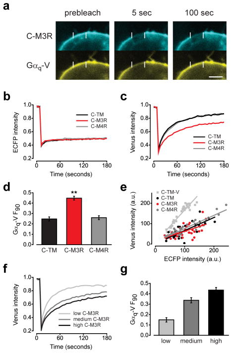
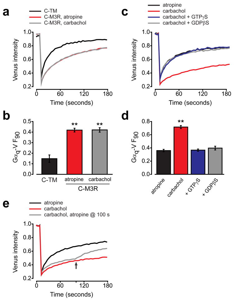
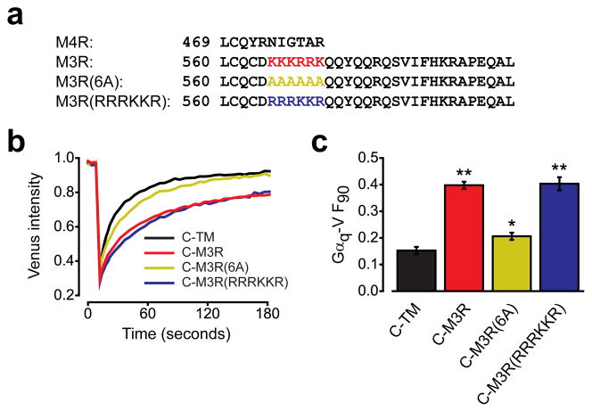
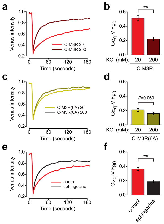
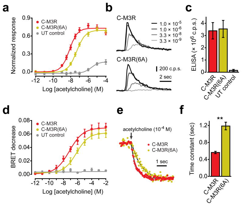
G protein-coupled receptors (GPCRs) transmit signals by forming active-state complexes with heterotrimeric G proteins. It has been suggested that some GPCRs also assemble with G proteins before ligand-induced activation and that inactive-state preassembly facilitates rapid and specific G protein activation. However, no mechanism of preassembly has been described, and no functional consequences of preassembly have been demonstrated. Here we show that M(3) muscarinic acetylcholine receptors (M3R) form inactive-state complexes with G(q) heterotrimers in intact cells. The M3R C terminus is sufficient, and a six-amino-acid polybasic sequence distal to helix 8 ((565)KKKRRK(570)) is necessary for preassembly with G(q). Replacing this sequence with six alanine residues prevents preassembly, slows the rate of G(q) activation and decreases steady-state agonist sensitivity. That other G(q)-coupled receptors possess similar polybasic regions and also preassemble with G(q) suggests that these GPCRs may use a common preassembly mechanism to facilitate activation of G(q) heterotrimers.


Conflict of interest statement

Competing financial interests

The authors declare no competing financial interests.

Figures

Figure 1
Figure 1. Immobile C-M3R decreases Gαq-V mobility
(a) HEK293 cells transiently expressing C-M3R, Gαq-V, Gβ1 and Gγ2 were biotin-avidin crosslinked and imaged before and after photobleaching of a 4 μm segment of the plasma membrane; scale bar=4 μm. Recovery of ECFP (b) and venus (c) fluorescence after photobleaching in cells expressing immobile C-TM (n=31), C-M3R (n=32) or C-M4R (n=33); traces are averages of all cells. (d) Unrecovered fluorescence 90 seconds after photobleaching (F90) for cells expressing C-TM, C-M3R and C-M4R; **, P<0.001 compared to C-TM. (e) ECFP and venus intensity at the plasma membrane for the same cells as panels b-d, and for cells expressing C-TM-V. C-TM, C-M3R and C-M4R were expressed in excess of Gαq-V. (f) Gαq-V fluorescence recovery after photobleaching in cells expressing low (n=16), medium (n=23) and high (n=17) levels of C-M3R and constant levels of Gαq-V. Average C-M3R fluorescence intensity in these three groups was 58 ± 3, 106 ± 3, and 150 ± 6 arbitrary units, respectively. Average Gαq-V fluorescence intensity was 85 ± 5, 86 ± 7, and 92 ± 8 a.u., respectively. (g) Unrecovered fluorescence in the same cells as panel f. Data represent mean values ± s.e.m..
Figure 2
Figure 2. Receptor activity does not affect Gαq-V mobility in intact cells
(a) Gαq-V fluorescence recovery after photobleaching in cells expressing C-TM (n=22), C-M3R in the presence of 10 μM atropine (n=44) or C-M3R in the presence of 50 μM carbachol (n=42). (b) Unrecovered fluorescence for the same cells as panel a; **, P<0.001 compared to C-TM. (c) Gαq-V fluorescence recovery in permeabilized, nucleotide-depleted cells expressing C-M3R in the presence of atropine (n=15), carbachol (n=15), or in the presence of carbachol with 0.3 mM GTPγS (n=15) or 0.3 mM GDPβS (n=24). (d) Unrecovered fluorescence for the same cells as panel c; **, P<0.001. (e) Gαq-V fluorescence recovery in permeabilized, nucleotide-depleted cells expressing C-M3R in the presence of atropine (n=20), carbachol (n=20), or in the presence of carbachol with atropine added at 100 seconds (arrow; n=20). Data represent mean values ± s.e.m..
Figure 3
Figure 3. The M3R C terminus is necessary and sufficient for preassembly with Gq
(a) Gαq-V fluorescence recovery in cells expressing C-TM (n=30), C-M3R (n=30), a chimeric C-M3R with the C terminus of M4R (C-M3R(M4ct); n=30) or C-M4R (n=24). (b) Unrecovered fluorescence for the same cells as panel a; **, P<0.001 compared to C-TM. (c) Gαq-V fluorescence recovery in cells expressing C-TM (n=16), C-M3R (n=18), a chimeric C-M4R with the C terminus of M3R (C-M4R(M3ct); n=37) or C-M4R (n=30). (d) Unrecovered fluorescence for the same cells as panel c; **, P<0.001 compared to C-TM. Unrecovered fluorescence for C-M3R and C-M4R(M3ct) were not significantly different (P=0.14). Data represent mean values ± s.e.m..
Figure 4
Figure 4. A polybasic region in the M3R C terminus is necessary for preassembly with Gq
(a) Alignment of the M4R and M3R C termini, together with mutants where amino acids 565–570 are replaced with alanines (6A) or have lysines and arginines exchanged for each other (RRRKKR). (b) Gαq-V fluorescence recovery in cells expressing C-TM (n=43), C-M3R (n=43), C-M3R(6A) (n=43) or C-M3R(RRRKKR) (n=20). (c) Unrecovered fluorescence for the same cells as panel b; **, P<0.001; *, P=0.047 compared to C-TM. Data represent mean values ± s.e.m..
Figure 5
Figure 5. Preassembly involves an electrostatic interaction and requires an electronegative plasma membrane
(a) Gαq-V fluorescence recovery in permeabilized cells expressing immobile C-M3R bathed in 20 mM KCl (n=19) or in 200 mM KCl (n=20). (b) Unrecovered fluorescence for the same cells as panel a; **, P<0.001. (c) Gαq-V fluorescence recovery in permeabilized cells expressing immobile C-M3R(6A) bathed in 20 mM KCl (n=21) or in 200 mM KCl (n=19). (d) Unrecovered fluorescence for the same cells as panel c. (e) Gαq-V fluorescence recovery in intact cells expressing immobile C-M3R in the absence (n=19) and presence (n=20) of 50 μM sphingosine. (f) Unrecovered fluorescence for the same cells as panel e; **, P<0.001. Data represent mean values ± s.e.m..
Figure 6
Figure 6. A polybasic region in the M3R C terminus is necessary for efficient activation of Gq
(a) Normalized aequorin luminescence (calcium) responses from cells expressing C-M3R (n=5), C-M3R(6A) (n=5) or untransfected control cells (UT control; n=4). (b) Exemplary luminescence traces from cells expressing C-M3R or C-M3R(6A) and exposed to a range of acetylcholine concentrations; c.p.s.=photon counts per second. (c) Cell surface expression of C-M3R (n=12) and C-M3R(6) (n=12) quantified by ELISA in intact cells, and compared to signals from untransfected controls (n=7). (d) Decreases in BRET between Gαq-Rluc8 and Gβ1γ2-V in cells expressing C-M3R (n=4), C-M3R(6A) (n=4) or untransfected control cells (n=4). (e) Normalized BRET responses from cells expressing C-M3R or C-M3R(6A) and exposed to 100 μM acetylcholine (arrow). Smooth lines are least squared fits to a single exponential function. (f) Fitted onset time constants from cells expressing C-M3R or C-M3R(6A) (n=4 each, 24 replicates per experiment); **, P<0.001. Data represent mean values ± s.e.m..

Comment in

  • Receptors: GPCR-G protein preassembly?
    Challiss RA, Wess J. Challiss RA, et al. Nat Chem Biol. 2011 Sep 19;7(10):657-8. doi: 10.1038/nchembio.665. Nat Chem Biol. 2011. PMID: 21931312 No abstract available.

References

    1. Gilman AG. G proteins: transducers of receptor-generated signals. Annu Rev Biochem. 1987;56:615–649. - PubMed
    1. De Lean A, Stadel JM, Lefkowitz RJ. A ternary complex model explains the agonist-specific binding properties of the adenylate cyclase-coupled beta-adrenergic receptor. J Biol Chem. 1980;255:7108–7117. - PubMed
    1. Oldham WM, Hamm HE. Structural basis of function in heterotrimeric G proteins. Q Rev Biophys. 2006;39:117–166. - PubMed
    1. Wess J, et al. Structural basis of receptor/G protein coupling selectivity studied with muscarinic receptors as model systems. Life Sci. 1997;60:1007–1014. - PubMed
    1. Tesmer JJ. The quest to understand heterotrimeric G protein signaling. Nat Struct Mol Biol. 2010;17:650–652. - PMC - PubMed

Publication types

Substances