. 2011 Aug 28;13(10):1252-8.
doi: 10.1038/ncb2320.

Dynamic maintenance of asymmetric meiotic spindle position through Arp2/3-complex-driven cytoplasmic streaming in mouse oocytes

Affiliations

Dynamic maintenance of asymmetric meiotic spindle position through Arp2/3-complex-driven cytoplasmic streaming in mouse oocytes

Kexi Yi et al. Nat Cell Biol. .

Abstract

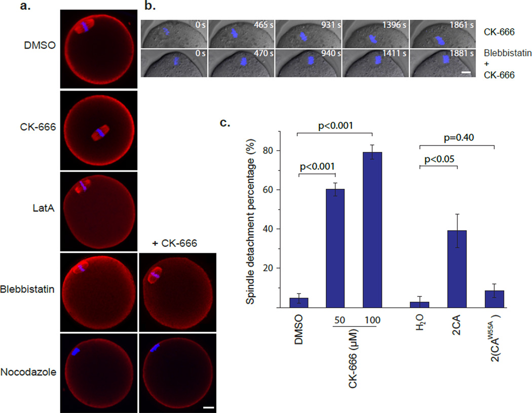
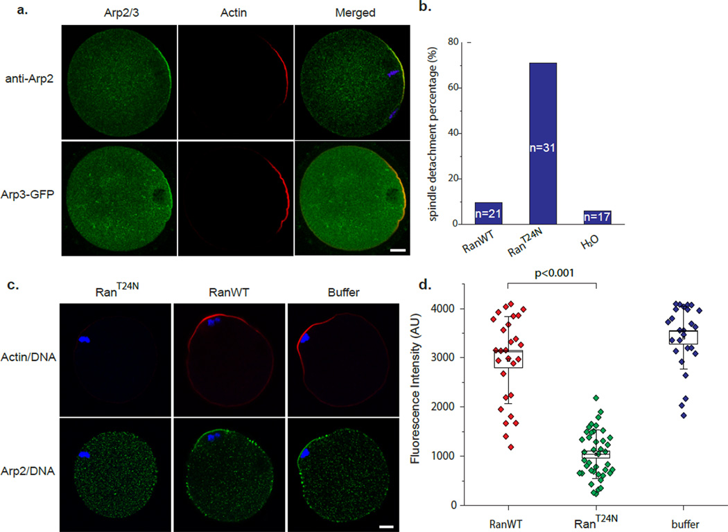
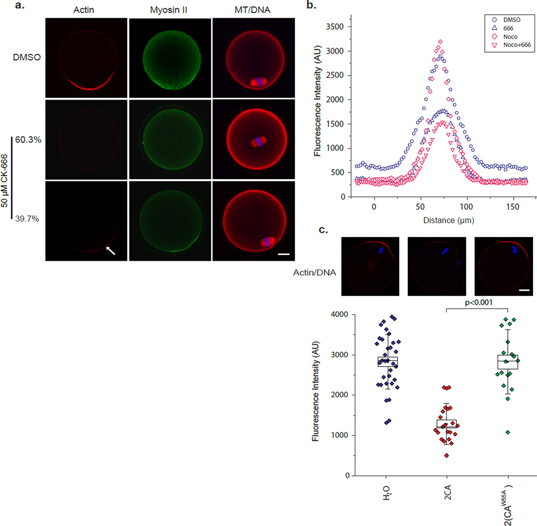
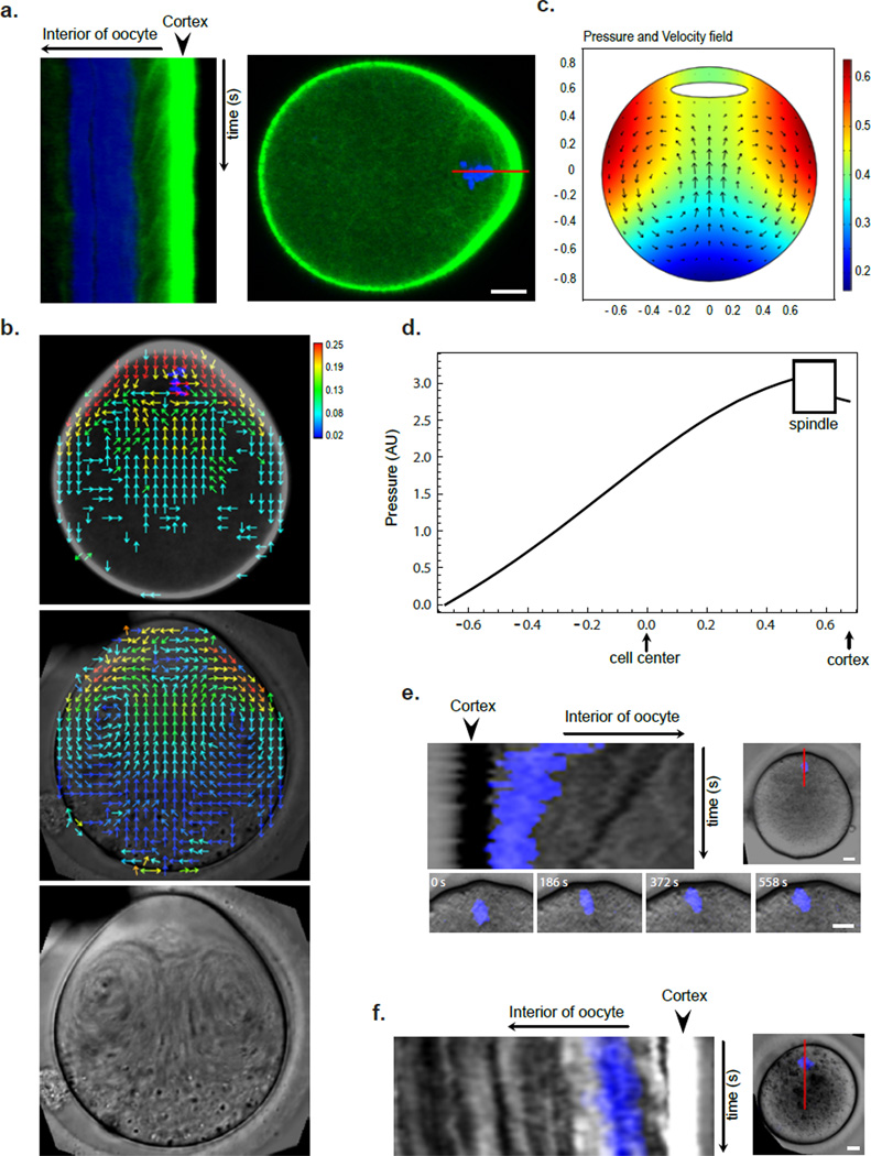
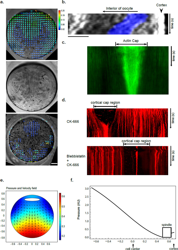
Mature mammalian oocytes are poised for completing meiosis II (MII) on fertilization by positioning the spindle close to an actomyosin-rich cortical cap. Here, we show that the Arp2/3 complex localizes to the cortical cap in a Ran-GTPase-dependent manner and nucleates actin filaments in the cortical cap and a cytoplasmic actin network. Inhibition of Arp2/3 activity leads to rapid dissociation of the spindle from the cortex. Live-cell imaging and spatiotemporal image correlation spectroscopy analysis reveal that actin filaments flow continuously away from the Arp2/3-rich cortex, driving a cytoplasmic streaming expected to exert a net pushing force on the spindle towards the cortex. Arp2/3 inhibition not only diminishes this actin flow and cytoplasmic streaming but also enables a reverse streaming driven by myosin-II-based cortical contraction, moving the spindle away from the cortex. Thus, the asymmetric MII spindle position is dynamically maintained as a result of balanced forces governed by the Arp2/3 complex.


Figures

Figure 1
Figure 1. Inhibition of Arp2/3 complex activity disrupts asymmetric MII spindle position
(a) Representative images of MII spindle position after different drug treatments. The Arp2/3 inhibitor CK-666 induced spindle detachment from the cortex towards the cell center. The bottom four panels show effects of blebbistatin and nocodazole on CK-666-induced spindle centralization. Scale bar: 10 µm. (b) Time-lapse imaging of chromosome movement in MII oocytes treated with 50 µM CK-666, or 50 µM CK-666 with 100 µM Blebbistatin. Scale bar: 10 µm. (c) Quantification of spindle detachment percentage after inhibiting Arp2/3 activity by either CK-666 or 2CA mRNA injection. Data are mean±s.e.m (3 experiments, 22–52 oocytes/experiment).
Figure 2
Figure 2. Ran signaling regulates cortical localization of Arp2/3 complex
(a) Cortical cap localization of Arp2/3 complex as determined by anti-Arp2 immuno-staining and Arp3-EGFP expression. In anti-Arp2 panel, blue shows the DAPI staining of chromatin. Scale bar: 10 µm. (b) Quantification of spindle detachment percentage after RanT24N mutant protein microinjection. (c) Confocal images showing Arp2 dislocalized in RanT24N-injected oocytes, but not in wild-type Ran (RanWT) or buffer injected oocytes. In this experiment, nocodazole was used to prevent chromosome detachment from the cortex. Scale bar: 10 µm. (d) Quantification of cortical actin cap intensities in Ran-injected oocytes in presence of nocodazole. Box range represents the standard error of the mean; whiskers are the standard deviation; the small square is the mean; and the line inside the box is the median.
Figure 3
Figure 3. The Arp2/3 complex is required for the majority of F-actin assembly in the cortical cap and for myosin-II ring maintenance
(a) Representative images showing actin and myosin-II localization by phalloidin staining and myosin II immuno-staining, respectively. All actin or myosin-II images were acquired in the same way so that their intensities can be compared. The images were pseudocolored after data acquisition, and the images shown in the same row were from the same oocyte. Note two class of staining patterns, representing 60.3% and 30.7% of total, are shown for CK-666-treated oocytes based on spindle position. (b) Quantification of cortical actin intensities in CK-666 treated and control oocytes (see online Methods). The intensity trace of each group is the mean from 10 oocytes. (c) Representative images and the quantification of cortical actin cap intensities in 2CA and 2(CAW55A)-injected oocytes. Box plots are as described in Fig. 2d legend. Scale bars: 10 µm.
Figure 4
Figure 4. Cytoplasmic streaming powered by Arp2/3 nucleated actin flow generates a net pushing force on spindle
(a) Kymograph showing continuous actin flow away from cortical cap. Movie duration: 1600 s. The right panel shows a still UtCH-GFP image and indicates the line along which the kymograph was generated. Scale bar: 10 µm. (b) Vector maps of actin flow (from a UtCH-GFP movie, top panel) and cytoplasmic streaming (from a DIC movie, middle panel) in MII oocyte obtained using STICS analysis. Heat bar unit: µm/sec. The lower panel is a time projection of the DIC movie showing a swirl pattern of cytoplasmic particles. Scale bar: 10 µm. (c) Pressure and velocity field maps from fluid dynamics simulation of MII oocytes with the source of flow near the cortical cap (see online Methods). (d) A plot of fluid pressure from the simulation in (c) as a function of the position along the axis through the spindle and cortical cap centers (y axis). Notice the higher pressure at the spindle surface away from the cortical cap than that at the side facing the cortex. (e) Kymograph showing chromatin (blue) movement toward the cortex after spindle disassembly by nocodazole. Movie duration: 2670 s. Notice the similar rate at which a cytolasmic particle moved (dark streak behind the chromatin). Scale bars: 10 µm. (f) Kymograph showing spindle migration back toward the cortex after drug wash-out in a CK-666 treated oocyte. Movie duration: 3450 s. Scale bar: 10 µm.
Figure 5
Figure 5. Myosin-II-dependent cortical cap contraction drives MII spindle away from the cortex in the absence of Arp2/3 activity
(a) Vector maps of the reverse cytoplasmic streaming in CK-666-treated oocyte (top panel) and blocking of this reverse streaming by blebbistatin (bottom panel). Heat bar unit: µm/sec. The middle panel is a time projection of the DIC movie showing a swirl pattern of cytoplamic particles in the CK-666-treated oocyte. Scale bar: 10 µm. (b) Kymograph showing the spindle/chromatin (blue) movement away from the cortex at a rate similar to that of cytoplasmic particles after CK-666 addition. Movie duration: 2070 s. Position of the line for kymograph generation is similar to that in Fig. 4e,f. (c) Kymograph generated along a line through the cortex of a UtCH-GFP expressing oocyte showing actin cap contraction after CK-666 addition. Movie duration: 2245 s. (d) Kymograph generated along a line through the cortex of a Texas red Con-A labeled oocyte showing cortical cap contraction after CK-666 addition along without (top panel) or with blebbistatin (bottom panel). The cap region shows low ConA staining as marked. Movie durations: 4395 s (top) and 4375 s (bottom). (e) Numerical simulation of fluid dynamics showing pressure and velocity fields in oocytes having a reverse cytoplasmic streaming due to cortical cap contraction. (f) A plot of fluid pressure from the simulation in (e) as a function of the position along the axis through spindle and cortical cap centers (y). Notice the higher pressure at the spindle surface facing the cortical cap than that away from the cortex.

Comment in

Similar articles

Cited by

References

    1. Sathananthan AH. Ultrastructure of the human egg. Hum Cell. 1997;10:21–38. - PubMed
    1. Webb M, Howlett SK, Maro B. Parthenogenesis and cytoskeletal organization in ageing mouse eggs. J Embryol Exp Morphol. 1986;95:131–145. - PubMed
    1. Kim NH, Moon SJ, Prather RS, Day BN. Cytoskeletal alteration in aged porcine oocytes and parthenogenesis. Mol Reprod Dev. 1996;43:513–518. - PubMed
    1. Maro B, Johnson MH, Webb M, Flach G. Mechanism of polar body formation in the mouse oocyte: an interaction between the chromosomes, the cytoskeleton and the plasma membrane. J Embryol Exp Morphol. 1986;92:11–32. - PubMed
    1. Longo FJ, Chen DY. Development of cortical polarity in mouse eggs: involvement of the meiotic apparatus. Dev Biol. 1985;107:382–394. - PubMed

Publication types

MeSH terms