. 2011 Aug 28;14(10):1267-75.
doi: 10.1038/nn.2922.

Drosophila Rae1 controls the abundance of the ubiquitin ligase Highwire in post-mitotic neurons

Affiliations

Drosophila Rae1 controls the abundance of the ubiquitin ligase Highwire in post-mitotic neurons

Xiaolin Tian et al. Nat Neurosci. .

Abstract

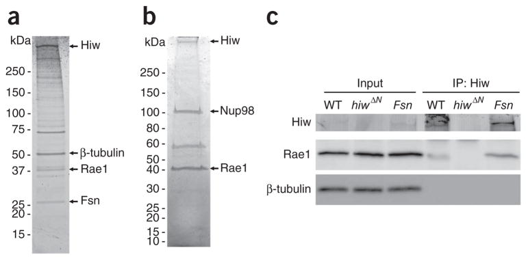
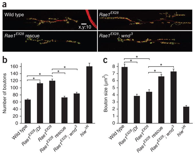
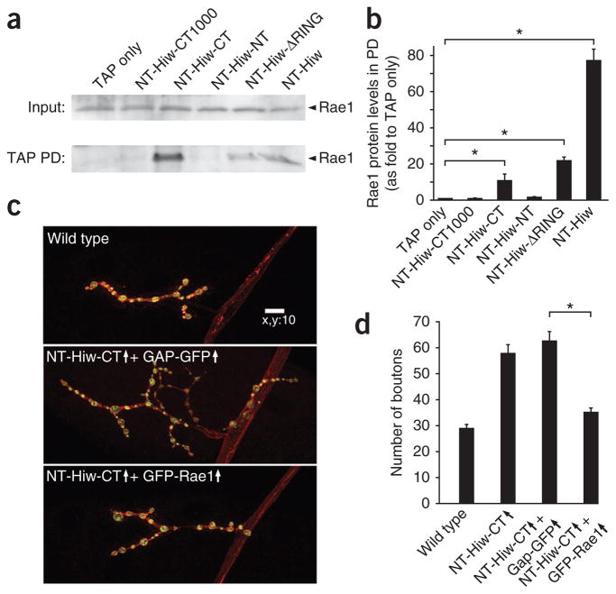
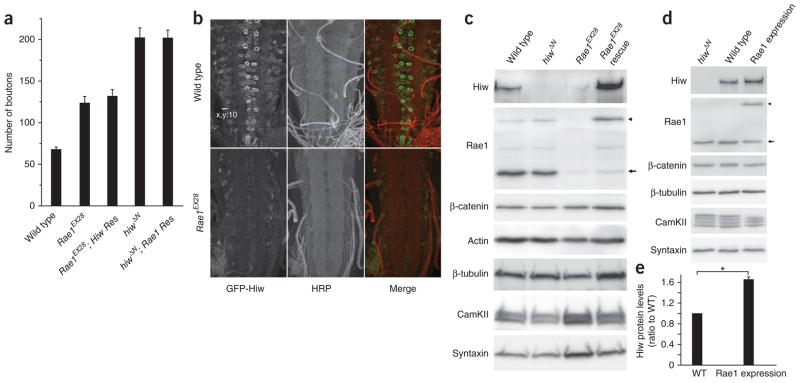
The evolutionarily conserved Highwire (Hiw)/Drosophila Fsn E3 ubiquitin ligase complex is required for normal synaptic morphology during development and axonal regeneration after injury. However, little is known about the molecular mechanisms that regulate the Hiw E3 ligase complex. Using tandem affinity purification techniques, we identified Drosophila Rae1 as a previously unknown component of the Hiw/Fsn complex. Loss of Rae1 function in neurons results in morphological defects at the neuromuscular junction that are similar to those seen in hiw mutants. We found that Rae1 physically and genetically interacts with Hiw and restrains synaptic terminal growth by regulating the MAP kinase kinase kinase Wallenda. Moreover, we found that the Rae1 is both necessary and sufficient to promote Hiw protein abundance, and it does so by binding to Hiw and protecting Hiw from autophagy-mediated degradation. These results describe a previously unknown mechanism that selectively controls Hiw protein abundance during synaptic development.


Conflict of interest statement

COMPETING FINANCIAL INTERESTS

The authors declare no competing financial interests.

Figures

Figure 1
Figure 1
Rae1 and Hiw interact with each other in neurons. (a) The Hiw-associated complex was purified by TAP (see Online Methods) and analyzed by one-dimensional SDS-PAGE gel followed by Sypro Ruby staining. Mass spectrometry identified the full-length Hiw, β-tubulin, Rae1 and Fsn, indicated by arrows. (b) The Rae1-associated complex purified from fly brains by TAP was analyzed by one-dimensional SDS-PAGE gel followed by Coomassie blue G-250 staining. Mass spectrometry identified Hiw, Nup98 and Rae1, indicated by arrows. (c) Larval brain lysate of wild-type (WT), hiw null mutant (hiwΔN) and Fsn mutant (Fsnf06595/Df(2R)7872) flies was subject to co-immunoprecipitation with antibody to Hiw (Hiw2b). Both the input and the immunoprecipitated complexes were analyzed by western blots with antibodies to Hiw or Rae1. The membrane was also blotted with antibody to β-tubulin to ensure equal amount of total proteins in the input samples. Full-length blots are presented in Supplementary Figure 8.
Figure 2
Figure 2
Generation of Rae1 mutant alleles. (a) The gene structure of Rae1. The black-arrowed boxes indicate exons. A transposable element, P{GawB}NP3499, located ~400 bp upstream of the first exon of Rae1 was used to generate imprecise excision mutants of Rae1. PCR analysis indicates that ~900 bp and a ~1.5-k bp DNA fragment in the Rae1 promoter region are missing in Rae1EX28 and Rae1EXB12, respectively. (b) Western blot analysis on Rae1 protein in the brain lysate of wild-type, Df(2R)5764/+, Rae1EX28/+, or late second instar Rae1EX28, Rae1EX28/Df(2R)5764(/Df) and Rae1EXB12 /Df(2R)5764 larvae. β-tubulin was used as the loading control. Full-length blots are presented in Supplementary Figure 9. (c) Quantification of Rae1 protein levels in the Rae1 heterozygous and homozygous mutant brains. Rae1 protein levels were first normalized to β-tubulin protein levels and are presented as percentage of wild-type level. *P < 0.05, **P < 0.001. Error bars denote s.e.m.
Figure 3
Figure 3
Rae1 is required to restrain synaptic terminal growth at the NMJ. (a) Representative confocal images of segment A3 muscle 6/7 synapses stained for both DVGLUT (green) and FasII (red), in late 2nd/early 3rd instar wild-type, Rae1EX28, Rae1EX28 presynaptic rescue (Rae1EX28; elav-Gal4/UASNTAP-Rae1) and Rae1EX28 wallenda suppression (Rae1EX28; wnd3) larvae. Scale bar represents 10 μm. (b,c) Quantification of bouton number (b) and size (c) in wild-type, Rae1EX28/Df, Rae1EX28, Rae1EX28 rescue, Rae1EX28; wnd3 suppression and hiwΔN larval NMJs (segment A3 muscle 6/7; n = 20, 21, 18, 20, 20 and 11, respectively, for bouton number; n = 271, 465, 548, 726, 486 and 826, respectively, for bouton size). There was no significant difference in either bouton number or bouton size between Rae1EX28 and Rae1EX28/Df (P > 0.1 for both comparisons). *P < 0.001. Error bars denote s.e.m.
Figure 4
Figure 4
Rae1 genetically interacts with hiw to restrain synaptic terminal growth. (a) Representative confocal images of muscle 4 synapses stained for both DVGLUT (green) and FasII (red) in wild-type, Rae1EX28/+, hiwND51, hiwND51; Rae1EX28/+, hiwΔN and hiwΔN; Rae1EX28/+ larvae. Scale bar represents 10 μm. (b) Quantification of bouton number at muscle 4 NMJs in wild-type, Rae1EX28/+, hiwND51, hiwND51; Rae1EX28/+, hiwΔN and hiwΔN; Rae1EX28/+ larvae (n = 27, 23, 42, 46, 23 and 25 cells, respectively). There was a significant increase in the number of boutons formed in hiwND51; Rae1EX28/+ compared with hiwND51 3rd instar larval NMJs (*P < 0.001). The number of boutons in the hiwΔN; Rae1EX28/+ double mutant was not significantly different from that in hiwΔN (**P > 0.5), demonstrating a lack of enhancement of the hiwΔN phenotype by Rae1EX28/+. Error bars denote s.e.m.
Figure 5
Figure 5
A structure and function analysis of Hiw functional domains. (a) Schematic presentation of NTAP-tagged (NT-) and HM-tagged (HM-) hiw transgenes encoding full-length, mutated or truncated Hiw proteins. Hiw functional domains are marked with colored boxes. The positions of amino-acid substitution in NT-HiwΔRING and the added amino acid residue necessary to maintain the reading frame in NT-Hiw-RCC1 and NT-Hiw-PHR are indicated. The right column summarizes the ability of the given hiw transgene to rescue the synaptic overgrowth phenotype in hiw mutants, to cause dominant negative overgrowth phenotype when expressed in wild-type background and to bind to Rae1. (b) Western blot analysis of fly brain lysate using antibody to TAP (PAP, Sigma) or Myc revealed the expression of wild-type and mutant hiw transgenes in predicted size. (c) Representative confocal images of muscle 4 synapses stained for DVGLUT (green) and FasII (red) in wild-type (+/hiwND8; +/+; GS-elav-Gal4/+), NT-Hiw overexpression (+/hiwND8; UAS-NT-hiw/+; GS-elav-Gal4/+), NT-Hiw-NT overexpression (+/hiwND8; UAS-NT-hiw-NT/+; GS-elav-Gal4/+), NT-Hiw-CT1000 overexpression (+/hiwND8; +/+; GS-elav-Gal4/UAS-NT-hiw-CT1000), NT-Hiw-PHR overexpression (+/hiwND8; +/+; GS-elav-Gal4/UAS-NT-hiw-PHR), HM-Hiw-HindIII overexpression (+/hiwND8; UAS-HW-hiw-HindIII/+; GS-elav-Gal4/+), NT-Hiw-RCC1 overexpression (NT-hiw-RCC1/hiwND8; +/+; GS-elav-Gal4/+), or NT-Hiw-CT overexpression (+/hiwND8; +/+; GS-elav-Gal4/UAS-NT-hiw-CT) 3rd larval NMJs. Overexpression is indicated by an up arrow. Scale bar represents 10 μm. (d) Quantification of the number of boutons at muscle 4 NMJs in wild-type larvae or larvae overexpressing wild-type or mutant hiw transgenes (n = 23, 24, 26, 27, 26, 21, 25 and 27, respectively; *P < 0.001). Error bars denote s.e.m.
Figure 6
Figure 6
Rae1 interacts with a fragment in the Hiw C-terminal region, and coexpression of Rae1 with NT-Hiw-CT suppresses the NT-Hiw-CT–induced dominant-negative overgrowth phenotype. (a) Indicated NTAP-tagged hiw transgenes (described in Fig. 5) and a TAP only transgene were expressed in neurons under the control of the BG380-Gal4 driver. Larval brain lysates from each sample were subject to IgG pulldown. Both the inputs and the IgG pulldown complexes were analyzed by western blot using antibody to Rae1. Rae1 was present in all of the pulldown complexes except for those from TAP only, NT-Hiw-CT1000 and NT-Hiw-NT. Full-length blots are presented in Supplementary Figure 10. (b) Quantification of the interaction between various Hiw transgenic proteins and Rae1. Rae1 intensities in TAP pulldown blots were normalized to both the intensities and molecular weights of the corresponding TAP pulldown proteins. (c) Representative confocal images of muscle 4 synapses stained for both DVGLUT (green) and FasII (red) in wild-type (BG380/Y;+/+; +/+), NT-Hiw-CT and GAP-GFP (BG380/Y; UAS-GAP-GFP/+; UAS-NT-hiw-CT/+), and NT-Hiw-CT and Rae1 (BG380/Y; +/+; UAS-NT-hiw-CT/UAS-GFP-Rae1) larvae. Scale bar represents 10 μm. (d) Quantification of number of boutons at muscle 4 NMJs in wild-type, NT-Hiw-CT overexpression (BG380/Y; +/+; UAS-NT-hiw-CT/+), NT-Hiw-CT and GAP-GFP, and NT-Hiw-CT and Rae1 (n = 24, 27, 32 and 26, respectively) larvae. Error bars denote s.e.m.; *P < 0.001.
Figure 7
Figure 7
Rae1 promotes Hiw abundance. (a) Neuronal expression of neither Hiw nor Rae1 rescues the synaptic terminal overgrowth phenotype caused by the loss of function of the other gene. Quantification of the number of boutons in wild-type, Rae1EX28, Rae1EX28; Hiw rescue (Res) (Rae1EX28; elav-Gal4/UAS-GFP-hiw), hiwΔN and hiwΔN; Rae1 rescue (hiwΔN; elav-Gal4/UAS-NTAP-Rae1) larval NMJs (segment A3 muscle 6/7; n = 12, 12, 12, 7 and 11, respectively). (b) Representative confocal images of wild-type (BG380-Gal4/+; +/+; UAS-GFP-hiw/+) or Rae1 mutant (BG380-Gal4/+; Rae1EX28; UAS-GFP-hiw/+) ventral ganglions with UAS-GFP-hiw trangene expressed in neurons. The larvae were stained with antibodies to horseradish peroxidase (HRP, red) and GFP (green). Scale bar represents 10 μm. (c) Western blots of total proteins extracted from wild-type, hiwΔN, Rae1EX28 and Rae1EX28 rescue (Rae1EX28; elav-Gal4/UAS-NTAP-Rae1) larval brains probed with indicated antibodies. Full-length blots are presented in Supplementary Figure 11. (d) Western blot analysis on total proteins extracted from hiwΔN, wild-type (C155-Gal4/+) and Rae1 expression (C155-Gal4/+; UAS-NTAP-Rae1/+) larval brains. Full-length blots are presented in Supplementary Figure 12. (e) Quantification of Hiw protein levels in the wild-type and the Rae1 expression larval brains (*P < 0.001, based on seven independent experiments). Arrows indicate a 38-kDa endogenous Rae1 protein and the arrowheads indicate the 58-kDa transgenic NTAP-Rae1 protein. Error bars denote s.e.m.
Figure 8
Figure 8
Rae1-Hiw interaction prevents autophagy-mediated degradation of Hiw protein. (a) Representative confocal images of segment A3 muscle 6/7 synapses stained for both DVGLUT (green) and FasII (red), in wild-type, Rae1EX28, Rae1EX28; atg1/+, Rae1EX28; atg2/+ and Rae1EX28; atg18/+ wandering larvae. Scale bar represents 10 μm. (b) Quantification of the number of boutons in wild-type, atg1/+, atg2/+, atg18/+, Rae1EX28, Rae1EX28; atg1/+, Rae1EX28; atg2/+ and Rae1EX28; atg18/+ larval NMJs (segment A3 muscle 6/7; n = 12, 12, 12, 12, 12, 16, 12 and 21, respectively). (c) Western blot analysis on total proteins extracted from wild-type, Rae1EX28, Rae1EX28; atg1/+, Rae1EX28; atg2/+ and Rae1EX28; atg18/+ larval brains. The neural form of β-catenin (82 kDa, see Online Methods) was used as an internal control of neuronal proteins. Full-length blots are presented in Supplementary Figure 13. (d) Quantification of Hiw protein levels in wild-type, Rae1EX28, Rae1EX28; atg1/+, Rae1EX28; atg2/+ and Rae1EX28; atg18/+ larval brains (based on six independent experiments). (e) Representative confocal images of muscle 4 synapses stained for both DVGLUT (green) and FasII (red) in wandering larvae of wild-type (C155-Gal4/+), Atg1 Gap-GFP (C155-Gal4/+; UAS-Gap-GFP/+; UAS-Atg16B/+), and Atg1 NTAP-Rae1 (C155-Gal4/+; UAS-NTAP-Rae1/+; UAS-Atg1/+). Scale bar represents 10 μm. (f) Quantification of the number of boutons at muscle 4 synapses in wild-type, Rae1 expression (C155-Gal4/+; UAS-NTAP-Rae1/+), Atg1 Gap-GFP and Atg1 NTAP-Rae1 wandering larvae (segment A2–4, n = 28, 27, 33 and 34, respectively). Error bars denote s.e.m. *P < 0.001, **P < 0.05.

Comment in

  • Maintaining a Highwire act.
    Spencer T. Spencer T. Nat Neurosci. 2011 Sep 27;14(10):1226. doi: 10.1038/nn1011-1226. Nat Neurosci. 2011. PMID: 21952265 No abstract available.

References

    1. Po MD, Hwang C, Zhen M. PHRs: bridging axon guidance, outgrowth and synapse development. Curr Opin Neurobiol. 2010;20:100–107. - PubMed
    1. DiAntonio A, et al. Ubiquitination-dependent mechanisms regulate synaptic growth and function. Nature. 2001;412:449–452. - PubMed
    1. Wan HI, et al. Highwire regulates synaptic growth in Drosophila. Neuron. 2000;26:313–329. - PubMed
    1. Uthaman SB, Godenschwege TA, Murphey RK. A mechanism distinct from highwire for the Drosophila ubiquitin conjugase bendless in synaptic growth and maturation. J Neurosci. 2008;28:8615–8623. - PMC - PubMed
    1. Schaefer AM, Hadwiger GD, Nonet ML. rpm-1, a conserved neuronal gene that regulates targeting and synaptogenesis in C. elegans. Neuron. 2000;26:345–356. - PubMed

Publication types

MeSH terms