. 2011 Aug 28;477(7364):354-8.
doi: 10.1038/nature10379.

In vitro centromere and kinetochore assembly on defined chromatin templates

Affiliations

In vitro centromere and kinetochore assembly on defined chromatin templates

Annika Guse et al. Nature. .

Abstract

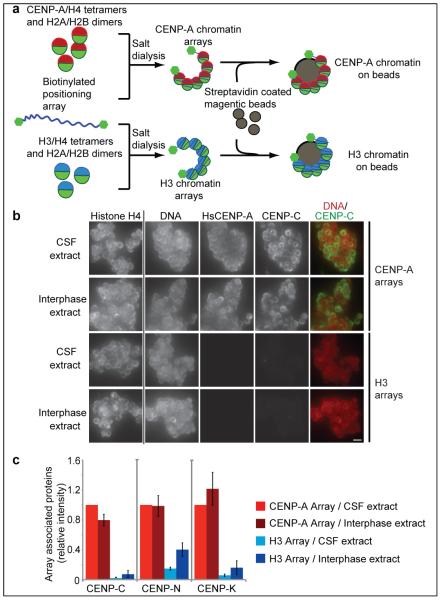
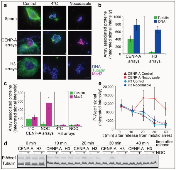
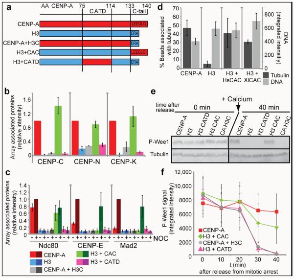
During cell division, chromosomes are segregated to nascent daughter cells by attaching to the microtubules of the mitotic spindle through the kinetochore. Kinetochores are assembled on a specialized chromatin domain called the centromere, which is characterized by the replacement of nucleosomal histone H3 with the histone H3 variant centromere protein A (CENP-A). CENP-A is essential for centromere and kinetochore formation in all eukaryotes but it is unknown how CENP-A chromatin directs centromere and kinetochore assembly. Here we generate synthetic CENP-A chromatin that recapitulates essential steps of centromere and kinetochore assembly in vitro. We show that reconstituted CENP-A chromatin when added to cell-free extracts is sufficient for the assembly of centromere and kinetochore proteins, microtubule binding and stabilization, and mitotic checkpoint function. Using chromatin assembled from histone H3/CENP-A chimaeras, we demonstrate that the conserved carboxy terminus of CENP-A is necessary and sufficient for centromere and kinetochore protein recruitment and function but that the CENP-A targeting domain--required for new CENP-A histone assembly--is not. These data show that two of the primary requirements for accurate chromosome segregation, the assembly of the kinetochore and the propagation of CENP-A chromatin, are specified by different elements in the CENP-A histone. Our unique cell-free system enables complete control and manipulation of the chromatin substrate and thus presents a powerful tool to study centromere and kinetochore assembly.


Figures

Figure 1
Figure 1. Reconstituted CENP-A chromatin supports centromere assembly in Xenopus egg extracts
(a) A schematic showing the reconstitution of CENP-A and H3 chromatin arrays and the attachment of the chromatin to magnetic beads via biotin end-labeled DNA. (b) Representative images comparing CENP-C binding to CENP-A and H3 chromatin arrays in CSF and interphase Xenopus extract. The left column shows the separate histone H4 staining used for normalization of the quantification, followed by staining for DNA, HsCENP-A and CENP-C. A merge image of the DNA (red) and CENP-C (green) channels is shown in the right column. Scale bar, 5 μm (c) Quantification of the array associated centromeric proteins CENP-C, CENP-N and CENP-K in CSF and interphase extracts, normalized to histone H4 levels. The levels are rescaled so that CENP-A arrays in CSF are set at 1. Error bars represent the standard error of the mean (SEM), n = 3 (p < 0.05 between CENP-A and H3 chromatin arrays for CENP-C, CENP-N and CENP-K).
Figure 2
Figure 2. CENP-A chromatin specifically recruits kinetochore proteins as a response to a mimic of kinetochore detachment from microtubules
(a) A schematic showing the experimental procedure. (b) Quantification of immunofluorescence analysis of CENP-C, Ndc80, CENP-E, Mad2, Rod or ZW10 recruitment to chromatin arrays with (+) and without (−) nocodazole (NOC). The levels are rescaled so that CENP-A arrays with (+) nocodazole are set at 1. Error bars represent SEM, n = 3 (p < 0.05 between − and + nocodazole for CENP-E, Mad2, Rod and ZW10 binding to CENP-A chromatin arrays). (c) Western blot analysis of CENP-C, Ndc80, Rod and ZW10 recruitment to CENP-A and H3 chromatin arrays with and without (+/−) nocodazole (NOC) in CSF and cycled egg extracts. H4 levels are shown as a loading control.
Figure 3
Figure 3. Kinetochores assembled on reconstituted CENP-A chromatin bind microtubules and generate a mitotic checkpoint signal
(a) Representative images of microtubule polymerization induced by sperm or reconstituted CENP-A and H3 chromatin. Microtubules (green) and Mad2 (magenta) levels are shown. Scale bar, 10μm (b) Quantification of tubulin and DNA associated with CENP-A and H3 chromatin beads. Error bars represent SEM, n = 5 (c) Quantification of tubulin and Mad2 levels associated with CENP-A and H3 chromatin beads after cold shock (4°C) and nocodazole (NOC) treatment. Error bars represent SEM, n = 5 (d) Western blot showing phospho-Wee1 (P-Wee1) levels as an indicator of the cell cycle stage and tubulin levels as a loading control. Samples from different time points after release from mitotic arrest (t 0′, t 10′, t 20′, t 30′, t 40′) are shown for CENP-A and H3 chromatin arrays, each incubated with nocodazole (+) or with DMSO (−) as a control. (e) Quantification of four independent experiments showing the phospho-Wee1 signal intensity (P-Wee1 signal) over time (min). Error bars represent SEM, n = 4.
Figure 4
Figure 4. The CENP-A C-terminus is required for centromere and kinetochore assembly in Xenopusegg extract
(a) A schematic showing the different CENP-A/H3 chimeras used in this study. The numbers on top represent the amino acid (AA) within HsCENP-A. (b) Quantification of immunofluorescence analysis of CENP-C, CENP-K and CENP-N recruitment to wild type and chimeric arrays. The relative amounts of each centromere protein bound to the arrays are shown relative to CENP-A arrays set to 1. Error bars represent SEM, n = 3 (p ≤ 0.05 for all proteins binding to CENP-A arrays compared to chimeric arrays except for the H3 arrays containing the CENP-A C-terminus). (c) Quantification of immunofluorescence analysis of Ndc80, CENP-E, Mad2 recruitment to chimeric chromatin arrays with (+) and without (−) nocodazole (NOC). Values are displayed relative to CENP-A arrays in the presence of nocodazole set to 1. Error bars represent SEM, n = 4. The efficiencies of recruitment of kinetochore proteins to CENP-A and H3+CAC arrays in nocodazole were not statistically distinguishable (p ≥ 0.26 for Ndc80, CENP-E and Mad2). (d) Quantification of microtubule binding to CENP-A, H3, H3+HsCAC and H3+XlCAC chromatin arrays represented as percentage of beads associated with tubulin levels above threshold (dark grey bars, left y-axis). Average DNA levels on chromatin beads are shown representing the levels of chromatin arrays bound to beads (light grey bars, right y-axis). Error bars represent SEM, n = 4 for CENP-A and H3 arrays, n = 5 for H3+HsCAC arrays and n = 2 for H3+XlCAC arrays. (e) Western blot analysis shows phospho-Wee1 (P-Wee1) levels as an indicator of the cell cycle stage at t 0′ (0 min) and t 40′ (40 min) after mitotic exit. Tubulin levels are shown as a loading control. (f) Quantification of the phospho-Wee1 signal intensity over time (P-Wee1). Error bars represent SEM, n = 5.

Comment in

References

    1. Cheeseman IM, Desai A. Molecular architecture of the kinetochore-microtubule interface. Nat Rev Mol Cell Biol. 2008;9:33–46. - PubMed
    1. Black BE, et al. Structural determinants for generating centromeric chromatin. Nature. 2004;430:578–582. - PubMed
    1. Black BE, Bassett EA. The histone variant CENP-A and centromere specification. Curr Opin Cell Biol. 2008;20:91–100. - PubMed
    1. Sekulic N, Bassett EA, Rogers DJ, Black BE. The structure of (CENP-A-H4)(2) reveals physical features that mark centromeres. Nature. 2010 - PMC - PubMed
    1. Carroll CW, Milks KJ, Straight AF. Dual recognition of CENP-A nucleosomes is required for centromere assembly. J Cell Biol. 2010;189:1143–1155. - PMC - PubMed

Publication types

MeSH terms