. 2010 Dec;88(6):899-911.
doi: 10.1002/cjce.20411.

BIOMIMETIC GRADIENT HYDROGELS FOR TISSUE ENGINEERING

Affiliations

BIOMIMETIC GRADIENT HYDROGELS FOR TISSUE ENGINEERING

Shilpa Sant et al. Can J Chem Eng. 2010 Dec.

Abstract

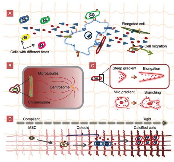
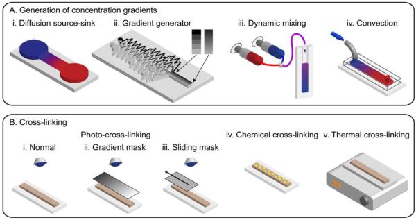
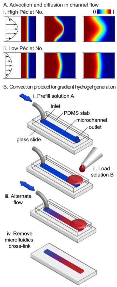
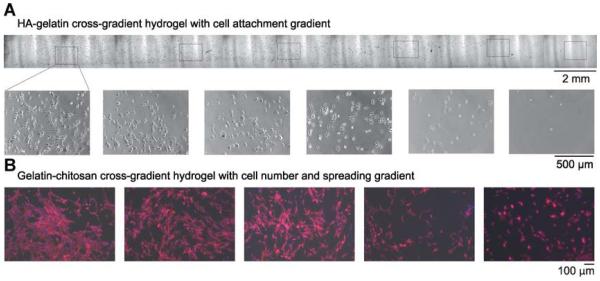
During tissue morphogenesis and homeostasis, cells experience various signals in their environments, including gradients of physical and chemical cues. Spatial and temporal gradients regulate various cell behaviours such as proliferation, migration, and differentiation during development, inflammation, wound healing, and cancer. One of the goals of functional tissue engineering is to create microenvironments that mimic the cellular and tissue complexity found in vivo by incorporating physical, chemical, temporal, and spatial gradients within engineered three-dimensional (3D) scaffolds. Hydrogels are ideal materials for 3D tissue scaffolds that mimic the extracellular matrix (ECM). Various techniques from material science, microscale engineering, and microfluidics are used to synthesise biomimetic hydrogels with encapsulated cells and tailored microenvironments. In particular, a host of methods exist to incorporate micrometer to centimetre scale chemical and physical gradients within hydrogels to mimic the cellular cues found in vivo. In this review, we draw on specific biological examples to motivate hydrogel gradients as tools for studying cell-material interactions. We provide a brief overview of techniques to generate gradient hydrogels and showcase their use to study particular cell behaviours in two-dimensional (2D) and 3D environments. We conclude by summarizing the current and future trends in gradient hydrogels and cell-material interactions in context with the long-term goals of tissue engineering.


Figures

Figure 1
Figure 1
Schematics of cell–cell contacts, cell–ECM interactions, and physicochemical gradients in vivo. A: Cell–cell contact and cell–ECM interactions generate chemical gradients that affect cell behaviours such as cell migration, cell elongation, and cell differentiation. B: Chromosomes generatea Ran-GTP gradient that organises the mitotic spindle during cell division. C: A steep gradient seen by one end of the cell results in elongation whereas a mild gradient seen by the whole cell results in branching. D: Mesenchymal stem cells (MSCs) sequentially differentiate into osteoids and calcified bone cells in response to the graded mechanical signals in the ECM.
Figure 2
Figure 2
Methods for generating gradient hydrogels. A: Gradient generation by (i) a source-sink diffusion device, (ii) a tree-like microfluidic gradient generator, (iii) mixing the input streams from syringe pumps, and (iv) microfluidic convection. B: Cross-linking by photopolymerisation using (i) a normal setup, (ii) a gradient mask, (iii) a sliding mask, and cross-linking by (iv) chemicals, and (v) heat.
Figure 3
Figure 3
Convection-driven gradient generation. A: Convection and diffusion in microchannel flows at (i) high and (ii) low Péclet numbers. B: Protocol for generating gradient hydrogels with convection. i: The channel is prefilled with solution A. ii: Solution B is loaded into the opposing port. iii: The flow is pumped back and forth in the channel until the gradient is the desired length. iv: The microfluidic system is removed and the gradient in prepolymer is cross-linked (Du et al., 2010).
Figure 4
Figure 4
A: Phase images (top: lower magnification; bottom: higher magnification) of smooth muscle cells (SMCs) cultured on a HA–gelatin cross-gradient hydrogel (Du et al., 2010). B: Effect of porous gelatin/chitosan cross-gradient on SMC behaviour. SMCs were cultured on the gradient hydrogel for 3 days. Fluorescence microscope images of SMCs show cytoskeletal organisation by F-actin staining (red) and nuclei by DAPI staining (blue) (He et al., 2010a).
Figure 5
Figure 5
Spatially regulated gene modification of fibroblasts within 3D collagen scaffolds. A: Schematic representation of fibroblast-seeded scaffolds containing spatial patterns of the Runx2 retrovirus (R2RV). The gradient was created by partially coating the proximal portion (left side) of the collagen scaffolds with poly-L-lysine (PLL) at a dipping speed of 170 μm/s. The scaffolds were then incubated in retroviral supernatant and seeded with cells. B: Confocal microscopy images of a graded distribution of FITC-labelled PLL (green). C: Confocal microscopy images of FITC-labelled PLL gradient colocalised with uniformly distributed cell nuclei (DAPI, blue). Scale bar: 2 mm. D: Immuno-histochemical staining for eGFP (pink) counterstained with haematoxylin (blue) revealed a gradient of Runx2-expressing cells. Scale bar: 2 mm (Phillips et al., 2008).
Figure 6
Figure 6
Differentiation of fibroblasts into myofibroblasts mediated by elasticity gradients in PEG hydrogels. Valvular interstitial cells (VICs) cultured on elasticity gradient hydrogels were immunostained for αSMA (green), an indicator of differentiation to myofibroblasts, as well as F-actin (red) and nuclei (blue). Images were taken at different representative positions along the gradient (elastic moduli noted on image). By day 3, a higher myofibroblastic differentiation was observed in cells on the high modulus side of the gradient (Kloxin et al., 2010).

References

    1. Abhyankar VV, Lokuta MA, Huttenlocher A, Beebe DJ. Characterization of a Membrane-Based Gradient Generator for Use in Cell-Signaling Studies. Lab Chip. 2006;6:389. - PubMed
    1. Ajdari A, Bontoux N, Stone HA. Hydrodynamic Dispersion in Shallow Microchannels: The Effect of Cross-Sectional Shape. Anal. Chem. 2006;78:387–392. - PubMed
    1. Amarie D, Glazier JA, Jacobson SC. Compact Microfluidic Structures for Generating Spatial and Temporal Gradients. Anal. Chem. 2007;79:9471–9477. - PMC - PubMed
    1. Annabi N, Nichol JW, Zhong X, Ji C, Koshy S, Khademhosseini A, Dehghani F. Controlling the Porosity and Microarchitecture of Hydrogels for Tissue Engineering. Tissue Eng. Pt. B Rev. 2010;16:371–383. - PMC - PubMed
    1. Ashe HL, Briscoe J. The Interpretation of Morphogen Gradients. Development. 2006;133:385–394. - PubMed

LinkOut - more resources