. 2012 Sep;32(9):2809-17.
doi: 10.1007/s00296-011-2067-8. Epub 2011 Aug 27.

Tenascin-C fragments are endogenous inducers of cartilage matrix degradation

Affiliations

Tenascin-C fragments are endogenous inducers of cartilage matrix degradation

Nidhi Sofat et al. Rheumatol Int. 2012 Sep.

Abstract

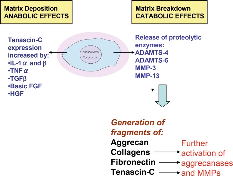
Cartilage destruction is a hallmark of osteoarthritis (OA) and is characterized by increased protease activity resulting in the degradation of critical extracellular matrix (ECM) proteins essential for maintaining cartilage integrity. Tenascin-C (TN-C) is an ECM glycoprotein, and its expression is upregulated in OA cartilage. We aimed to investigate the presence of TN-C fragments in arthritic cartilage and establish whether they promote cartilage degradation. Expression of TN-C and its fragments was evaluated in cartilage from subjects undergoing joint replacement surgery for OA and RA compared with normal subjects by western blotting. The localization of TN-C in arthritic cartilage was also established by immunohistochemistry. Recombinant TN-C fragments were then tested to evaluate which regions of TN-C are responsible for cartilage-degrading activity in an ex vivo cartilage explant assay measuring glycosaminoglycan (GAG) release, aggrecanase and matrix metalloproteinase (MMP) activity. We found that specific TN-C fragments are highly upregulated in arthritic cartilage. Recombinant TN-C fragments containing the same regions as those identified from OA cartilage mediate cartilage degradation by the induction of aggrecanase activity. TN-C fragments mapping to the EGF-L and FN type III domains 3-8 of TN-C had the highest levels of aggrecan-degrading ability that was not observed either with full-length TN-C or with other domains of TN-C. TN-C fragments represent a novel mechanism for cartilage degradation in arthritis and may present new therapeutic targets for the inhibition of cartilage degradation.


Figures

Fig. 1
Fig. 1
Western blot analysis of cartilage extracts from patients using an anti-TN-C antibody. A total of 1 μg protein was run per well and membranes developed with alkaline phosphatase (AP) substrate for 1 min each. Sample identification: 1 29 (F), lower leg, amputation; 2 17 (F), lower limb, osteosarcoma; 3 45 (M), lower limb, amputation; 4 72 (M), hip OA; 5 68 (M), hip OA; 6 72 (M), knee OA; 7 75 (F), knee OA; 8 71 (M), hip OA; 9 70 (F), knee OA; 10 66 (F), knee OA; 11 45 (M), hip, RA; 12 69 (F), hip, RA. b Equivalent samples used as in (a) with 1 μg protein per well stained with Coomassie Brilliant Blue
Fig. 2
Fig. 2
Domain composition of TN-C fragments identified from arthritic cartilage. Human TN-C has a modular structure with repeating units: the TA attachment (heptad) domain at the N-terminus, 14.5 EGF-L repeats, 8–17 FN type III repeats (conserved type III repeats numbered 1–8 are present in all isoforms with splice variants labelled by their designated letter format) and a fibrinogen (FBG) globe at the C-terminus. The table shows the composition of TN-C fragments isolated from arthritic cartilage in our study
Fig. 3
Fig. 3
Distribution of TN-C in human OA cartilage. ad are images from 2 OA subjects stained with anti-TN-C antibody. eh are the same OA subjects stained with an isotype control IgG mouse antibody. Zones of cartilage are delineated using the following abbreviations—S superficial, M midzone, D deep zone. Bars indicate section size of 10 μM or 50 μM
Fig. 4
Fig. 4
TN-C fragments induce cartilage degradation in normal articular cartilage. a Recombinant TN-C fragments used for cartilage stimulation were run on SDS–PAGE. All proteins were loaded at 1 μg each and stained with silver. b TN-C fragments at 1 μM were used to stimulate porcine cartilage explants for 2 days. The culture medium was harvested and tested for the presence of GAG using the DMMB dye assay (n = 3). c, d Equal volumes of conditioned media (50 μl) from the experiment shown in b were deglycosylated and western blots performed for aggrecanase-cleaved neoepitopes using the BC-3 antibody (c) and the BC-14 antibody (d). Loading controls were medium alone, c, used as a negative control and IL-1α, which served as a positive control
Fig. 5
Fig. 5
a Porcine cartilage was stimulated with recombinant TN-C fragments with a concentration range of 0.01–1 μM. After 2 days, conditioned medium was harvested and GAG release measured using the DMMB assay (n = 3). b Normal human cartilage (obtained from a 69-year-old subject with a femoral neck fracture) was stimulated with recombinant TN-C fragments at 1 μM each. Conditioned medium was harvested after 2 days and GAG release measured using the DMMB assay (n = 3)
Fig. 6
Fig. 6
Scheme for role of TN-C and TN-C fragments in OA pathology

References

    1. Altman RD. Classification of disease: osteoarthritis. Semin Arthritis Rheum. 1991;20:40–47. doi: 10.1016/0049-0172(91)90026-V. - DOI - PubMed
    1. Nuki G. Osteoarthritis: a problem of joint failure. Z Rheumatol. 1999;58:142–147. doi: 10.1007/s003930050164. - DOI - PubMed
    1. Mankin HJ, Lippiello L. Biochemical and metabolic abnormalities in articular cartilage from osteoarthritic human hips. J Bone Joint Surg Am. 1971;52:424–434. - PubMed
    1. Goldring SR, Goldring MB. The role of cytokines in cartilage matrix degeneration in osteoarthritis. Clin Orthop Relat Res. 2004;427:S27–S36. doi: 10.1097/01.blo.0000144854.66565.8f. - DOI - PubMed
    1. Nagase H, Kashiwagi M. Aggrecanases and cartilage matrix degradation. Arthritis Res Ther. 2003;5:94–103. doi: 10.1186/ar630. - DOI - PMC - PubMed

Publication types

MeSH terms