

Models of neurodegenerative disease - Alzheimer's anatomical and amyloid plaque imaging
- PMID: 21874485
- PMCID: PMC3866378
- DOI: 10.1007/978-1-61779-219-9_16
Models of neurodegenerative disease - Alzheimer's anatomical and amyloid plaque imaging
Abstract
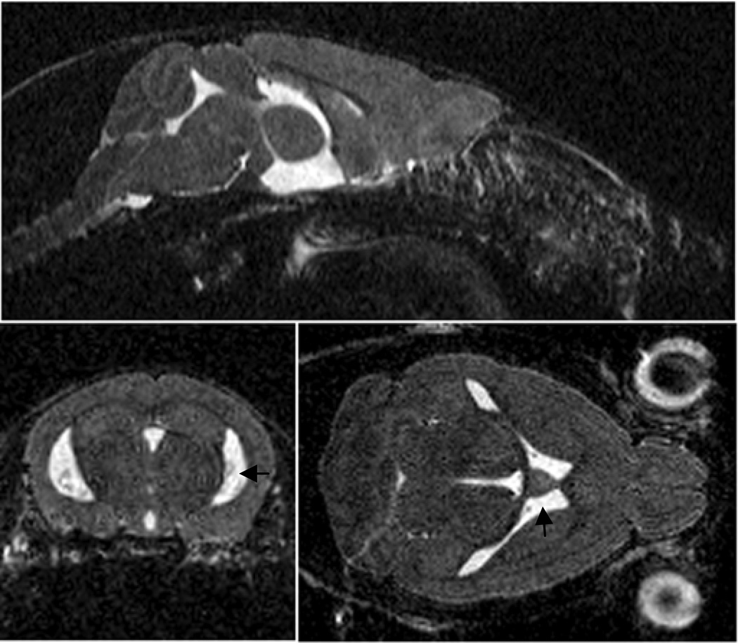
Alzheimer's disease (AD) is an important social and economic issue for our societies. The development of therapeutics against this severe dementia requires assessing the effects of new drugs in animal models thanks to dedicated biomarkers. According to the amyloid cascade hypothesis, β-amyloid deposits are at the origin of most of the lesions associated with AD. These extracellular deposits are therefore one of the main targets in therapeutical strategies. Aβ peptides can be revealed histologically with specific dyes or antibodies, or by magnetic resonance microscopy (μMRI) that uses their association with iron as a source of signal. The microscopic size of the lesions necessitates the development of specific imaging protocols. Most protocols use T (2)-weighted sequences that reveal the aggregates as hypointense spots. This chapter describes histological methods that reveal amyloid plaques with specific stains and MR-imaging protocols for in vivo and ex vivo MR imaging of AD mice.
Figures
Similar articles
-
Detection of amyloid plaques targeted by bifunctional USPIO in Alzheimer's disease transgenic mice using magnetic resonance microimaging.PLoS One. 2013;8(2):e57097. doi: 10.1371/journal.pone.0057097. Epub 2013 Feb 27. PLoS One. 2013. PMID: 23468919 Free PMC article.
-
Imaging beta amyloid aggregation and iron accumulation in Alzheimer's disease using quantitative susceptibility mapping MRI.Neuroimage. 2019 May 1;191:176-185. doi: 10.1016/j.neuroimage.2019.02.019. Epub 2019 Feb 7. Neuroimage. 2019. PMID: 30739060
-
Contrast Enhanced Magnetic Resonance Imaging of Amyloid-β Plaques in a Murine Alzheimer's Disease Model.J Alzheimers Dis. 2023;93(2):411-419. doi: 10.3233/JAD-220198. J Alzheimers Dis. 2023. PMID: 37038807 Free PMC article.
-
Amyloid imaging using high-field magnetic resonance.Magn Reson Med Sci. 2010;9(3):95-9. doi: 10.2463/mrms.9.95. Magn Reson Med Sci. 2010. PMID: 20885081 Review.
-
PET imaging of amyloid in Alzheimer's disease.Lancet Neurol. 2004 Sep;3(9):519-27. doi: 10.1016/S1474-4422(04)00853-1. Lancet Neurol. 2004. PMID: 15324720 Review.
Cited by
-
Whole-Brain Single-Cell Imaging and Analysis of Intact Neonatal Mouse Brains Using MRI, Tissue Clearing, and Light-Sheet Microscopy.J Vis Exp. 2022 Aug 1;(186):10.3791/64096. doi: 10.3791/64096. J Vis Exp. 2022. PMID: 35969091 Free PMC article.
-
Challenges of multimorbidity of the aging brain: a critical update.J Neural Transm (Vienna). 2015 Apr;122(4):505-21. doi: 10.1007/s00702-014-1288-x. Epub 2014 Aug 5. J Neural Transm (Vienna). 2015. PMID: 25091618 Review.
-
Chemically-defined camelid antibody bioconjugate for the magnetic resonance imaging of Alzheimer's disease.MAbs. 2017 Aug/Sep;9(6):1016-1027. doi: 10.1080/19420862.2017.1342914. Epub 2017 Jun 28. MAbs. 2017. PMID: 28657418 Free PMC article.
-
Effects and Mechanism of Particulate Matter on Tendon Healing Based on Integrated Analysis of DNA Methylation and RNA Sequencing Data in a Rat Model.Int J Mol Sci. 2022 Jul 25;23(15):8170. doi: 10.3390/ijms23158170. Int J Mol Sci. 2022. PMID: 35897746 Free PMC article.
-
The role of iron in neurodegenerative disorders: insights and opportunities with synchrotron light.Front Pharmacol. 2014 Aug 19;5:191. doi: 10.3389/fphar.2014.00191. eCollection 2014. Front Pharmacol. 2014. PMID: 25191270 Free PMC article. Review.
References
-
- Hyman BT, Marzloff K, Arriagada PV. The lack of accumulation of senile plaques or amyloid burden in Alzheimer's disease suggests a dynamic balance between amyloid deposition and resolution. J. Neuropath. Exp. Neur. 1993;52:594–600. - PubMed
-
- Jack CR, Jr, Lowe VJ, Weigand SD, Wiste HJ, Senjem ML, Knopman DS, Shiung MM, Gunter JL, Boeve BF, Kemp BJ, Weiner M, Petersen RC. Serial PIB and MRI in normal, mild cognitive impairment and Alzheimer's disease: implications for sequence of pathological events in Alzheimer's disease. Brain. 2009;132:1355–1365. - PMC - PubMed
-
- Hardy J, Selkoe DJ. The amyloid hypothesis of Alzheimer's disease: progress and problems on the road to therapeutics. Science. 2002;297:353–356. - PubMed
-
- Blennow K, de Leon MJ, Zetterberg H. Alzheimer's disease. Lancet. 2006;368:387–403. - PubMed
Publication types
MeSH terms
Grants and funding
LinkOut - more resources
Full Text Sources
Medical
