Review
. 2012 Jul;9(6):746-58.
doi: 10.2174/156720512801322564.

N-methyl D-aspartate (NMDA) receptor antagonists and memantine treatment for Alzheimer's disease, vascular dementia and Parkinson's disease

Affiliations
Review

N-methyl D-aspartate (NMDA) receptor antagonists and memantine treatment for Alzheimer's disease, vascular dementia and Parkinson's disease

David Olivares et al. Curr Alzheimer Res. 2012 Jul.

Abstract

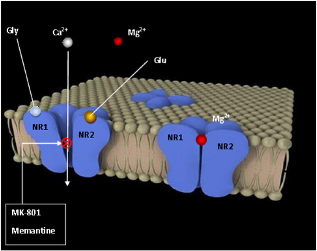
Memantine, a partial antagonist of N-methyl-D-aspartate receptor (NMDAR), approved for moderate to severe Alzheimer's disease (AD) treatment within the U.S. and Europe under brand name Namenda (Forest), Axura and Akatinol (Merz), and Ebixa and Abixa (Lundbeck), may have potential in alleviating additional neurological conditions, such as vascular dementia (VD) and Parkinson's disease (PD). In various animal models, memantine has been reported to be a neuroprotective agent that positively impacts both neurodegenerative and vascular processes. While excessive levels of glutamate result in neurotoxicity, in part through the over-activation of NMDARs, memantine-as a partial NMDAR antagonist, blocks the NMDA glutamate receptors to normalize the glutamatergic system and ameliorate cognitive and memory deficits. The key to memantine's therapeutic action lies in its uncompetitive binding to the NMDAR through which low affinity and rapid off-rate kinetics of memantine at the level of the NMDAR-channel preserves the physiological function of the receptor, underpinning memantine's tolerability and low adverse event profile. As the biochemical pathways evoked by NMDAR antagonism also play a role in PD and since no other drug is sufficiently effective to substitute for the first-line treatment of L-dopa despite its side effects, memantine may be useful in PD treatment with possibly fewer side effects. In spite of the relative modest nature of its adverse effects, memantine has been shown to provide only a moderate decrease in clinical deterioration in AD and VD, and hence efforts are being undertaken in the design of new and more potent memantine-based drugs to hopefully provide greater efficacy.


Conflict of interest statement

The authors declare that they do not have any conflicts of interest.

Figures

Fig. (1)
Fig. (1)
Schematic illustration of APP protein and its Aβ product after cleavage by α-, β- and γ-secretases. β- and γ-secretase cleaves on the N- and C-terminal ends of the Aβ region respectively. γ-Secretase cleavage yields a 39–43 amino acid product. Long and more fiblillogenic 42–43 amino acid Aβ species are implicated in AD pathogenesis and may seed the formation of Aβ40 fibrils. Mutations in the APP gene and in genes encoding proteins known as presenilins increase the production of long Aβ. Presenilins-1 and −2 is thought function as γ-secretases (for a review, see [142]).
Fig. (2)
Fig. (2)
Depiction of the tetrameric NMDAR at rest (right) and activated after depolarization and binding of agonists glycine and glutamate, suppressing the magnesium channel blockade (left), where antagonists MK-801 and memantine have their allosteric binding site.
Fig. (3)
Fig. (3)
Schematic structure of eight NR1 receptor isoforms (NR1A–H). Exons 5, 21 and 22 encode three splice cassettes named N1, C1 and C2. Carboxy-terminals variants are generated by splicing out of cassettes C1 and/or C2; and amino-terminal variant, by splicing out of N1. If C2 is excised, the first stop codon is suppressed, resulting in a new open reading frame that encodes the sequence named C2’ [143].
Fig. (4)
Fig. (4)
Classical model of normal activity of the basal ganglia and malfunctioning in PD (reviewed in Ref. [114]). Thin arrows indicate downregulation and thick arrows upregulation. Blue arrows show glutamatergic activatory efferents, red arrows indicate inhibitory GABAergic efferents and green arrows, activatory/inhibitory projections. (A) The ‘direct pathway’ is comprised of striatal neurons with D1 activatory dopaminergic receptors and their GABAergic efferent projections to the globus pallidus internus (GPi) which together the SNpr transmit inhibitory signals via GABAergic output to the thalamic ventral anterior (VA) nucleus. The ‘indirect pathway’ is comprised of striatal neurons with D2 inhibitory dopaminergic receptors. The striatum (composed of caudate and putamen) projects GABAergic output to the globus pallidus externus (GPe). From this point, they synapse to the nucleus subthalamicus (STN), from which glutamatergic activatory projections reach the GPi and the substantia nigra (SNpc/SNpr). The subthalamic nucleus also gets excitatory input directly from the cortex and induces the GPi to increase GABA release in the VA. While GPe keeps it in check, the SNpc dopamine binds D2 receptors to inhibit this pathway, blocking the inhibition of the subthalamic circuit by GPe. Additionally, the striatum and the SNpc receive glutamatergic efferents from the neocortex. (B) Loss of dopaminergic afferents (broken green arrows) entails a dis-repression of striatal D2 neurons leads to over-activity of their GABAergic projections to the GPe, and this in turn decreases its GABAergic efferent activity to the STN. As a consequence, STN glutamatergic projections to the GPi render over-active, increasing GABAergic output from the GPi to the VA. On the contrary, the D1 striatal neurons are underactive, therefore the GABAergic output to the GPi and to the SNpr are reduced and consequently the GABAergic output from both to the VA is increased. As a consequence in both cases, the loss of dopaminergic projections causes a failure to desinhibit the thalamocortical output, leading to bradykinesia, a typical symptom in PD. (C) (D) Possible targets at the basal ganglia level of NMDAR antagonists amantadine and memantine. Broken arrows mean suppression of output projections. A putative target is the STN (C), overactive in PD by blocking the subthalamic stimulation of GPi and a normalization of downstream thalamocortical connections. Other target could be the indirect pathway from the striatum (D), triggering a dis-repression of SNpr and leading to a normalization of STN activity and therefore a thalamic-cortical neuron well-functioning.

References

    1. Care TSCoTAiH. Dementia - Etiology and Epidemiology. A Systematic Review. 2008;1:755.
    1. Lahiri DK, Rogers JT, Greig NH, Sambamurti K. Rationale for the development of cholinesterase inhibitors as anti-Alzheimer agents. Curr Pharm Des. 2004;10(25):3111–3119. - PubMed
    1. Delacourte A. [From physiopathology to treatment of Alzheimer’s disease] Rev Neurol (Paris) 2006;162(10):909–912. - PubMed
    1. Joray S, Herrmann F, Mulligan R, Schnider A. Mechanism of disorientation in Alzheimer’s disease. Eur Neurol. 2004;52(4):193–197. - PubMed
    1. Esposito F, Rochat L, Van der Linden AC, Lekeu F, Quittre A, Charnallet A, et al. Apathy and executive dysfunction in Alzheimer disease. Alzheimer Dis Assoc Disord. 2010;24(2):131–137. - PubMed

Publication types

MeSH terms