

An immunologic portrait of cancer
- PMID: 21875439
- PMCID: PMC3175185
- DOI: 10.1186/1479-5876-9-146
An immunologic portrait of cancer
Abstract
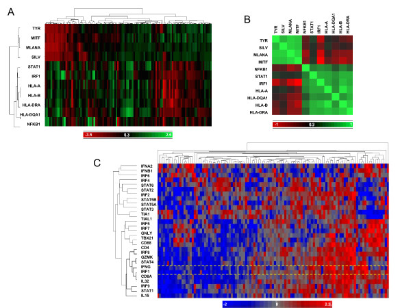
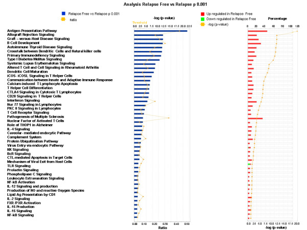
The advent of high-throughput technology challenges the traditional histopathological classification of cancer, and proposes new taxonomies derived from global transcriptional patterns. Although most of these molecular re-classifications did not endure the test of time, they provided bulk of new information that can reframe our understanding of human cancer biology. Here, we focus on an immunologic interpretation of cancer that segregates oncogenic processes independent from their tissue derivation into at least two categories of which one bears the footprints of immune activation. Several observations describe a cancer phenotype where the expression of interferon stimulated genes and immune effector mechanisms reflect patterns commonly observed during the inflammatory response against pathogens, which leads to elimination of infected cells. As these signatures are observed in growing cancers, they are not sufficient to entirely clear the organism of neoplastic cells but they sustain, as in chronic infections, a self-perpetuating inflammatory process. Yet, several studies determined an association between this inflammatory status and a favorable natural history of the disease or a better responsiveness to cancer immune therapy. Moreover, these signatures overlap with those observed during immune-mediated cancer rejection and, more broadly, immune-mediated tissue-specific destruction in other immune pathologies. Thus, a discussion concerning this cancer phenotype is warranted as it remains unknown why it occurs in immune competent hosts. It also remains uncertain whether a genetically determined response of the host to its own cancer, the genetic makeup of the neoplastic process or a combination of both drives the inflammatory process. Here we reflect on commonalities and discrepancies among studies and on the genetic or somatic conditions that may cause this schism in cancer behavior.
Figures
References
-
- Alizadeh AA, Eisen MB, Davis RE, Ma C, Lossos IS, Rosenwald A, Bedrick JC, Sabet H, Tran T, Xin Y. et al.Distinct types of diffuse large B-cell lymphoma identified by gene expression profiling. Nature. 2000;403:467–578. - PubMed
-
- Bittner M, Meltzer P, Chen Y, Jiang E, Seftor E, Hendrix M, Radmacher M, Simon R, Yakhini Z, Ben-Dor A. et al.Molecular classification of cutaneous malignant melanoma by gene expression: shifting from a countinuous spectrum to distinct biologic entities. Nature. 2000;406:536–840. doi: 10.1038/35020115. - DOI - PubMed
Publication types
MeSH terms
LinkOut - more resources
Full Text Sources
Other Literature Sources
Research Materials
