. 2011 Nov 24:196:49-65.
doi: 10.1016/j.neuroscience.2011.08.032. Epub 2011 Aug 19.

Neuronal localization of M2 muscarinic receptor immunoreactivity in the rat amygdala

Affiliations

Neuronal localization of M2 muscarinic receptor immunoreactivity in the rat amygdala

A J McDonald et al. Neuroscience. .

Abstract

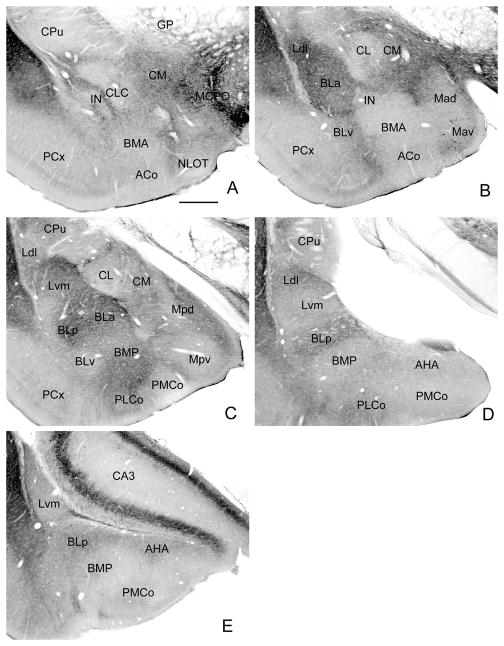
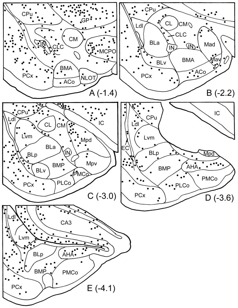
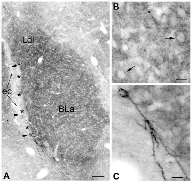
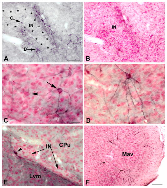
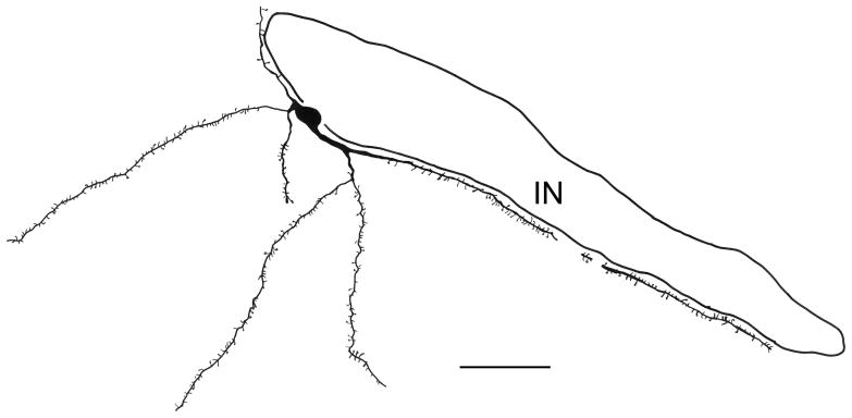
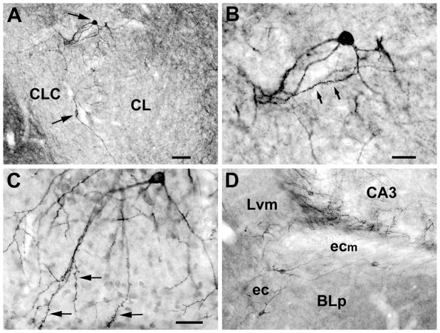
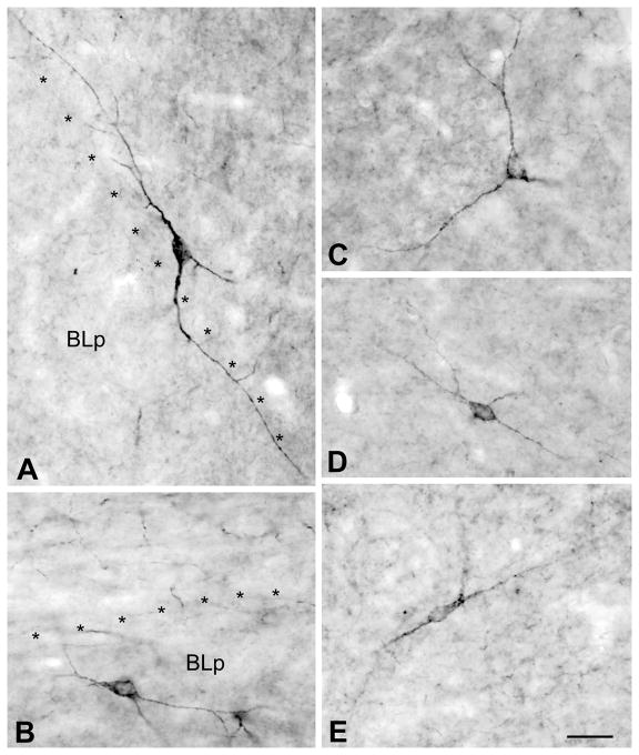
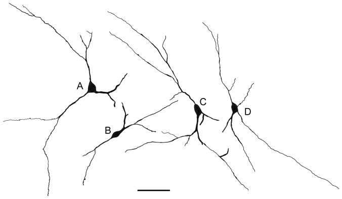
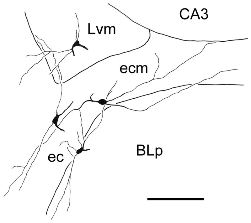
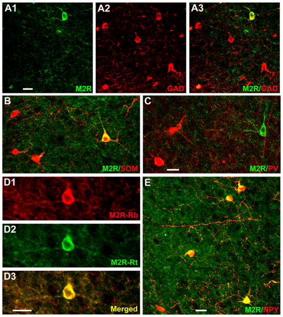
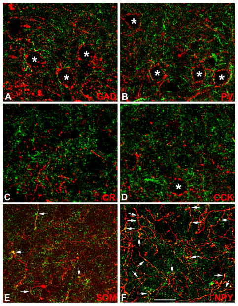
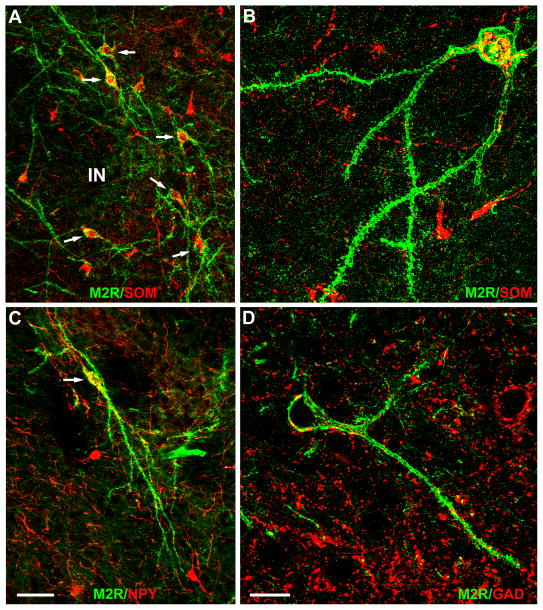
Muscarinic cholinergic neurotransmission in the amygdala is critical for memory consolidation in emotional/motivational learning tasks, but little is known about the neuronal distribution of different receptor subtypes. Immunohistochemistry was used in the present investigation to localize the m2 receptor (M2R). Differential patterns of M2R-immunoreactivity (M2R-ir) were observed in the somata and neuropil of the various amygdalar nuclei. Neuropilar M2R-ir was strongest in rostral portions of the basolateral nuclear complex (BLC). M2R-positive (M2R+) somata were seen in low numbers in all nuclei of the amygdala. Most M2R+ neurons associated with the BLC were in the lateral nucleus and external capsule. These cells were nonpyramidal neurons that contained glutamatic acid decarboxylase (GAD), somatostatin (SOM), and neuropeptide Y (NPY), but not parvalbumin (PV), calretinin (CR), or cholecystokinin (CCK). Little or no M2R-ir was observed in GAD+, PV+, CR+, or CCK+ axons in the BLC, but it was seen in some SOM+ axons and many NPY+ axons. M2R-ir was found in a small number of spiny and aspiny neurons of the central nucleus that were mainly located along the lateral and ventral borders of its lateral subdivision. Many of these cells contained SOM and NPY. M2R+ neurons were also seen in the medial nucleus, including a distinct subpopulation of neurons that surrounded its anteroventral subdivision. The latter neurons were negative for all neuronal markers analyzed. The intercalated nuclei (INs) were associated with two types of large M2R+ neurons, spiny and aspiny. The small principal neurons of the INs were M2R-negative. The somata and dendrites of the large spiny neurons, which were actually found in a zone located just outside of the rostral INs, expressed SOM and NPY, but not GAD. These findings indicate that acetylcholine can modulate a variety of discrete neuronal subpopulations in various amygdalar nuclei via M2Rs, especially neurons that express SOM and NPY.


Figures

Fig. 1
Fig. 1
Low power photomicrographs of M2R immunoreactivity at five anteroposterior levels of the amygdala in one representative brain (see Fig. 2 for the bregma levels of these coronal sections as well as nuclear borders drawn after the sections were Nissl counterstained). Scale bar = 0.5 mm. Abbreviations: ACo, anterior cortical nucleus; AHA, amygdalohippocampal area; BLa, anterior basolateral nucleus; BLp, posterior basolateral nucleus; BLv, ventral basolateral nucleus; BMA, anterior basomedial nucleus; BMP, posterior basomedial nucleus; CA3; cornu ammonis field 3; CL, lateral central nucleus, CLC, lateral capsular central nucleus; CM, medial central nucleus; CPu, caudate-putamen; GP, globus pallidus; IC, internal capsule; IN, intercalated nucleus; Ldl, laterodorsal lateral nucleus; Lvm, ventromedial lateral nucleus; Mad, anterodorsal medial nucleus; Mav, anteroventral medial nucleus; MCPO, magnocellular preoptic nucleus; Mpd, posterodorsal medial nucleus; Mpv, posteroventral medial nucleus; NLOT, nucleus of the lateral olfactory tract; PCx, piriform cortex; PLCo, posterolateral cortical nucleus; PMCo, posteromedial cortical nucleus.
Fig. 2
Fig. 2
Drawings showing the locations of M2R+ neurons (dots) at the five levels of the amygdala of the brain shown in Fig. 1. Bregma levels are taken from the atlas by Paxinos and Watson (1997). Neurons were plotted from three adjacent sections at each level, including the sections shown in Fig. 1. Nuclear borders were drawn from these sections after coverslips were removed and sections were counterstained with pyronin Y (see Experimental Procedures). See Fig. 1 for scale bar and abbreviations. EC, external capsule.
Fig. 3
Fig. 3
A) Higher power photomicrograph of the portion of the basolateral nuclear complex shown in Fig. 1B. Note dense neuropilar M2R-ir in the anterior subdivision of the basolateral nucleus (BLa) and the dorsolateral subdivision of the lateral nucleus (Ldl). The lateral paracapsular portion of the intercalated nucleus (asterisks), located between the BLa and the external capsule (EC), exhibits no M2R-ir; it was identified following subsequent counterstaining of the section. Large arrows indicate two medium-sized aspiny M2R+ neurons located in the external capsule. A large spiny “SPIN” neuron (see text) is located at the ventral border of the intercalated nucleus (arrowhead); its dendrites avoid entering the intercalated nucleus. B) High power photomicrograph of a field in the BLa showing neuropilar M2R-ir. Note that some perikarya of M2R-negative putative pyramidal cells appear to be surrounded by M2R+ puncta (arrows). C) High power photomicrograph of the SPIN neuron shown in A. Note spines on the dendrites in the lower portion of the field. Scale bars = 100 μm in A, 20 μm in B and C.
Fig. 4
Fig. 4
A) Higher power photomicrograph of the portion of the rostral intercalated nucleus shown in Fig. 1A. The same field is shown after pyronin Y counterstaining in B. Asterisks indicate the borders of the intercalated nucleus. Arrows indicate M2R+ neurons shown at higher power in C and D after counterstaining. The upper arrow indicates a large aspiny M2R+ neuron that is located in the intercalated nucleus; several more lightly-stained large aspiny M2R+ neurons are seen above and to left of this cell. The M2R+ zone surrounding the intercalated nucleus is mainly filled with dendrites and cell bodies of large spiny M2R+ neurons (“SPIN” neurons), one of which is indicated by the lower arrow. C) Higher power photomicrograph of the large aspiny M2R+ neuron shown in A after counterstaining (arrow). Note that it is larger than the small principal neurons of the intercalated nucleus which surround it. The perikaryon of a large M2R-negative neuron of the intercalated nucleus is also seen in this field (arrowhead). D) Higher power photomicrograph of the M2R+ SPIN neuron shown in A after counterstaining; its spiny dendrites avoid entering the intercalated nucleus above. E) An M2R+ SPIN neuron (arrowhead) associated with one of the medial paracapsular nuclei (IN) located between the ventromedial subdivision of the lateral nucleus (Lvm) and the caudate-putamen (CPu) at the bregma −3.3 level. Note that its dendrites extend along the border of the IN and into the Lvm, but avoid entering the IN. Also note the lack of neuropil staining in the IN. See Fig. 5 for a drawing of this neuron. F) Higher power photomicrograph of the M2R+ neurons surrounding the anteroventral subdivision of the medial nucleus at the level of Figs. 1B and 2B after counterstaining. Scale bars = 100 μm in A (B is the same magnification), 25 μm in C (D is the same magnification), 50 μm in E, and 100 μm in F.
Fig. 5
Fig. 5
Camera lucida drawing of the SPIN neuron shown in Fig. 4E. It was located adjacent to one of the medial paracapsular intercalated nuclei (IN) and was reconstructed from two adjacent sections. Scale bar = 50 μm.
Fig. 6
Fig. 6
A) Photomicrographs of two spiny M2R+ neurons (arrows) located along the lateral and dorsal edges of the lateral subdivision of the central nucleus (CL). The ventral neuron is bitufted and located at the border between CL and the lateral capsular subdivision of the central nucleus (CLC). The dorsal neuron is multipolar and located between the central nucleus and the caudate-putamen. B) Higher power view of the dorsal neuron in A, showing a spiny dendritic segment (arrows). C) M2R+ neuron in the anteroventral subdivision of the medial nucleus (Mav) in a counterstained section. Its cell body is located in the ventral part of the cellular layer, and its dendrites, and those of other M2R+ Mav neurons, extend ventrally into the molecular layer. Note complex dendritic appendages on the portions of the dendrites in the molecular layer (arrows). D) Several M2R+ neurons in the region of the external capsule (ec) and its medial extension (ecm) between the ventromedial lateral nucleus (Lvm) and BLp. See Fig. 9 for a drawing of these neurons. Scale bars = 65 μm in A, 25 μm in B, 30 μm in C, and 50 μm in D.
Fig. 7
Fig. 7
Photomicrographs of M2R+ nonpyramidal cells in the basolateral nuclear complex and external capsule. A) M2R+ neuron in the external capsule. Asterisks indicate the border separating BLp from the external capsule, which is located dorsolateral to the nucleus. B) M2R+ neuron in the posterior subdivision of the basolateral nucleus (BLp). Asterisks indicate the border separating BLp from the medial extension of the external capsule, which is located dorsal to the nucleus. C–E) M2R+ neurons in the lateral nucleus. Scale bar = 25 μm for all photomicrographs.
Fig. 8
Fig. 8
Camera lucida drawings of representative M2R+ nonpyramidal neurons cells in the lateral nucleus and external capsule. A) The M2R+ neuron in the lateral nucleus shown in Fig. 7C. B) The M2R+ neuron in the lateral nucleus shown in Fig. 7E. C) The M2R+ neuron in the external capsule shown in Fig. 7A. D) M2R+ neuron in the lateral nucleus. Scale bar = 50 μm.
Fig. 9
Fig. 9
Camera lucida drawing of M2R+ nonpyramidal neurons cells in the lateral nucleus (Lvm) and external capsule (ec), including its medial extension between the lateral nucleus and the BLp (ecm), that was reconstructed from 3 adjacent sections. One of the sections is depicted in Fig. 6D. Scale bar = 100 μm.
Fig. 10
Fig. 10
Immunofluorescence photomicrographs of M2R+ nonpyramidal cells in the lateral amygdalar nucleus. A1) An M2R+ neuron (green) is seen in the upper portion of this field, and the edge of another M2R+ neuron is seen in the lower left corner. A2) GAD+ neurons in the same field as A1 (red). A3) Merged image of A1 and A2. Coexpression of M2R and GAD is indicated in yellow. Note yellow labeling of both M2R+ neurons seen in A1. B) M2R+ structures (green) and SOM+ neurons (red) in a merged image. The single M2R+ neuron in this field co-expresses SOM (yellow). C) M2R+ neuron (green) and PV+ neurons (red) in a merged image. The single M2R+ neuron in this field does not express PV. D) The rabbit M2R antibody (D1) and the rat M2R antibody (D2) produce virtually identical patterns of labeling in both perikarya and the neuropil of the lateral nucleus (D3) and all other forebrain regions investigated. E) M2R+ structures (green) and NPY+ neurons (red) in a merged image. All four M2R+ neurons in this field co-express NPY (yellow). Scale bars: 25μm in A and E, 20μm in C and D (B is at the same magnification as C).
Fig. 11
Fig. 11
Immunofluorescence photomicrographs of M2R neuropilar imunoreactivity (green) and axons expressing markers for interneurons (red) in the basolateral amygdala. Yellow indicates colocalization. Asterisks indicate unlabeled somata of putative pyramidal cells. A) M2R/GAD in the BLa. B) M2R/PV in the BLa. C) M2R/CR in the BLa. D) M2R/CCK in the lateral nucleus. E) M2R/SOM in the BLa. Arrows indicate several M2R+/SOM+ axon terminals. F) M2R/NPY in the BLa. Arrows indicate some of the many M2R+/NPY+ axon terminals in this field. Note little or no M2R-ir in GAD+, PV+, CR+, or CCK+ axons. Scale bar = 25 μm.
Fig. 12
Fig. 12
Immunofluorescence photomicrographs of M2R+ SPIN neurons. A). Six SPIN neurons (arrows) exhibit robust M2R-ir in their somata and dendrites (green). All 6 express SOM-ir that is mainly confined to their somata (red). Areas of co-expression within the cytoplasm of these neurons are indicated by yellow. Note that these 6 M2R+/SOM+ neurons surround a portion of the intercalated nucleus (IN) located just rostral to the BLa. B). Higher power photomicrograph of a M2R+ SPIN neuron co-expressing SOM. C) An M2R+ SPIN neuron (green) co-expresses NPY in its soma. D) M2R-ir (green) and GAD-ir (red) in a field that contains an M2R+ SPIN neuron. The SPIN neuron does not contain GAD, but GAD+ puncta (putative axon terminals) contact the cell body and dendrites of the SPIN neuron. In some cases these red GAD+ puncta appear yellow due to their partial overlap with the green M2R-ir in the contacted SPIN neuron. Scale bars = 50 μm in C (A is at the same magnification), 20μm in D (B is at the same magnification).

Similar articles

Cited by

References

    1. Alheid GF, deOlmos JS, Beltramino CA. The Rat Nervous System. San Diego: Academic Press; 1995. Amygdala and Extended Amygdala; pp. 495–578.
    1. Amaral DG, Bassett JL. Cholinergic innervation of the monkey amygdala: an immunohistochemical analysis with antisera to choline acetyltransferase. J Comp Neurol. 1989;281:337–361. - PubMed
    1. Apergis-Schoute J, Pinto A, Paré D. Muscarinic control of long-range GABAergic inhibition within the rhinal cortices. J Neurosci. 2007;27:4061–4071. - PMC - PubMed
    1. Ben-Ari Y, Zigmond RE, Shute CC, Lewis PR. Regional distribution of choline acetyltransferase and acetylcholinesterase within the amygdaloid complex and stria terminalis system. Brain Res. 1977;120:435–444. - PubMed
    1. Busti D, Geracitano R, Whittle N, Dalezios Y, Ma ko M, Kaufmann W, Sätzler K, Singewald N, Capogna M, Ferraguti F. Different fear states engage distinct networks within the intercalated cell clusters of the amygdala. J Neurosci. 2011;31:5131–5144. - PMC - PubMed

Publication types