. 2011 Sep 5;194(5):779-87.
doi: 10.1083/jcb.201104019. Epub 2011 Aug 29.

GCC185 plays independent roles in Golgi structure maintenance and AP-1-mediated vesicle tethering

Affiliations

GCC185 plays independent roles in Golgi structure maintenance and AP-1-mediated vesicle tethering

Frank C Brown et al. J Cell Biol. .

Abstract

GCC185 is a long coiled-coil protein localized to the trans-Golgi network (TGN) that functions in maintaining Golgi structure and tethering mannose 6-phosphate receptor (MPR)-containing transport vesicles en route to the Golgi. We report the identification of two distinct domains of GCC185 needed either for Golgi structure maintenance or transport vesicle tethering, demonstrating the independence of these two functions. The domain needed for vesicle tethering binds to the clathrin adaptor AP-1, and cells depleted of GCC185 accumulate MPRs in transport vesicles that are AP-1 decorated. This study supports a previously proposed role of AP-1 in retrograde transport of MPRs from late endosomes to the Golgi and indicates that docking may involve the interaction of vesicle-associated AP-1 protein with the TGN-associated tethering protein GCC185.


Figures
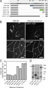
Figure 1.
Figure 1.
Maintenance of Golgi ribbon structure requires GCC185 residues 1,332–1,438. (A) A schematic diagram of GCC185 highlighting the residues predicted to form coiled coils (shown in gray). Truncation constructs used in the depletion and rescue experiments are shown. In blue is a region needed for MPR vesicle tethering; in green is a region needed for maintenance of proper Golgi ribbon structure. Dashed lines represent deleted sequences. (B) Cells were treated with GCC185 siRNA for 72 h. After 48 h of siRNA treatment, cells were transfected with plasmids encoding the indicated siRNA-resistant myc-tagged rescue proteins. (left) Golgi structure assessed using mouse anti-GM130 and Alexa Fluor 488 goat anti–mouse antibodies. (right) GCC185 rescue construct expression detected using chicken anti-myc and Cy3 goat anti–chicken antibodies. Approximate cell outlines are indicated. Bars, 10 µm. (C) Quantification of rescue. The formation of a Golgi ribbon was scored visually by other laboratory members from blinded datasets; the data represent the mean of two experiments in which a total of >45 cells were counted for each construct. Error bars represent SEM from independent experiments. The scoring error between laboratory members was less than a few percent. (D) An immunoblot of the indicated GCC185 constructs (asterisks) after 48 h of expression in HeLa cells. Proteins were detected with anti-myc tag antibody. Three background bands are seen in all lanes. Molecular mass marker mobility is shown at the right in kilodaltons.
Figure 2.
Figure 2.
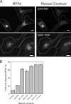
GCC185 residues 890–1,030 are needed for MPR vesicle tethering. (A) Cells were treated with GCC185 siRNA for 72 h. After 24 h of siRNA treatment, cells were transfected with plasmids encoding the indicated myc-tagged proteins. (left) MPR localization determined using 2G11 mouse anti-MPR and Alexa Fluor 488 goat anti–mouse antibodies. (right) GCC185 rescue construct expression detected using chicken anti-myc and Cy3 goat anti–chicken antibodies. Approximate cell outlines are indicated. Bars, 10 µm. (B) Quantification of rescue experiments. MPR dispersal was scored visually using blinded datasets; the data represent the mean of at least two experiments in which a total of >45 cells were counted for each condition. Error bars represent SEM from independent experiments.
Figure 3.
Figure 3.
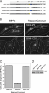
Coiled-coil residues 939–1,031 are required for MPR vesicle tethering. (A) A schematic diagram of GCC185 and the truncation constructs used in the depletion and rescue experiments. In blue is a coiled-coil region needed for MPR vesicle consumption; in yellow is an adjacent putative unstructured region. (B) GCC185 depletion and rescue with the indicated constructs as described in Fig. 2. Approximate cell outlines are indicated. Bars, 10 µm. (C) Quantification of rescue. MPR dispersal was scored visually using blinded datasets; the data represent the mean of at least two experiments in which a total of >100 cells were counted for each condition. Error bars represent SEM from independent experiments. (D) An immunoblot of the indicated GCC185 constructs after 48 h of expression in HeLa cells. Proteins were detected with anti-myc tag antibody. Molecular mass marker mobility is shown in kilodaltons.
Figure 4.
Figure 4.
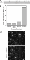
A Rab9 binding site in coiled-coil 3 is dispensable for vesicle tethering. (A) A schematic diagram of GCC185. Depicted in orange is a coiled-coil region containing a Rab9 binding site (Hayes et al., 2009). (B) GCC185-depleted cells were rescued and analyzed as described in Fig. 2. The quantification of rescue is shown. MPR dispersal was scored visually from blinded datasets; the data represent the mean of at least two experiments in which a total of >45 cells were counted for each condition. Error bars represent SEM from independent experiments. (C) Representative images. MPR localization (top) and GCC185 rescue with Δ862–882 (bottom) are shown. Approximate cell outlines are indicated. Bar, 10 µm.
Figure 5.
Figure 5.
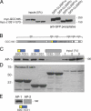
The clathrin adaptor AP-1 binds the GCC185 coiled-coil domain required for vesicle tethering. (A) CLASP coimmunoprecipitation with GCC185. HeLa cells expressing GFP–CLASP1-α or GFP–CLASP2-γ with either full-length myc-tagged GCC185 or myc-tagged GCC185 lacking residues 1,331–1,573 were lysed and incubated with NHS-Sepharose–coupled GFP-binding protein. Bound proteins were analyzed by SDS-PAGE and immunoblotting with anti-myc tag antibody. Left lanes, inputs (5%); right lanes, eluted samples. (B) A schematic diagram of GCC185 as shown in Fig. 3. (C) Binding of AP-1 to the indicated GCC185 constructs. Reactions contained 10 µM GST-GCC185 constructs and ∼15 nM AP-1 purified from bovine brain membranes. Proteins were collected on glutathione-Sepharose, eluted with glutathione, and resolved by SDS-PAGE. Bound AP-1 was visualized by immunoblotting using 100/3 mouse anti–AP-1 γ-adaptin antibody (Sigma-Aldrich). (D) GCC185 constructs from reactions in B were visualized using Ponceau S staining. (E) 4 µM GST-tagged GCC185 890–1,031 was incubated with 390 nM of purified bovine AP-1 or 520 nM AP-2. Bound material was collected on glutathione-Sepharose beads, washed with 30 vol of binding buffer, and eluted with glutathione. Bound AP-2 was detected using 100/2 anti–α-adaptin antibody (Sigma-Aldrich). (C and E) In blue are residues 939–1,031; in yellow are residues 890–938, which represent a second region that is predicted to be unstructured. Molecular mass marker mobility is shown in kilodaltons.
Figure 6.
Figure 6.
AP-1 decorates transport vesicles that accumulate in cells depleted of GCC185. (A and B) Deconvolved micrographs of cells either mock transfected (A) or transfected with GCC185 siRNA (B) labeled for MPR and AP-1. MPR localization was visualized using 2G11 mouse anti-MPR and Alexa Fluor 647 goat anti-IgG2a mouse antibodies. AP-1 was visualized using 100/3 mouse anti–AP-1 and Alexa Fluor 555 goat anti-IgG2b mouse antibodies. Approximate cell outlines are indicated. Bars, 10 µm. (C and D) Enlargement of the boxed regions in control (A) or GCC185-depleted (B) cells. Bars, 2.5 µm. (E) Quantification of the percentage of peripheral MPR-containing vesicles that also contain AP-1. For GCC185-depleted cells, 490 MPR+ vesicles were counted from five cells from two independent experiments. For control cells, 76 MPR+ peripheral vesicles were counted from four cells from three independent experiments. Error bars represent SEM.
Figure 7.
Figure 7.
AP-1 and GCC185 occupy distinct TGN microdomains. Endogenous AP-1 or Golgin 245 and GCC185 were localized by deconvolution immunofluorescence microscopy. Bar, 5 µm.

References

    1. Aivazian D., Serrano R.L., Pfeffer S.R. 2006. TIP47 is a key effector for Rab9 localization. J. Cell Biol. 173:917–926 10.1083/jcb.200510010 - DOI - PMC - PubMed
    1. Braulke T., Bonifacino J.S. 2009. Sorting of lysosomal proteins. Biochim. Biophys. Acta. 1793:605–614 10.1016/j.bbamcr.2008.10.016 - DOI - PubMed
    1. Burguete A.S., Fenn T.D., Brunger A.T., Pfeffer S.R. 2008. Rab and Arl GTPase family members cooperate in the localization of the golgin GCC185. Cell. 132:286–298 10.1016/j.cell.2007.11.048 - DOI - PMC - PubMed
    1. Cai H., Reinisch K., Ferro-Novick S. 2007. Coats, tethers, Rabs, and SNAREs work together to mediate the intracellular destination of a transport vesicle. Dev. Cell. 12:671–682 10.1016/j.devcel.2007.04.005 - DOI - PubMed
    1. Carroll K.S., Hanna J., Simon I., Krise J., Barbero P., Pfeffer S.R. 2001. Role of Rab9 GTPase in facilitating receptor recruitment by TIP47. Science. 292:1373–1376 10.1126/science.1056791 - DOI - PubMed

Publication types

MeSH terms